Skull and Spine
4.0(1)
Card Sorting
1/49
Earn XP
Description and Tags
Study Analytics
Name | Mastery | Learn | Test | Matching | Spaced |
|---|
No study sessions yet.
50 Terms
1
New cards
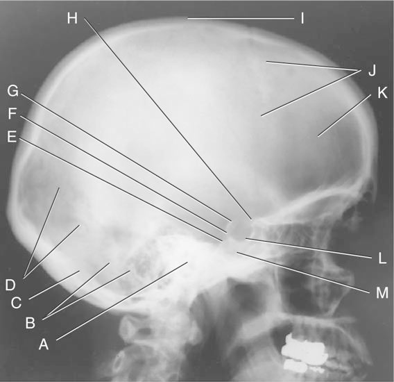
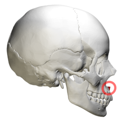
Lateral skull: the structure H is the:
a. Sella turcica
b. Dorsum Sellae
c. anterior clinoid process
d. occipital bone
a. Sella turcica
b. Dorsum Sellae
c. anterior clinoid process
d. occipital bone
C
2
New cards
For RPO and LPO positions for sacroiliac joints:
a. The central ray must be angled 25 degrees cephalad
b. Image the joint nearest the image receptor
c. The part must be angled at 10 to 15 degrees to coincide with the angle of the joints
d. Image the joint farthest from the image receptor
a. The central ray must be angled 25 degrees cephalad
b. Image the joint nearest the image receptor
c. The part must be angled at 10 to 15 degrees to coincide with the angle of the joints
d. Image the joint farthest from the image receptor
d
3
New cards
For the AP axial (Towne) projection for the skull, the central ray is directed:
a. 25 degrees to the IOML
b. 30 degrees to the IOML
c. 37 degrees to the OML
d.30 degrees to the OML
a. 25 degrees to the IOML
b. 30 degrees to the IOML
c. 37 degrees to the OML
d.30 degrees to the OML
d
4
New cards
For the AP projection of the coccyx: a. The central ray should be directed 25 degrees caudad, entering 4 inches superior to the symphysis pubis
b. The central ray should be directed 10 degrees caudad, entering 2 inches superior to the symphysis pubis
c. The central ray should be directed 25 degrees caudad, entering 2 inches superior to the symphysis pubis
d. The central ray should be directed 10 degrees cephalad, entering 2 inches superior to the symphysis pubis
b.
5
New cards
The intervertebral foramina of the thoracic spine are demonstrated with the
a. midsagittal plane 90 degrees to the IR.
b. coronal plane 90 degrees to the IR.
c. coronal plane 70 degrees to the IR.
d. midsagittal plane 70 degrees to the IR.
a. midsagittal plane 90 degrees to the IR.
b. coronal plane 90 degrees to the IR.
c. coronal plane 70 degrees to the IR.
d. midsagittal plane 70 degrees to the IR.
b
6
New cards
When the AP axial projection of the skull is performed:
1. The OML is parallel to the cassette
2. The central ray is directed through the foramen magnum 37 degrees to the OML
3. The central ray is directed through the foramen magnum 30 degrees to the IOML
4. The MSP is parallel to the plane of the film
a. All are true
b. None are true
c. 1, 2
d. 1, 3, 4
1. The OML is parallel to the cassette
2. The central ray is directed through the foramen magnum 37 degrees to the OML
3. The central ray is directed through the foramen magnum 30 degrees to the IOML
4. The MSP is parallel to the plane of the film
a. All are true
b. None are true
c. 1, 2
d. 1, 3, 4
b
7
New cards
When the SMV projection of the skull is performed, the IOML:
a. Is placed parallel to the plane of the cassette
b. Is not used in positioning for the SMV
c. Is placed parallel to the central ray
d. Is placed perpendicular to the plane of the cassette
a. Is placed parallel to the plane of the cassette
b. Is not used in positioning for the SMV
c. Is placed parallel to the central ray
d. Is placed perpendicular to the plane of the cassette
a
8
New cards
For the AP axial (Towne) projection for the skull, the central ray is directed:
a. 30 degrees to the IOML
b. 37 degrees to the OML
c. 25 degrees to the IOML
d. 30 degrees to the OML
a. 30 degrees to the IOML
b. 37 degrees to the OML
c. 25 degrees to the IOML
d. 30 degrees to the OML
d
9
New cards
For the PA axial (Haas) projection of the skull, the CR is directed:
a. 30 degrees caudad to the IOML
b. 25 degrees cephalic to the OML
c. 25 degrees caudad to the OML 30 d. degrees cephalic to the IOM
a. 30 degrees caudad to the IOML
b. 25 degrees cephalic to the OML
c. 25 degrees caudad to the OML 30 d. degrees cephalic to the IOM
b
10
New cards
With the patient in the PA position and the OML and CR perpendicular to the IR, the resulting radiograph will demonstrate the petrous pyramids
a. above the orbits.
b. in the lower third of the orbits.
c. completely within the orbits.
d. below the orbits.
a. above the orbits.
b. in the lower third of the orbits.
c. completely within the orbits.
d. below the orbits.
c
11
New cards
The spongy area of the skull caverna is called
a. pterion.
b. bregma.
c. diploe.
d. lambda.
a. pterion.
b. bregma.
c. diploe.
d. lambda.
c
12
New cards
The zygomatic process projects off of which of the following:
a. occipital bone
b. frontal bone
c. sphenoid bone
d. temporal bone
a. occipital bone
b. frontal bone
c. sphenoid bone
d. temporal bone
d
13
New cards
The cross table lateral skull projection on a trauma patient reveals air-fluid levels in the sphenoid sinus. This is an indication of which of the following a:
a. concussion
b. hemangioma
c. subdural hematoma
d. basal skull fracture
a. concussion
b. hemangioma
c. subdural hematoma
d. basal skull fracture
d
14
New cards
In the lateral projection for the Sella turcica, the CR is directed:
a. 3/4 inch anterior and 3/4 inch inferior to the EAM
b. 5 degrees cephalic and 1 inch above the EAM
c. 2 inches superior to the EAM
d. 3/4 inch anterior and 3/4 inch superior to the EAM
a. 3/4 inch anterior and 3/4 inch inferior to the EAM
b. 5 degrees cephalic and 1 inch above the EAM
c. 2 inches superior to the EAM
d. 3/4 inch anterior and 3/4 inch superior to the EAM
d
15
New cards
You have placed the patient prone with the face resting on the zygoma, nose and chin. The CR is perpendicular to the plane of the film and enters the skull at approximately 1 inch superior and posterior to the top of the ear attachment. This projection will best demonstrate the:
a. lacrimal bones
b. sphenoid strut superior
c. orbital fissure
d. optic canal
a. lacrimal bones
b. sphenoid strut superior
c. orbital fissure
d. optic canal
d
16
New cards
Lateral deviation of the nasal septum may be best demonstrated in the
a. AP axial (Grashey-Towne method) projection.
b. PA axial (Caldwell method) projection.
c. lateral projection.
d. parietoacanthal (Waters method) projection.
a. AP axial (Grashey-Towne method) projection.
b. PA axial (Caldwell method) projection.
c. lateral projection.
d. parietoacanthal (Waters method) projection.
d
17
New cards
When positioning the patient for the Rhese method, which of the following is correct:
a. the central ray (CR) is directed at an angle of 53 degrees caudal
b. the mid-sagittal plane forms an angle of 53 degrees from the plane of the film
c. the central ray (CR) is directed at an angle of 53 degrees cephalic
d. the coronal plane forms and angle of 53 degrees from the plane of the film
\
b
18
New cards
Which of the sinuses are developed at birth:
a. frontal and sphenoid
b. frontal only
c. maxillary and frontal
d. maxillary only
a. frontal and sphenoid
b. frontal only
c. maxillary and frontal
d. maxillary only
d
19
New cards
To visualize one side of the body, ramus, coronoid process and mandibular condyle free from superimposition from the other side, which of the following do you do:
a. use a 25 degree caudal tube angle when the head is in the lateral position
b. use a 25 degree cephalic tube angle when the head is in the lateral position
c. rotate the head so the nose, zygoma and chin tilt
d. the head towards the shoulder farthest away from the film
a. use a 25 degree caudal tube angle when the head is in the lateral position
b. use a 25 degree cephalic tube angle when the head is in the lateral position
c. rotate the head so the nose, zygoma and chin tilt
d. the head towards the shoulder farthest away from the film
b
20
New cards
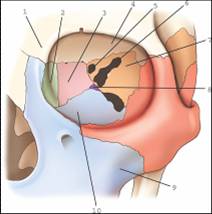
Waters - the structure illustrated as # 13 is the:
a. Frontal process of zygoma
b. Zygomatic arch
c. Zygomaticofrontal suture
d. Maxillary sinus
e. Transverse process and foramen transversarium of C1
f. Body of mandible
g. Infraorbital foramen
h. Frontal sinus Coronoid process of mandible
I. Nasal septum
J. Odontoid process of C2
k. Ethmoid sinus
L. Zygomatic process of frontal bone
m. Inferior orbital rim
\
b.
21
New cards
Townes - What suture is demonstrated as # 7 is?
a. coronal
b. lambdoid
c. sphenofrontal
d. sagittal
a. coronal
b. lambdoid
c. sphenofrontal
d. sagittal
b
22
New cards
What is the angle formed by the intervertebral foramina and the midsagittal plane in the thoracic spine?
a. 15 degrees.
b. 70 degrees.
c. 90 degrees.
d. 45 degrees.
a. 15 degrees.
b. 70 degrees.
c. 90 degrees.
d. 45 degrees.
c
23
New cards
When the AP oblique projection is performed for the cervical vertebrae, the central ray is directed:
a. 25 to 30 degrees cephalad
b. 5 to 10 degrees cephalad
c. 15 to 20 degrees cephalad
d. 15 to 20 degrees caudad
a. 25 to 30 degrees cephalad
b. 5 to 10 degrees cephalad
c. 15 to 20 degrees cephalad
d. 15 to 20 degrees caudad
c
24
New cards
The CR will parallel the intervertebral foramina in which of the following projections?
1. Lateral cervical spine
2. Lateral thoracic spine
3. Lateral lumbar spine
a. 1, 2, and 3
b. 2 and 3 only
c. 1 only
d. 1 and 2 only
1. Lateral cervical spine
2. Lateral thoracic spine
3. Lateral lumbar spine
a. 1, 2, and 3
b. 2 and 3 only
c. 1 only
d. 1 and 2 only
b
25
New cards
What is a defect in the pars articularis?
a. Hangman's fracture
b. Spondylolysis
c. Spina bifida
d. Jefferson's fracture
a. Hangman's fracture
b. Spondylolysis
c. Spina bifida
d. Jefferson's fracture
b
26
New cards
A kyphotic curve is formed by which of the following?
1. Sacral vertebrae
2. Thoracic vertebrae
3. Lumbar vertebrae
a.1 only
b. 3 only
c. 1 and 2 only
d.1 and 3 only
1. Sacral vertebrae
2. Thoracic vertebrae
3. Lumbar vertebrae
a.1 only
b. 3 only
c. 1 and 2 only
d.1 and 3 only
c
27
New cards
For the lateral projection of L5-S1: 1.The patient is in lateral position 2.The hips and knees are extended 3.The cassette is centered at the level of the transverse plane that passes midway between the iliac crests and the ASIS 4. A cylinder cone may be used to greatly reduce the production of scatter radiation 5.The central ray is directed to a point 1.5 inches anterior to the palpated spinous process of L5
a. 1, 2, 3, 5
b. 1, 3, 4, 5
c. 1, 2
d. 1, 2, 3, 4, 5
a. 1, 2, 3, 5
b. 1, 3, 4, 5
c. 1, 2
d. 1, 2, 3, 4, 5
b
28
New cards
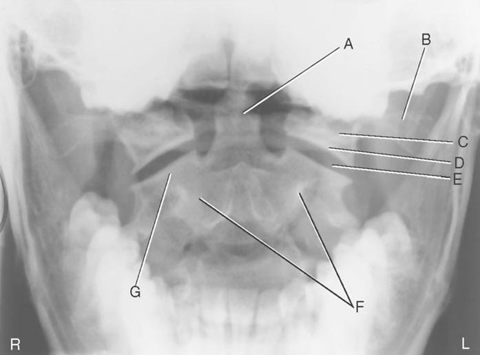
The structure designated as A is the:
a. Right superior articular
b. Left zygapophyseal joint surface of C2
c. Body of C2
d. Left transverse process of C1
e. Centrally located dens
f. Left lateral mass of C1
g. Inferior articular surface of C1
e
29
New cards
The bone labeled 2 is the:
a. ethmoid.
b. maxilla.
c. lacrimal.
d. palatine.
a. ethmoid.
b. maxilla.
c. lacrimal.
d. palatine.
c
30
New cards
The structure designated as # 5 is the:
a. Superior articular process (facet)
b. Inferior articular process (facet)
c. Pars intucularis
d. Pedicle
e. Transverse process of second lumbar vertebravertebra
f. lamina
\
a. Superior articular process (facet)
b. Inferior articular process (facet)
c. Pars intucularis
d. Pedicle
e. Transverse process of second lumbar vertebravertebra
f. lamina
\
b
31
New cards
Which of the following aspect of the vertebral column curves concave posteriorly:
1. Cervical
2. Thoracic
3. Lumbar
a. 2 and 3
b. 1 and 3
c. 1 and 2
d. 1, 2 and 3
1. Cervical
2. Thoracic
3. Lumbar
a. 2 and 3
b. 1 and 3
c. 1 and 2
d. 1, 2 and 3
b
32
New cards
Which projection is necessary if the top of T1 and the C7-T1 interspace is not clearly demonstrated on the lateral projection, dorsal decubitus position of the cervical spine?
a. Lateral projection of the thoracic spine
b. Lateral projection, swimmer's technique
c. AP projection, open-mouth position
d. AP axial C-spine
a. Lateral projection of the thoracic spine
b. Lateral projection, swimmer's technique
c. AP projection, open-mouth position
d. AP axial C-spine
b
33
New cards
To show the right sacroiliac joint, the patient is positioned
a. 25-30 degrees LPO.
b. 25-30 degrees RPO.
c. 30-40 degrees LPO.
d. 30-40 degrees RPO.
a. 25-30 degrees LPO.
b. 25-30 degrees RPO.
c. 30-40 degrees LPO.
d. 30-40 degrees RPO.
a
34
New cards
The SID for a lateral cervical spine must be a minimum of how many inches?
a. 60-72 inches
b. 40 inches
c. 40-60 inches
d. 48 inches
a. 60-72 inches
b. 40 inches
c. 40-60 inches
d. 48 inches
a
35
New cards
The inferior part of the bony septum of the nose is formed by the:
a. vomer
b. maxillary bones
c. palatine bones
c. inferior
d. nasal conchae
a. vomer
b. maxillary bones
c. palatine bones
c. inferior
d. nasal conchae
a
36
New cards
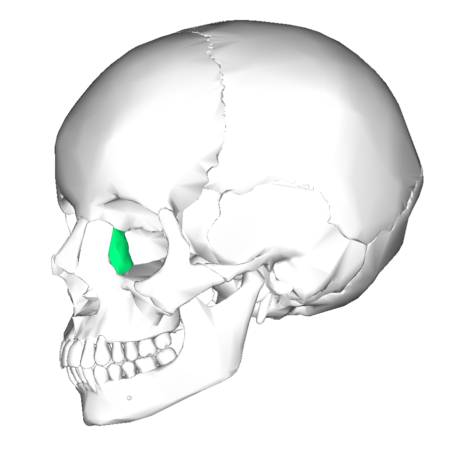
Which of the following bones is shown in green on the image provided.
a. lacrimal
b. palatine
c. nasal
d. vomer
\
a. lacrimal
b. palatine
c. nasal
d. vomer
\
a
37
New cards
Which facial bones are clearly demonstrated on the parietoacanthial projection (Waters method)? 1.) orbits 2.) maxillae 3.) zygomatic arches
a. 1, 2, and 3
b. 2 and 3
c. 1 and 3
d. 1 and 2
a. 1, 2, and 3
b. 2 and 3
c. 1 and 3
d. 1 and 2
a
38
New cards
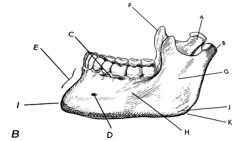
The portion of the mandible identified as F in the figure shown is the:
a. body
b. Coronoid Process
c. Condyloid process
d. ramus
\
a. body
b. Coronoid Process
c. Condyloid process
d. ramus
\
b
39
New cards
Which of the following results in fracture(s) of one or more of the bones of the face?
a. blow-out fractures
b. zygomatic bone fracture
c. condylar fractures
d. LeFort fractures
a. blow-out fractures
b. zygomatic bone fracture
c. condylar fractures
d. LeFort fractures
d
40
New cards
Which aspect of the mandible is best demonstrated on the Waters method?
a. Mentum
b. Body
c. Ramus
d. Condyloid process
a. Mentum
b. Body
c. Ramus
d. Condyloid process
d
41
New cards
The area circled in red and colored in red in the image shown is the
a. inferior orbital fissure
b. infraorbital foramen
c. anterior nasal spine
d. inferior nasal concha
\
a. inferior orbital fissure
b. infraorbital foramen
c. anterior nasal spine
d. inferior nasal concha
\
c
42
New cards
On the image shown, blue represents the ethmoid bon
True
False
True
False
True
43
New cards
Which of the following represents the mandibular condyles:
G
H
F
K
E
L
G
H
F
K
E
L
K
44
New cards
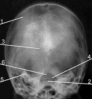
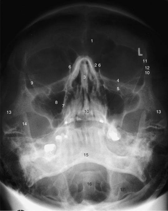
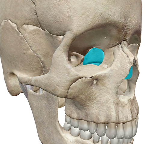
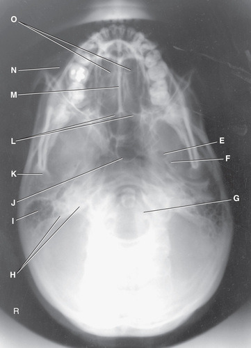
On this image of the skull, the dorsum sella is indicated by number:
a.
b. 4
c. 3
d. 6
e. 5
f. 2
g. 1
a.
b. 4
c. 3
d. 6
e. 5
f. 2
g. 1
f. 2
45
New cards
On this image shown, the Foramen spinosum is indicated by number:
a. 15
b. 18
c. 14
d. 17
e. 6
f. 16
\
a. 15
b. 18
c. 14
d. 17
e. 6
f. 16
\
f. 16
46
New cards
which cranial bone contains the cribriform plate? a.
Temporal
b. Occipital
c. Sphenoid
d. Ethmoid
Temporal
b. Occipital
c. Sphenoid
d. Ethmoid
d
47
New cards
The petromastoid portion is part of which bone?
a. occipital
b. temporal
c. ethmoid
d. sphenoid
a. occipital
b. temporal
c. ethmoid
d. sphenoid
b
48
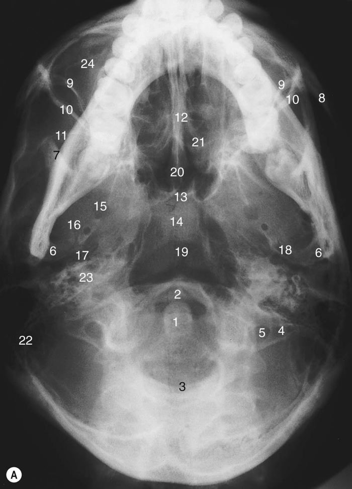
New cards
The area where the parietal bone, squamosal suture, and greater wing of the sphenoid bone articulate is termed:
a. pterion
b. asterion
c. bregma
d. lambda
a. pterion
b. asterion
c. bregma
d. lambda
a
49
New cards
Fontanelles, as known as "soft spot" in babies are soft membranous gaps (sutures) between the cranial bones and number a total of ____ in an infant.
a. eight
b. four
c. two
d. six
a. eight
b. four
c. two
d. six
d
50
New cards
What is the only paranasal sinus not contained within a cranial bone?
a. maxillary
b. Sphenoid
c. Frontal
d. Ethmoid
a. maxillary
b. Sphenoid
c. Frontal
d. Ethmoid
a