Lecture 30: Emotion, Risk, and Reward
1/16
There's no tags or description
Looks like no tags are added yet.
Name | Mastery | Learn | Test | Matching | Spaced | Call with Kai |
|---|
No analytics yet
Send a link to your students to track their progress
17 Terms
skin conductance response
Skin conductance response is a product of electrodermal activity
Activation of eccrine sweat glands – controlled solely by the sympathetic nervous system – increases electrical conductance of the skin surface
Characteristic onset latency and peak latency
Measures emotional arousal
Identifies stimulus-evoked arousal responses that arise subconsciously
James-Lange feedback theory
feedback circuit whereby sensory stimuli lead to autonomic physiological
responses, which then produce a conscious feeling
Cannon-Bard theory of emotion
parallel processing circuit whereby
sensory stimuli are directed to the
cortex to produce a conscious feeling, and to the hypothalamus to produce an autonomic physiological response
Functional lateralization
Callosotomy performed to alleviate epileptic seizures
Image projected onto right
visual field: Patient can identify the
object using both verbal and
non-verbal cues
Image projected onto left
visual field: Patient is unable to identify
the object verbally, but able to identify the object non-verbally
If the projected image has emotional significance, the patient experiences altered emotions even though they
cannot recognize the image
Asymmetric smiles
Facial expressions are often more quickly and fully expressed by the left facial musculature than the right
Since the left lower face is controlled by the right hemisphere, most individuals are emotionally “left-faced” in the same sense that most individuals are right-handed
Duchenne smile
Emotion-driven contraction of the
orbicularis oculi, together with the zygomaticus major
It is initiated by motor areas in the anterior cingulate cortex
Cingulate motor cortex mediates non-conscious emotion-driven facial movements
Emotion can produce motor activity
independent of somatomotor cortex
Fear acquisition
Dissociation between fear learning and declarative memory
Simple forms of fear conditioning can be dissociated from declarative memory
Fearful stimuli can be conditioned subliminally, without conscious awareness in healthy individuals
Subconscious fear
amygdala becomes active in response to fearful stimuli even if the subject is unaware of the fearful stimulus
During a binocular rivalry task in which subjects are presented with houses,
neutral faces, and fearful faces, the amygdala exhibits greater activity for fearful than for neutral faces even when the subject reports seeing only the house
Posttraumatic Stress Disorder
Deficits of fear extinction
When a stimulus that previously predicted a threat becomes “safe,” PTSD patients continue to exhibit
greater skin conductance response to the now-safe stimulus
greater amygdala activation during the extinction test, as well as hypoactivation of the vmPFC
This pattern is indicative of persistent hyperreactivity to threats, and difficulty engaging executive control circuits to suppress acquired fears when they are no longer appropriate to express
Normal fear extinction depends on the vmPFC
damage to vmPFC
lack the somatic marker predicting risky actions, leading to non-optimal choices and poor decision-making
Supports James-Lange Theory
Interoception
conscious or subconscious cortical representation of the body’s physiological state
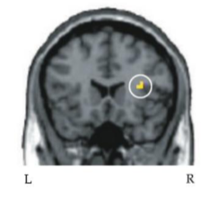
Activity of the insular cortex correlates with a subject’s ability to detect their own heartbeat
Damage to insular cortex impairs the ability to recognize certain emotions, especially disgust
Emotional effects on attentional blink
amygdala appears able to override the attentional blink, allowing emotional stimuli to reach awareness more readily
Individuals with bilateral damage to the amygdala do not exhibit this emotional facilitation
stress and consolidation
Those who experienced the cold stress had better recall of emotionally-charged image
Dopaminergic projections from the VTA
Dopaminergic neurons of the VTA have
projections to the septal nuclei and nucleus accumbens via the mesolimbic pathway, and to diffuse cortical areas via the mesocortical pathway
Several lines of evidence link these dopaminergic projections to reward processing
drugs of abuse
Nearly all drugs of abuse exert their addictive influence through alterations of the mesolimbic pathway
These drugs either cause VTA neurons to release more DA, block the reuptake of DA, or indirectly modulate the activity of neurons in the VTA or nucleus accumbens
gambling addiction
nucleus accumbens activates to winning plays but also in response to near-misses (right)
brain responds in a way that reinforces the problem behavior even when the reward is not delivered
The magnitude of activation is greater in individuals with gambling problems
Parkinson’s patients who are treated with dopamine agonists have a higher risk of developing gambling problems
These patients can also exhibit other uncontrolled compulsions and addictive behaviors
Dopamine
activity of dopamine neurons encode
errors in reward prediction
Events that are “better than expected” cause high activity in VTA neurons
Events that are “worse than expected” inhibit activity in VTA neurons
Events that occur “as expected” produce no change in activity, even if the event still provides hedonic reward