Cortex and cerebellum
1/31
There's no tags or description
Looks like no tags are added yet.
Name | Mastery | Learn | Test | Matching | Spaced | Call with Kai |
|---|
No analytics yet
Send a link to your students to track their progress
32 Terms
What do Corticothalamic connections do? what does eeg show during active thinking?
Regulate thalamic activity and can exert a flexible control of thalamocortical inputs
During active thinking, electroencephalography (EEG) reveals a strong appearance of gamma range oscillation from around 20–50 Hz.
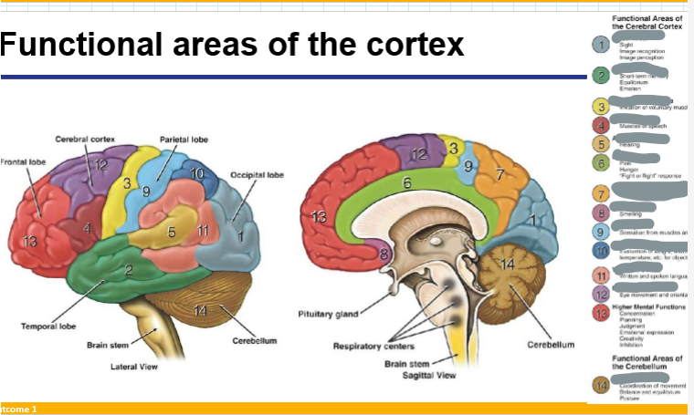
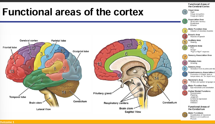
What are the main functions of the cerebral cortex?
Conscious awareness, thought, memory, sensation, movement, and higher-order association.
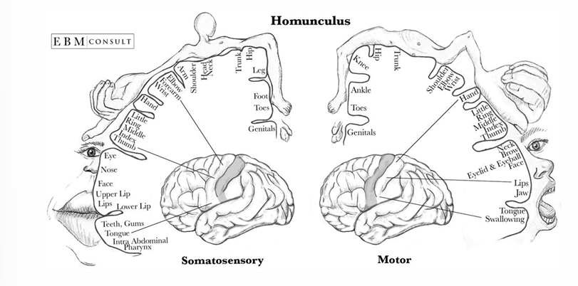
Draw out the motor and somatosensory cortical maps
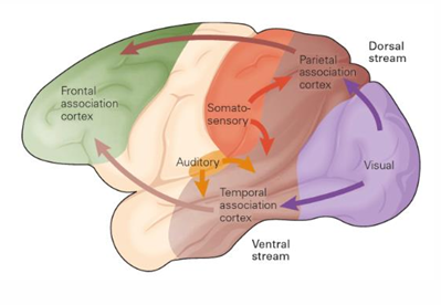
What are the dorsal and ventral streams of sensory processing?
Dorsal Stream ("Where"/"How"): Projects from the occipital lobe to the parietal lobe; processes spatial location and guides action.
Ventral Stream ("What"): Projects from the occipital lobe to the temporal lobe; involved in object identification and recognition
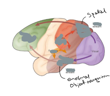
Processing stream
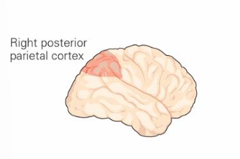
What is hemispatial neglect and what causes it?
A condition where a patient fails to attend to stimuli on one side of space, typically the left. It is caused by damage to the right parietal cortex
What is visual object agnosia and which brain region is typically affected?
It is the inability to recognize visually presented objects despite intact visual acuity. Damage to the ventral stream (occipitotemporal cortex)/medioventral temporal cortex is commonly implicated.
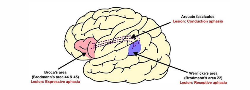
Compare Broca’s and Wernicke’s aphasia in terms of location and symptoms.
what is conduction lesion
Broca’s aphasia (left inferior frontal gyrus): Non-fluent speech, good comprehension.
Wernicke’s aphasia (left posterior superior temporal gyrus): Fluent but meaningless speech, poor comprehension
Conduction lesion- arcuate fasciculus which joins broca’s and wenicke’s
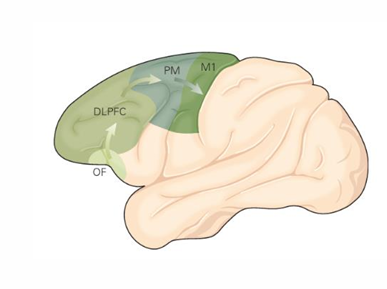
What 4 things is the frontal cortex involved in
Speech formation (Broca's area)
Gaze (frontal eye fields)
Working memory (dorsolateral prefrontal cortex)
Risk processing (ventromedial prefrontal cortex)
explain these hemorrhages
Epidural
Subdural
Subarachnoid
Epidural- beside dura so between skull and dura mater
Arterial bleed between skull and dura mater; appears as a lens-shaped (biconvex) mass on imaging.
Subdural- below the dural so between the dura and arachnoid
Venous bleed between dura and arachnoid mater; appears as a crescent-shaped mass.
Subarachnoid- below the arachnoid so below dura and arachnoid
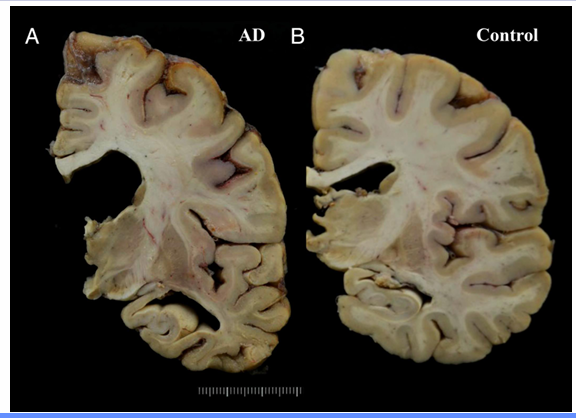
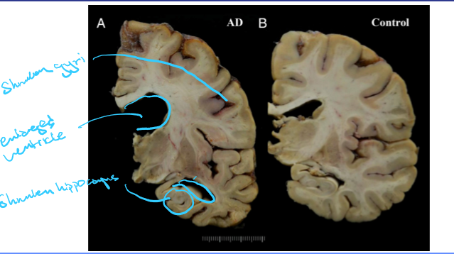
What are three features of the cortex of someone with Alzheimer’s
Shrunken gyri
Enlarged ventricle
Shrunken hippocampus
What is cortical plasticity
The brain’s ability to change and adapt its structure and function in response to experience, learning, or injury.
eg After losing a finger, nearby areas in the somatosensory cortex expand to represent adjacent fingers more strongly., phantom limb
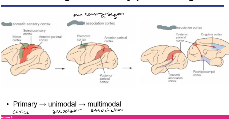
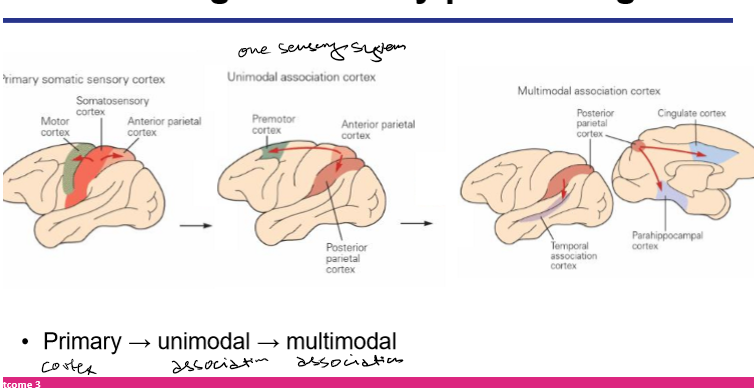
How does sensory info from primary sensory cortices get to frontal cortex
Sensory information goes from their primary sensory cortices to unimodal sensory areas and then multimodal cortical areas (via dorsal and ventral streams) before reaching at the frontal cortex
What 5 things is the cerebellum involved in
• Maintenance of equilibrium
• Posture and muscle tone
• Coordination of movement
• Hearing
• Facial sensations
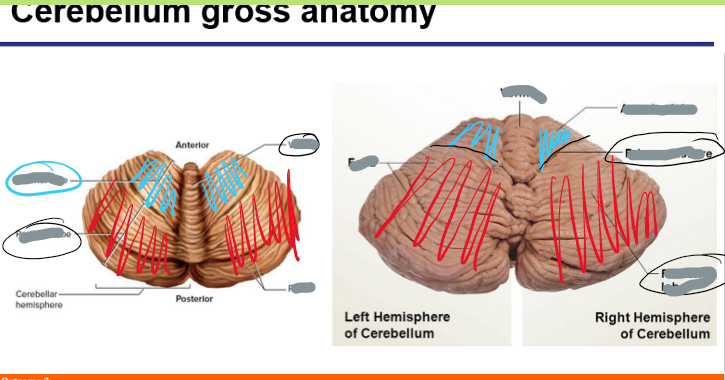
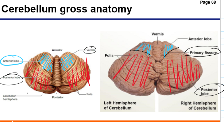
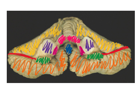
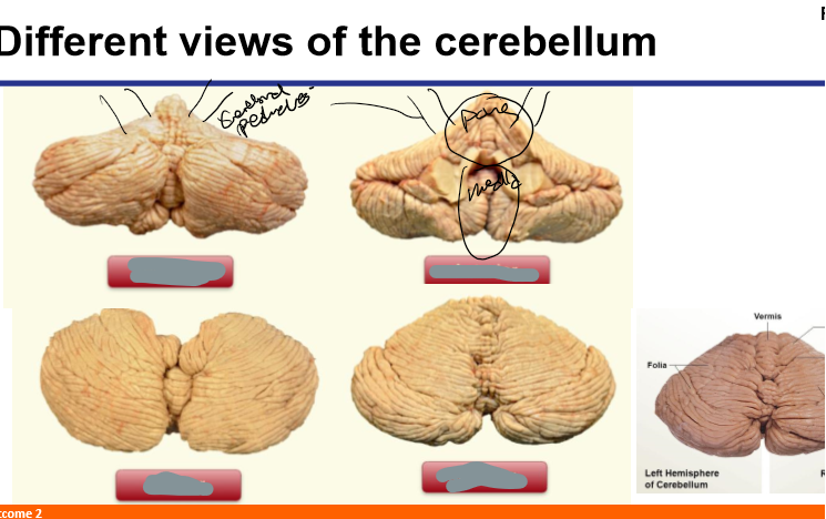
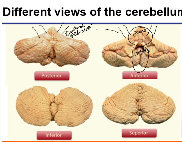
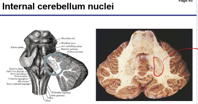
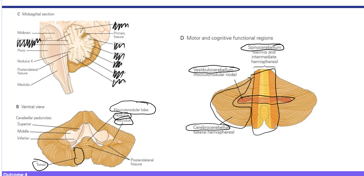
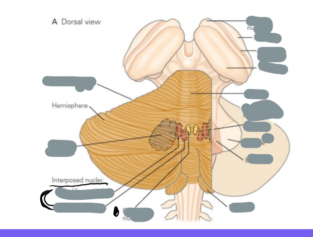
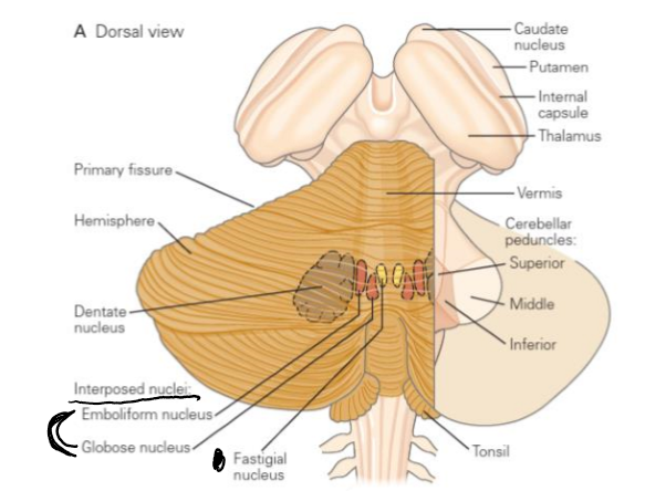
Label the 8 structures
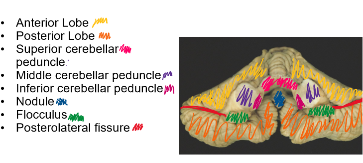
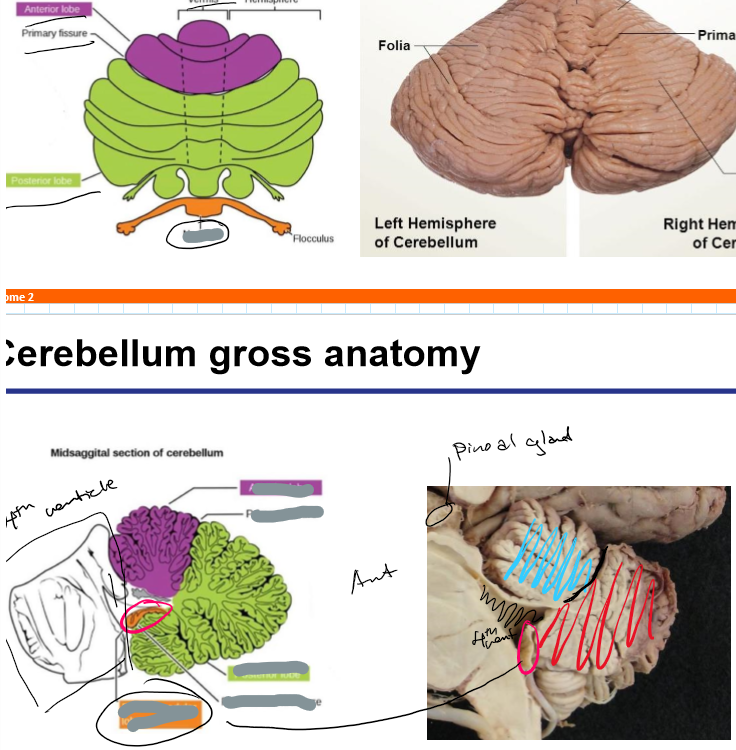
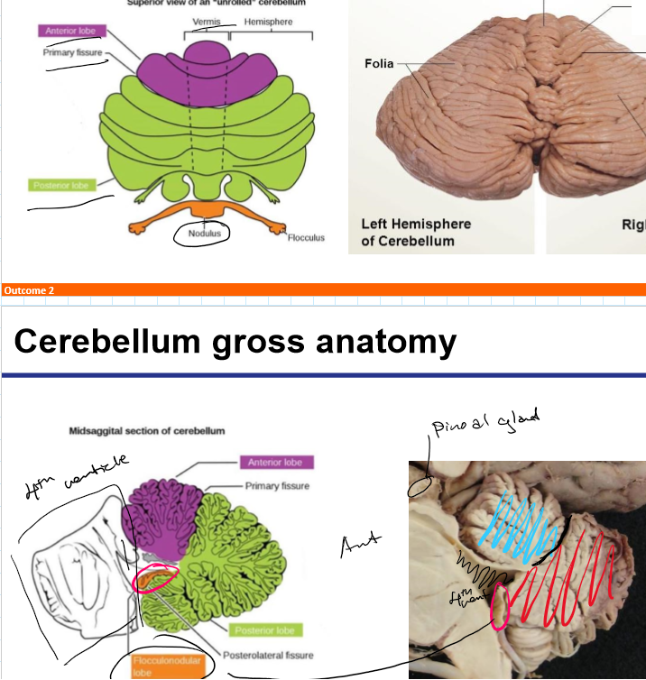
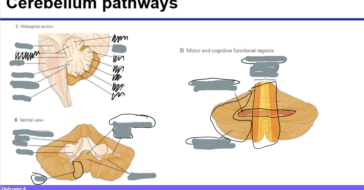
What are the three anatomical lobes of the cerebellum and their main functions?
Anterior lobe: Coordinates limb movements
Posterior lobe: Fine motor coordination and planning
Flocculonodular lobe: Balance and eye movements
What are the four deep cerebellar nuclei and their roles?
Dentate- planning, initiation and control of voluntary movements (ventral: non-motor, dorsal: motor)
Emboliform- part of spinocerebellum tract, that regulates the precision of limb movements
Globose- part of descending motor tracts involved in motor control
Fastigial- vestibular and saccadic eye movements
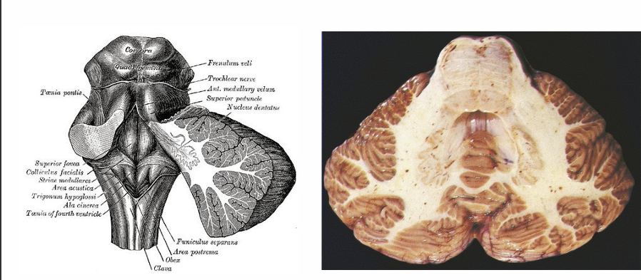
Where is the dentate nucleus located
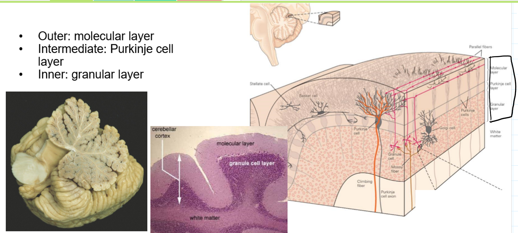
What are the 3 cell layers of the cerebellum
MPG
Outer: molecular layer
Intermediate: Purkinje cell layer
Inner: granular layer
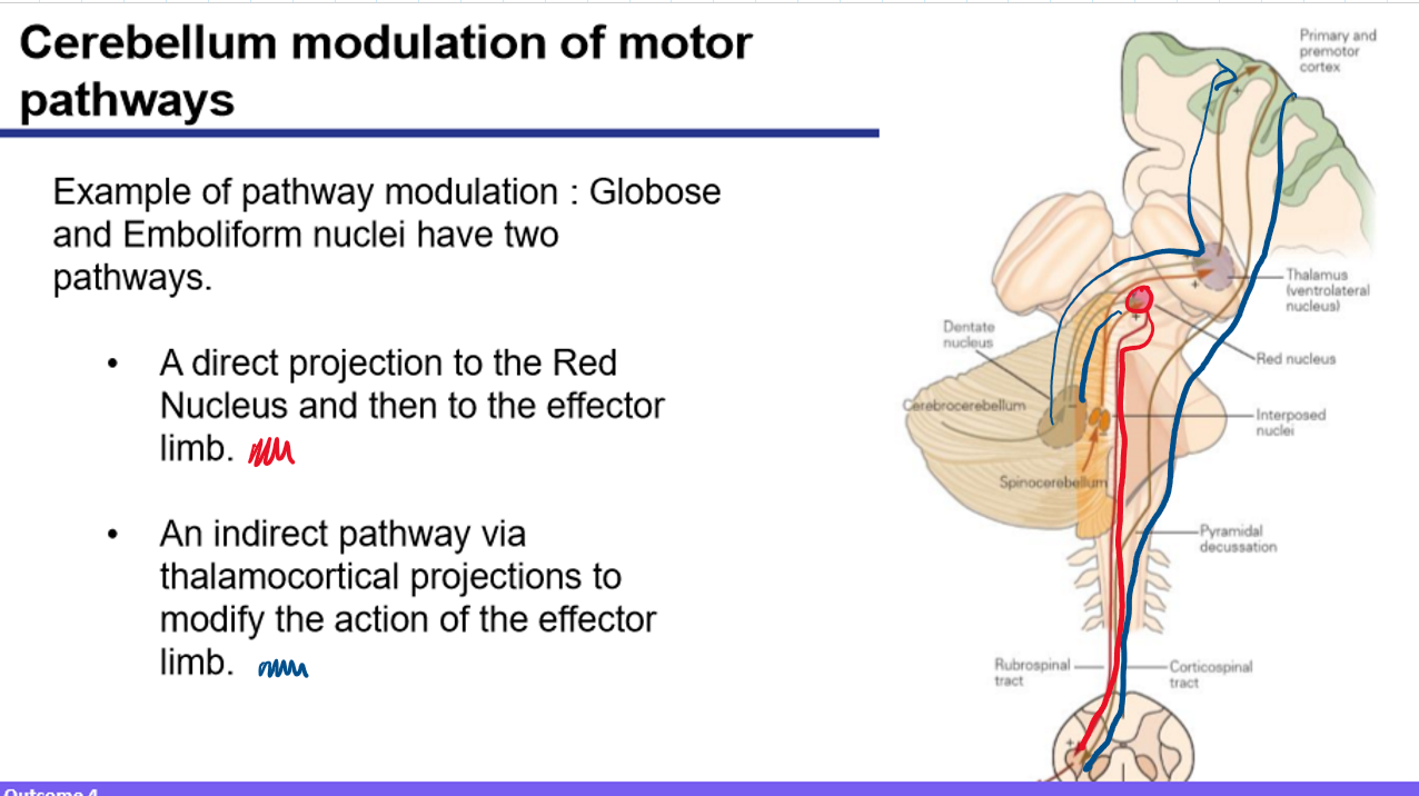
How do the Globose and Emboliform nuclei modulate limb movement?
Direct and indirect pathway
They have two pathways:
Direct pathway → Projects to the Red Nucleus, which sends output via the rubrospinal tract to the effector limb.
Indirect pathway → Projects to the thalamus, which then influences the motor cortex to modify action.
What is the function of the Flocculonodular lobe in motor control?
It receives input from the vestibular apparatus and sends output to modulate balance and eye movements, especially via the vestibulospinal tract.
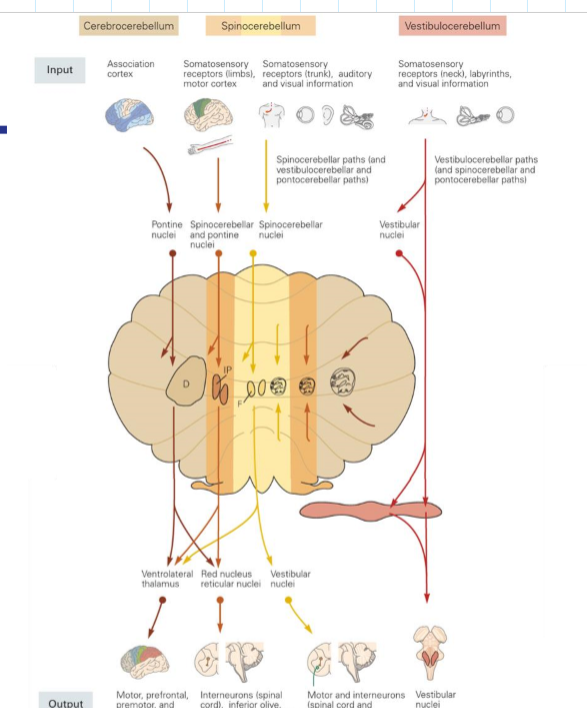
Describe the cerebrocerebellar pathway and its function
Cortex → pontine nuclei → middle cerebellar peduncle → lateral cerebellum → dentate nucleus → thalamus → motor cortex
Function: muscular coordination and control
Q14: What is the spinocerebellar pathway and its role?
Spinal proprioceptive input → cerebellum via inferior and superior peduncles → interposed nuclei → red nucleus and spinal tracts, globose, emboliform and fastigal
Function: Monitors and adjusts posture, balance, and limb position.
Explain the vestibulocerebellar pathway.
Input from vestibular system → flocculonodular lobe → fastigial nucleus → vestibular, red nucleus and reticular nuclei