B1.1 - Cell structures
1/28
There's no tags or description
Looks like no tags are added yet.
Name | Mastery | Learn | Test | Matching | Spaced | Call with Kai |
|---|
No analytics yet
Send a link to your students to track their progress
29 Terms
Which of Light microscopes and Electron are optical and which isn’t ?
Light Microscopes - Optical
Electron Microscopes - Non - Optical
Why do we use microscopes ?
Many Biological structures are too small to be seen by the naked eye so scientists use optical microscopes to see tissue, cells and organelle
Summary of how optical microscopes work:
Light is directed through a thin layer of biological material that is supported on a glass slide
Then light is focused through several lenses so an image is visible through the eyepiece
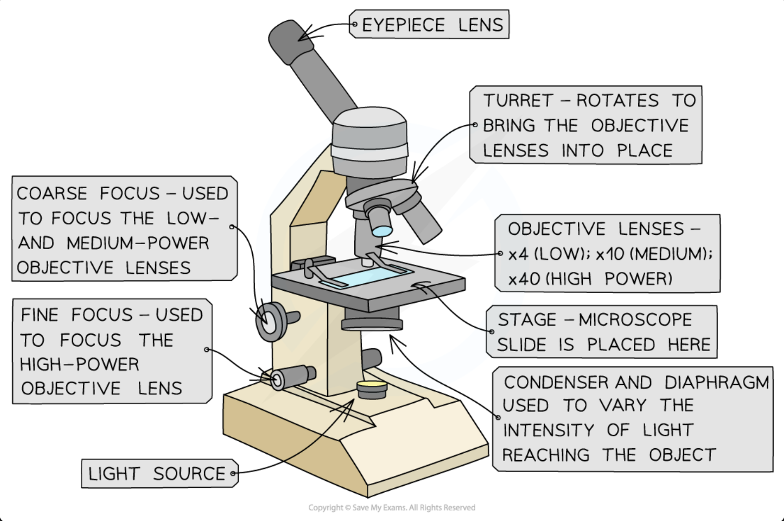
Name the components of a Light (optical) Microscope:
The eyepiece lens
The objective lenses
The stage
The light source (can be external also)
The coarse and fine focus
What other external apparatus is used when prepping a light microscope?
Forceps
Scissors
Scalpel
Coverslip
Slides
Pipette
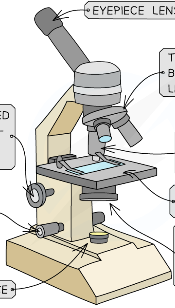
What are each of these labels referring to?
What is the setup - prep - calibration of a light microscope?
Specimens must be placed on a slide to be observed under the light microscope.
Is carefully done to avoid damaging specimen + structures within it
How to prep a liquid specimen for a light Microscope?
Add few drops of sample to slide using a pipette
Place coverslip on top and gently press down to remove air bubbles
Wear gloves to prevent cross contamination
How to prep a solid specimen for a light Microscope
Cut a small sample of tissue
Then peel a very thin layer of cells from tissue using scalpel/forceps and place on a slide
(Some samples may need chemicals to make tissue rigid (kill))
Place coverslip on top and gently press down to remove air
(Some tissues may need a stain to make structures visible, (if transparent/colourless))
ex: methylene blue to stain cheek cells and iodine to stain onion cells
How to prevent dehydration of a tissue?
Thin layers of material placed on slide can dry up rapidly
So adding a drop of water to specimen (beneath coverslip) can prevent cell damage by dehydration
How to view a specimen on Light Microscope?
Always start with low power objective lens:
Easier to find what you’re looking for
This helps prevent damage to lens or coverslip if stage is raised too high
Why is an image unclear/blurry on Light microscope?
Switch to low power objective lens and use coarse focus to get a clearer image
Consider if sample is thin enough for light to pass through to see structures
Could be cross-contamination with foreign cells
Limitations of Light Microscopy:
Size of cells/structures may appear inconsistent in different specimen slides: cell structures are 3d and different tissue samples will have been cut at different planes resulting in inconsistencies when viewed on a 2D slide
Optical Microscopes aren’t the most powerful so some structures can’t be seen
Treatment of specimens when preparing slides could alter cell structures
How to get smaller units and how to get bigger units in unit conversion?
to get smaller units you x
to get bigger units you /
1m = 1000mm
1mm = 1000µm
1µm = 1000nm
Calculating Magnification formula and triangle: (with sizes)
Magnification = Image size / Actual Size

Calculating Magnification of light microscope: (with lens)
Magnification of light microscope = Magnification of eyepiece lens x Magnification of objective lens (picture the triangle with this formula asw)
Do m to mm to micrometres to nanometers
1m = 1000mm
1mm = 1000µm
1µm = 1000nm
What are organisms made of?
made of cells which are the smallest unit with the basic properties of life
Name the 2 types of cellular organisms
eukaryotes or prokaryotes
Define a Eukaryote:
A eukaryote is a larger and more complex organism than Pro-K whose cells contain a nucleus and membrane‑bound organelles to help efficiently carry out functions within the cell (ex: animal and plant cells)
Define a Prokaryote:
Only Unicellular and is an organism whose cells do not have a nucleus or membrane‑bound organelles (ex: bacteria)
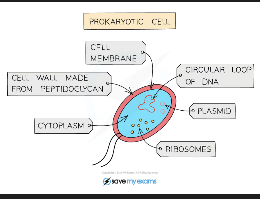
What’s the Prokaryote cell structure like?
KEY: NO NUCLEUS IN PROKARYOTE
Cytoplasm - contains gel like substance where most chemical reactions take place
Cell Membrane - encloses the cytoplasm, acts as a selective barrier, controlling what goes in + out of cell.
Receptors in membrane help prokaryotes sense their environment
Cell Wall - The outside of the cell is surrounded by a cell wall which Helps provide strength and maintain the shape of the cell
One long circular chromosome in Pro-K which floats free in cytoplasm (no nucleus), controls cell activities and replication
Plasmids - Small loops of DNA called plasmids may be present: - Can be exchanged between bacteria and give additional benefits ex: antibiotic resistance
Ribosomes - For protein Synthesis
Label this Animal Cell
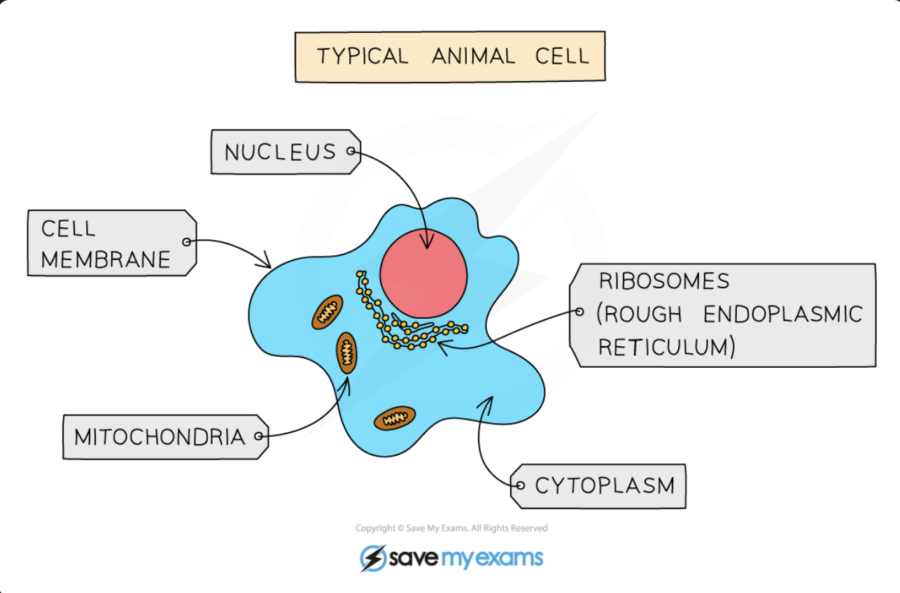
Name the Structures and Functions in Animal Cell: (also what do Animal cells have similar to Pro-K and what do animal cells have that Pro-K don’t have)
(b) Cytoplasm - Gel-like substances composed of water and dissolved solutes , supports internal cell structures, site of many chemical reactions including anaerobic respirations
(b) Cell membrane - Holds cell together separating inside of cell from outside , controls which substances can enter and leave cell
(a only) Nucleus - Contains Genetic Material (DNA) which controls the activities of the cell
(a only) Ribosomes - Site of Protein Synthesis, Found in cytoplasm
(a only) Mitochondria - Site of most of the reactions involved in aerobic respiration, cells with high metabolism (carry out many different cell reactions) have significantly higher numbers of mitochondria than cells with lower metabolism
Label this Plant cell:
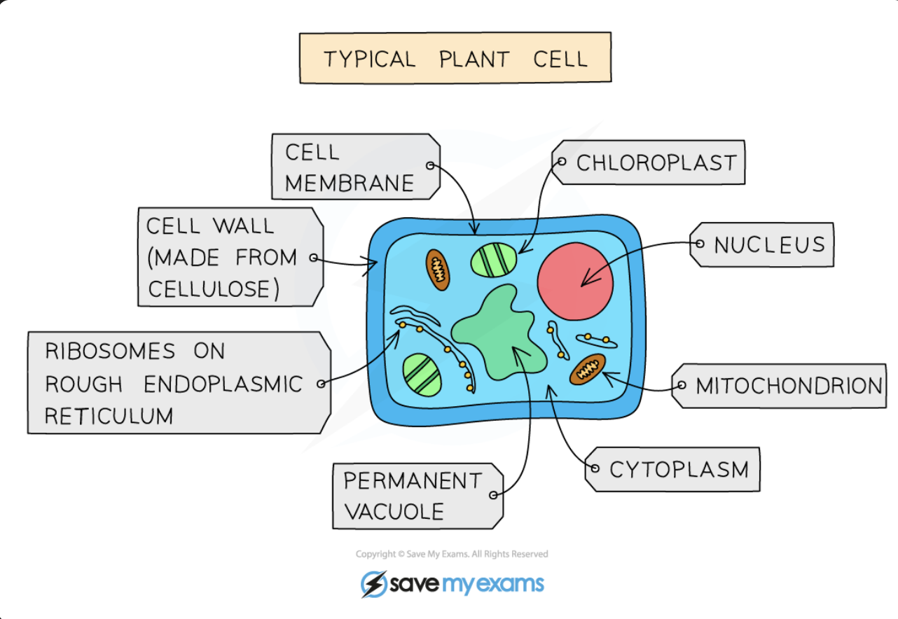
Name the Structures and Functions in Plant Cell: (also what do Animal cells have that Plant cells don’t have) (all other sub-cellular structures both animal and plant have)
(p only) Cell Wall - Rigid Cell wall made of cellulose to provide additional support for the cell, defining its shape
(p only) Chloroplasts - Site of Photosynthesis providing food for plants, they contain green chlorophyll pigment absorbs light energy needed for the reaction to occur
(p only) Vacuole - Contain cell sap, used for storage of certain material, also helps support cell shape
(b) Cytoplasm - Gel-like substances composed of water and dissolved solutes , supports internal cell structures, site of many chemical reactions including anaerobic respirations
(b) Cell membrane - Holds cell together separating inside of cell from outside , controls which substances can enter and leave cell
(b) Nucleus - Contains Genetic Material (DNA) which controls the activities of the cell, controls cell division
(b) Ribosomes - Site of Protein Synthesis, Found in cytoplasm
(b) Mitochondria - Site of most of the reactions involved in aerobic respiration, cells with high metabolism (carry out many different cell reactions) have significantly higher numbers of mitochondria than cells with lower metabolism
Why are Electron microscopes?
Light microscopes have limited resolution and magnification
Electron microscopes help address this limitation with beams of electrons, not light, to visualise specimens
Wavelength of electron beams are much smaller than visible light which give electron microscopes a much higher resolution and magnification. so we can see smaller structures in detail
Why aren’t Electron Microscopes found in schools?
Very expensive and not portable
Electron Microscopes vs Light Microscopes: (differences)
EM:
Large + once installed, can’t be moved
Vacuum needed
Complicated sample prep
Over x500 000 Mag
Resolution of 0.5nm
Specimens are dead
LM:
Small + easy to carry
No Vacuum needed
Easy sample prep
up to x2000 Mag
Resolution of 200nm
Specimens can be living/dead