Lab 1: Histology
1/67
Name | Mastery | Learn | Test | Matching | Spaced | Call with Kai |
|---|
No analytics yet
Send a link to your students to track their progress
68 Terms
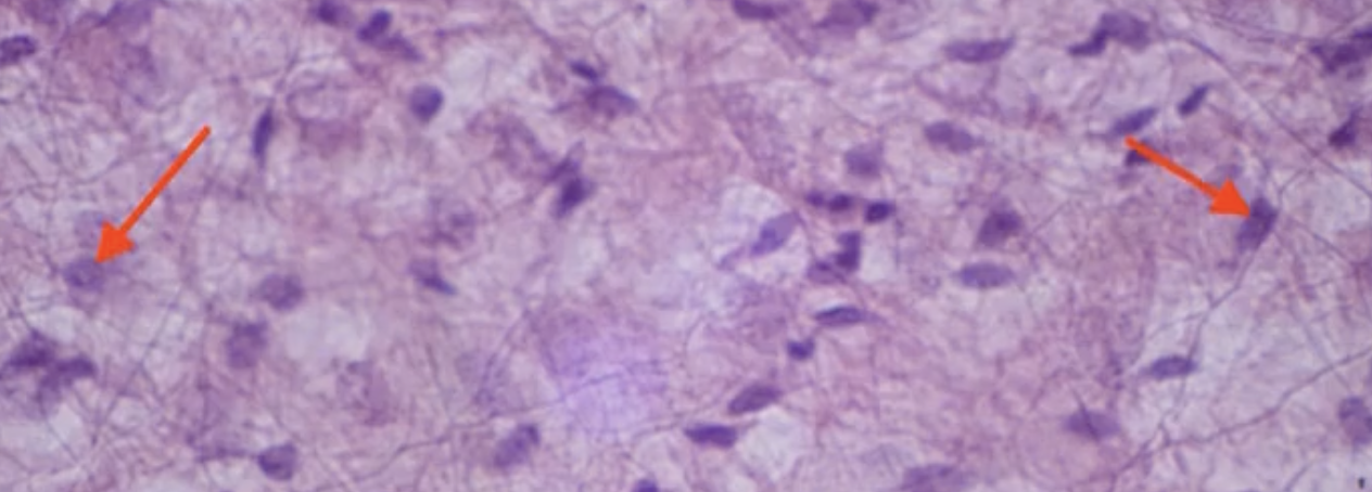
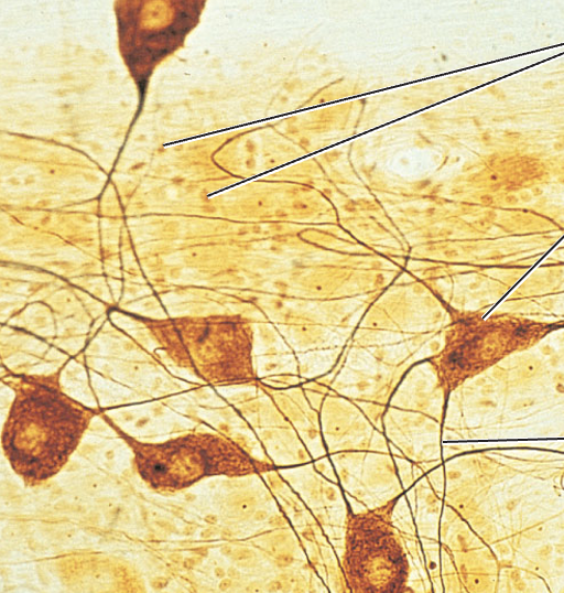
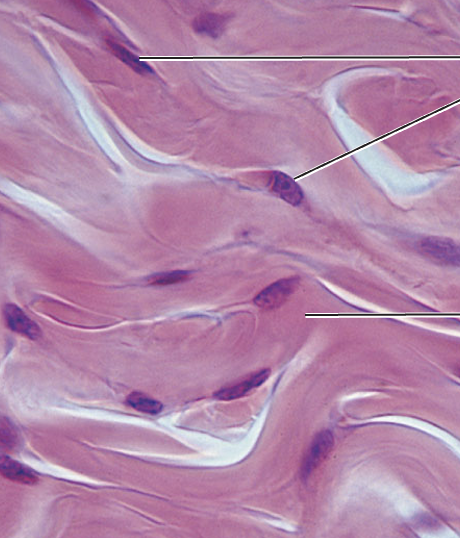
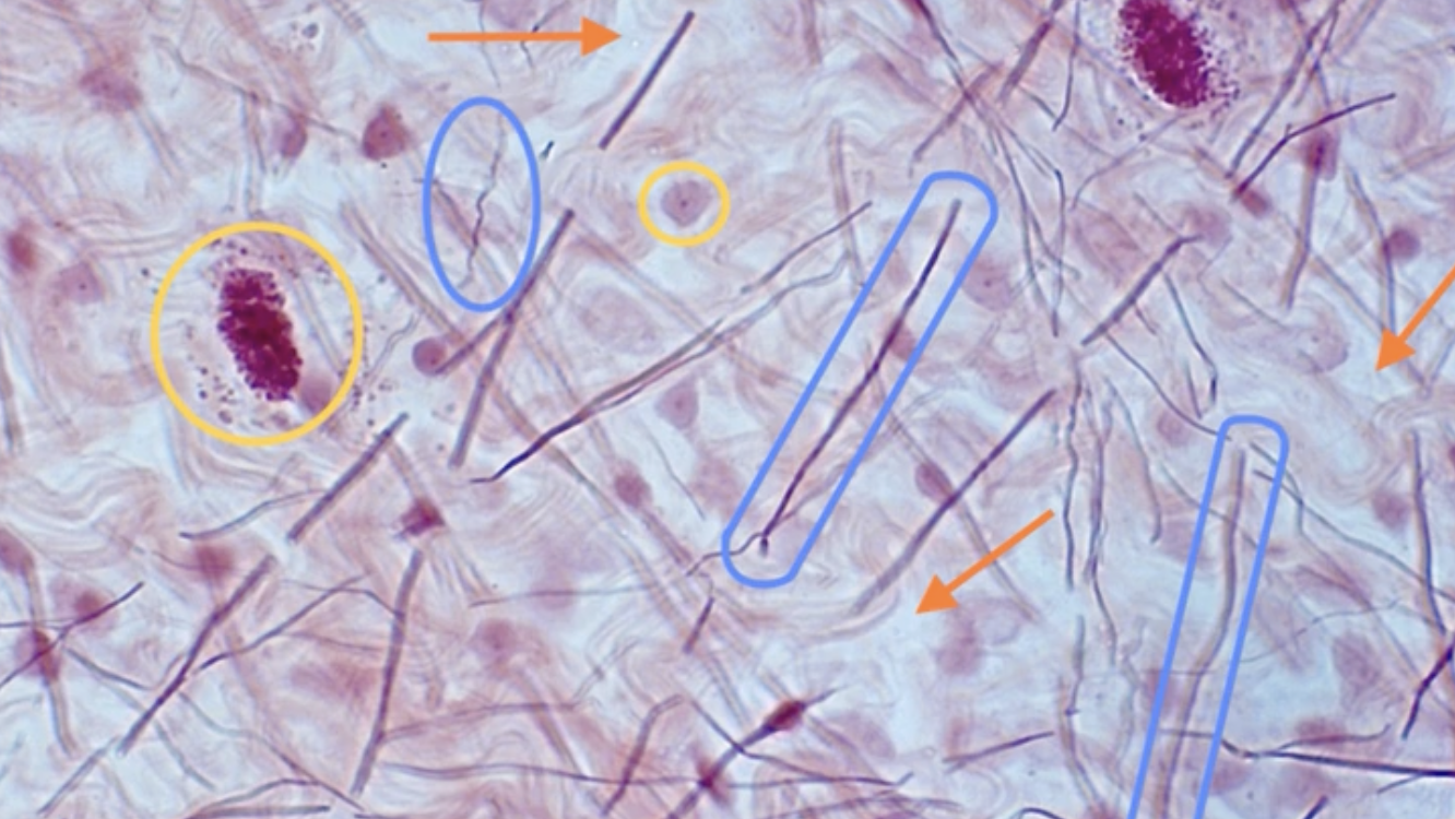
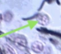
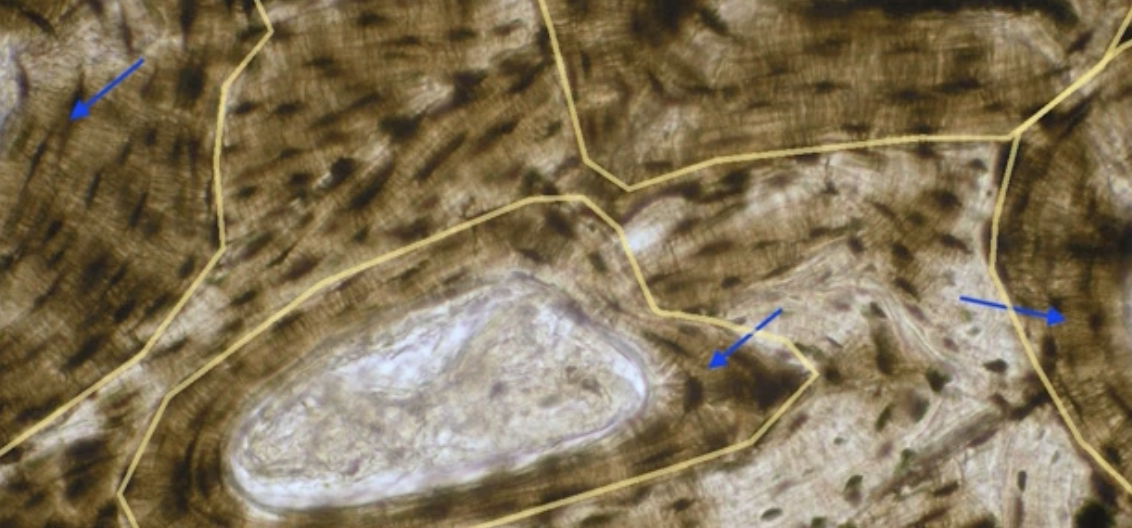
What are the arrows pointing to?
Fibroblasts
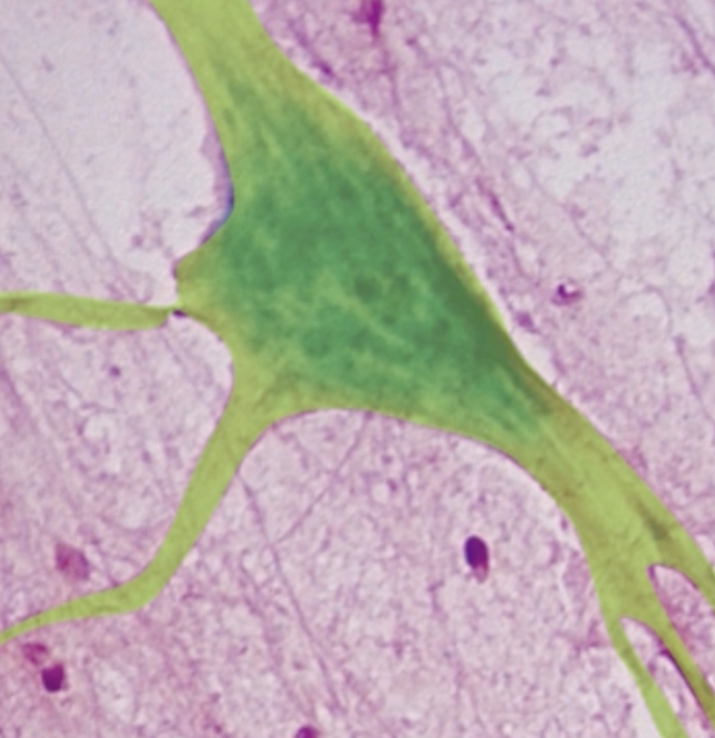
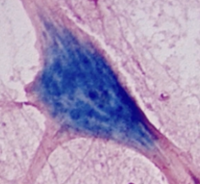
What type of cell is highlighted in green?
Neurons
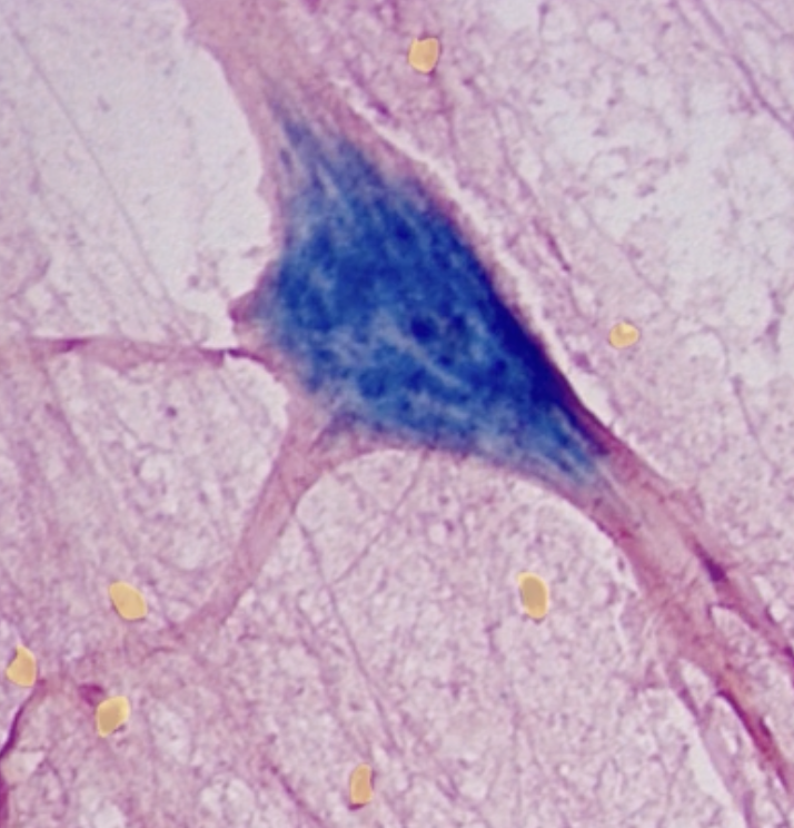
What type of cell is highlighted in yellow?
Glial
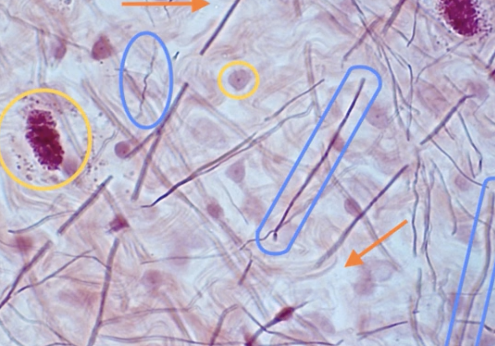
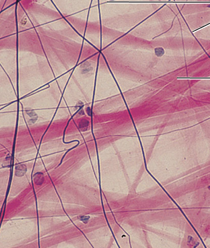
What is circled in blue?
Fibers
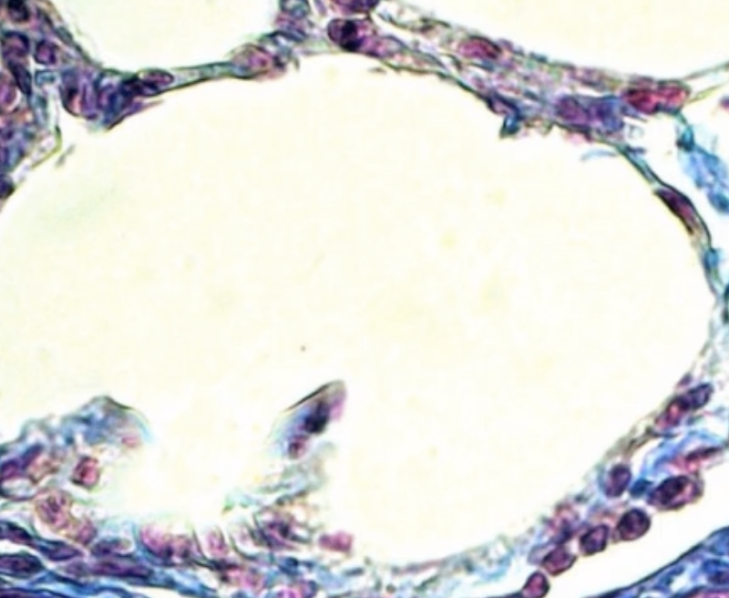
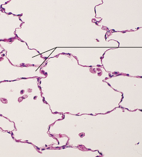
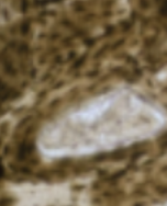
What type of tissue does this image depict?
Simple squamous epithelial
What tissue type does this image depict?
Simple squamous epithelial
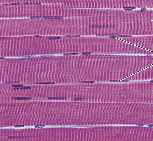
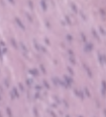
What tissue type does this image depict?
Skeletal muscle

What tissue type does this image depict?
Cardiac muscle
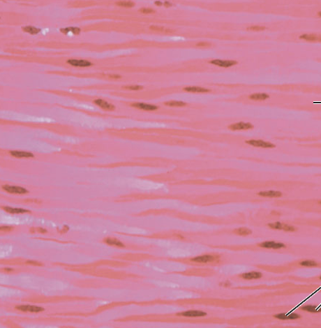
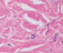
What tissue type does this image depict?
Smooth muscle
What tissue type does this image depict?
Nervous
What tissue type does this image depict?
Simple squamous epithelial
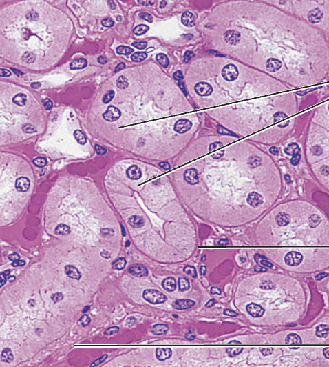
What tissue type does this image depict?
Simple cuboidal epithelium
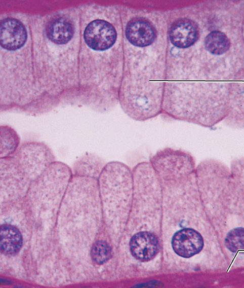
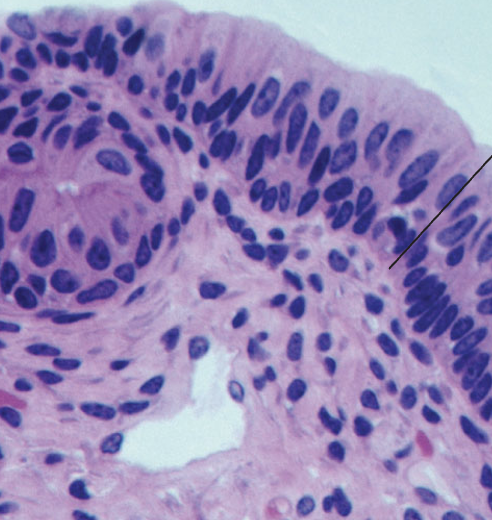
What tissue type does this image depict?
Simple columnar epithelial
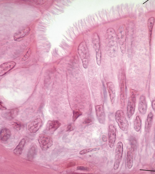
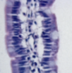
What tissue type does this image depict?
Pseudostratified columnar epithelial
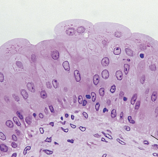
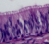
What tissue type does this image depict?
Stratified squamous epithelial
What tissue type does this image depict?
Stratified cuboidal epithelial
What tissue type does this image depict?
Stratified columnar epithelial
What tissue type does this image depict?
Transitional epithelial
What tissue type does this image depict?
Loose areolar connective
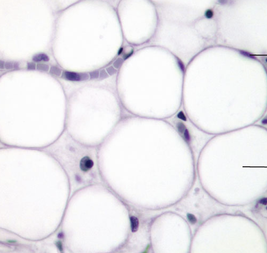
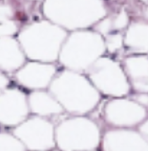
What tissue type does this image depict?
Loose adipose connective
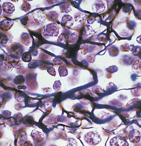
What tissue type does this image depict?
Loose reticular connective
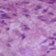
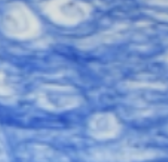
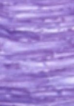
What tissue type does this image depict?
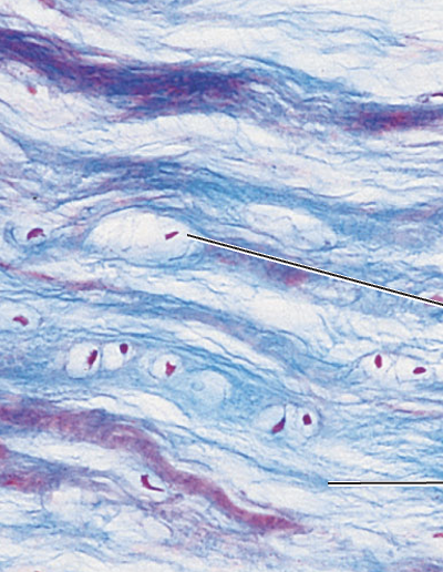
Dense irregular connective
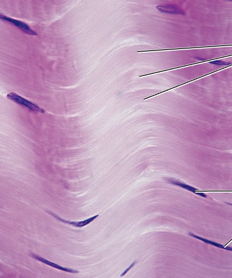
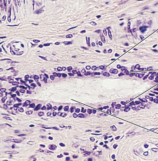
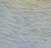
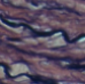
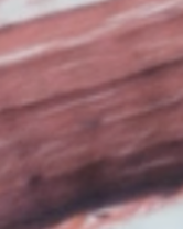
What tissue type does this image depict?
Dense regular connective
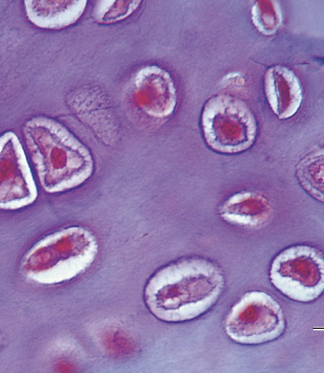
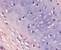
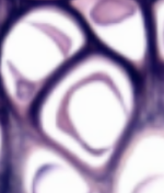
What tissue type does this image depict?
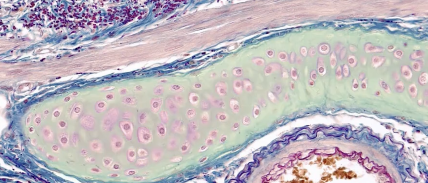
Hyaline cartilage connective
What tissue type does this image depict?
Elastic cartilage connective
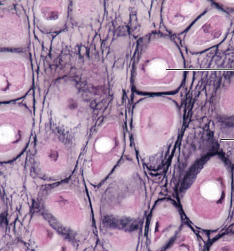
What tissue type does this image depict?
Fibrocartilage connective
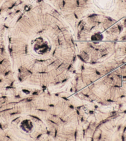
What tissue type does this image depict?
Bone connective
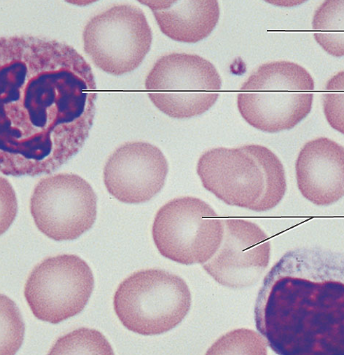
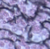
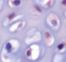
What tissue type does this image depict?
Blood connective
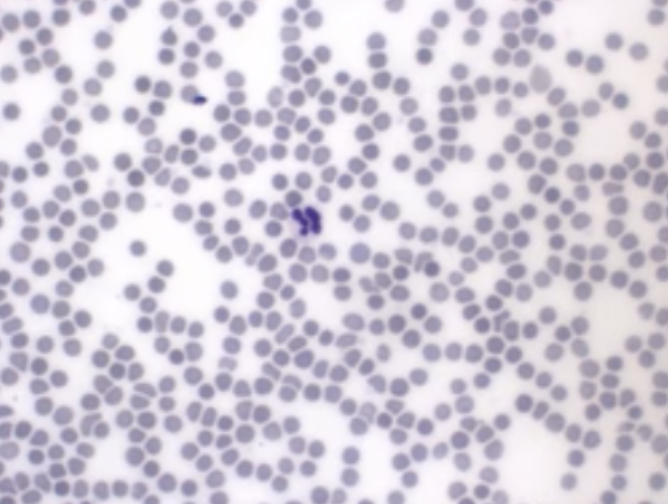
What tissue type does this image depict?
Blood connective

What tissue type does this image depict?
Simple cuboidal epithelial
What tissue type does this image depict?
Simple columnar epithelial
What tissue type does this image depict?
Pseudostratified columnar epithelial

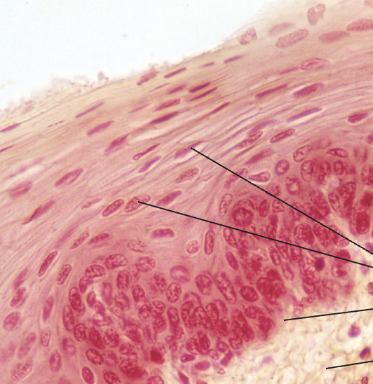
What tissue type does this image depict?
Keratinized stratified squamous epithelial

What tissue type does this image depict?
Stratified squamous epithelial

What tissue type does this image depict?
Stratified cuboidal epithelial

What tissue type does this image depict?
Stratified columnar epithelial

What tissue type does this image depict?
Transitional epithelial
Which type of tissue does this image depict?
Hyaline cartilage connective
What tissue type does this image depict?
Loose areolar connective
What tissue type does this image depict?
Loose adipose connective
What tissue type does this image depict?
Loose reticular connective
Which type of tissue does this image depict?
Dense regular connective
Which type of tissue does this image depict?
Dense irregular connective
Which type of tissue does this image depict?
dense elastic connective
Which type of tissue does this image depict?
Hyaline cartilage connective
Which type of tissue does this image depict?
Elastic cartilage connective
Which type of tissue does this image depict?
Fibrocartilage connective
Which type of tissue does this image depict?
Bone connective
Which type of tissue does this image depict?
Skeletal muscle
Which type of tissue does this image depict?
Cardiac muscle
Which type of tissue does this image depict?
Smooth muscle
Which type of tissue does this image depict?
Nervous
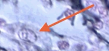
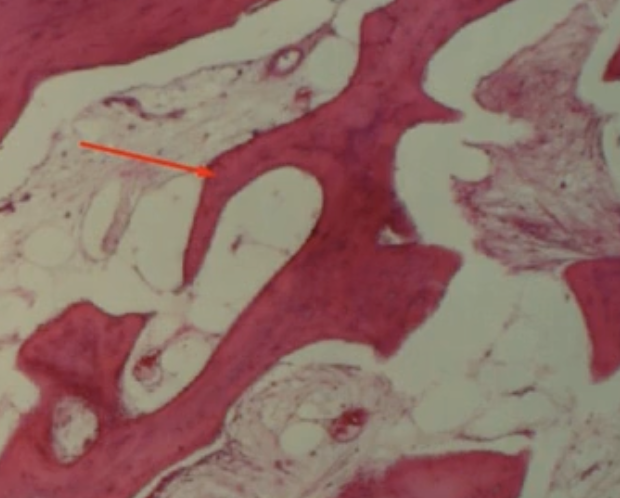
What is the orange arrow pointing to?
Ground substance
Which cells have the suffix blast?
Arrow

Which cells have the suffix cyte?
Squares
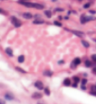
What type of cell is the arrow pointing to?
Mast cells
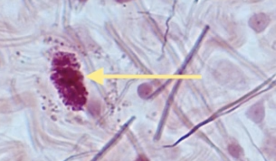
What cell is the arrow pointing to?
White blood cells
What cell is the arrow pointing to?
Macrophages
What does the highlighted green part of cartilage represent?
Ground substance

What cells are circled in orange in cartilage?
Chondroblasts
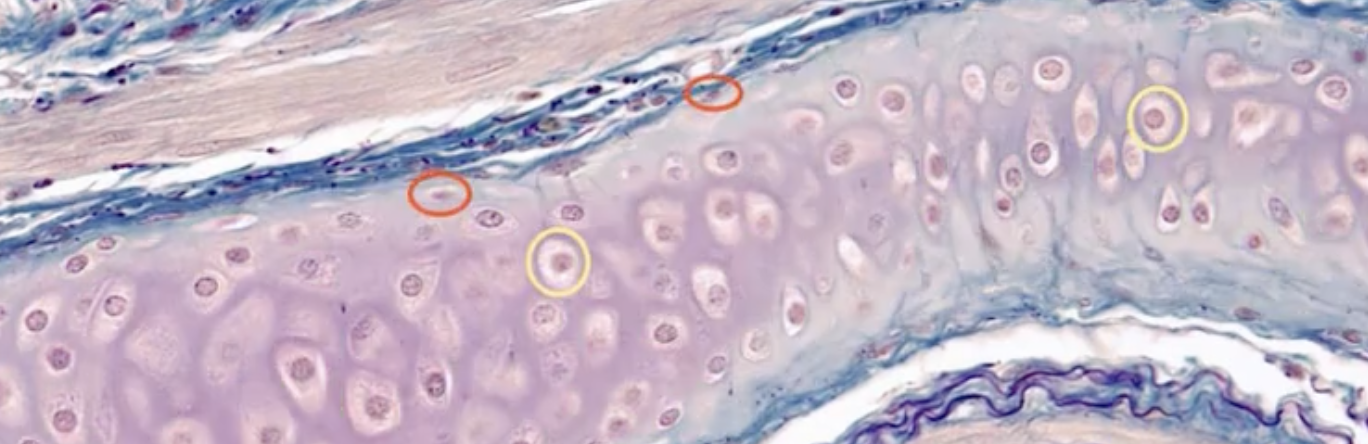
What cells are circled in yellow in cartilage?
Chondrocytes
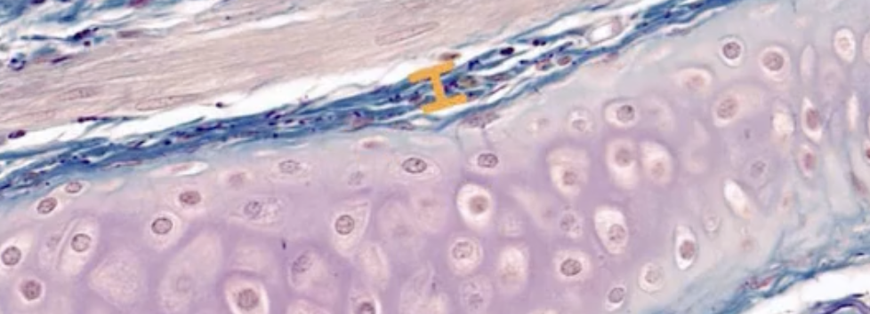
What part of the cartilage has a orange bar?
Perichondrium

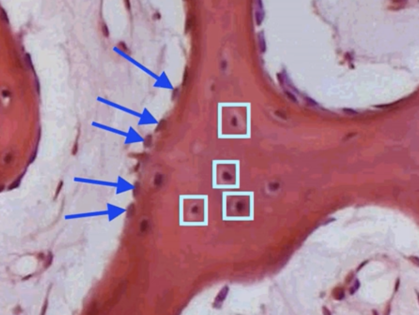
What part of bone are the dark blue arrows pointing to?
Osteoblasts
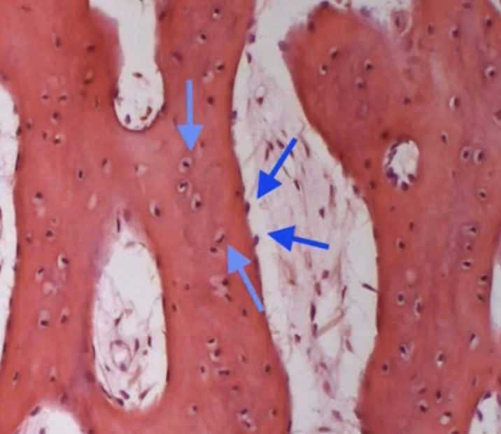
What part of bone are the light blue arrows pointing to?
Osteoclasts
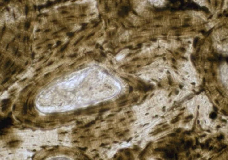
What type of bone tissue does the picture depict?
Compact
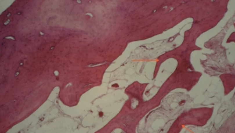
What type of bone tissue does the picture depict?
Spongey
What are the blue arrows of compact bone pointing to?
Osteocytes
What are the red arrows of spongey bone pointing to?
Trabeculae