lab practical 2
1/81
There's no tags or description
Looks like no tags are added yet.
Name | Mastery | Learn | Test | Matching | Spaced | Call with Kai |
|---|
No analytics yet
Send a link to your students to track their progress
82 Terms
classification of bones; what are the four type and how are they determined
the four classes of bone are determined by shape
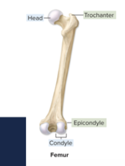
long bones: greater in length than width; ex: femur, humerus
short bones: length nearly equal to width; ex: carpals and tarsals
flat bones: flat, thin surfaces, may be slightly curved; ex: cranial bones
irregular bones: elaborate, sometimes complex shapes; ex: vertebrae
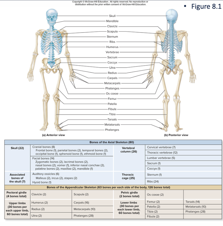
axial skeleton
composed of bones along central body axis; form the longitudinal axis of the body from skull to the end of the vertebral column
skull, vertebral column, and thoracic cage
appendicular skeleton
bones of upper and lower limbs & girdles of bones attach limbs to axial skeleton
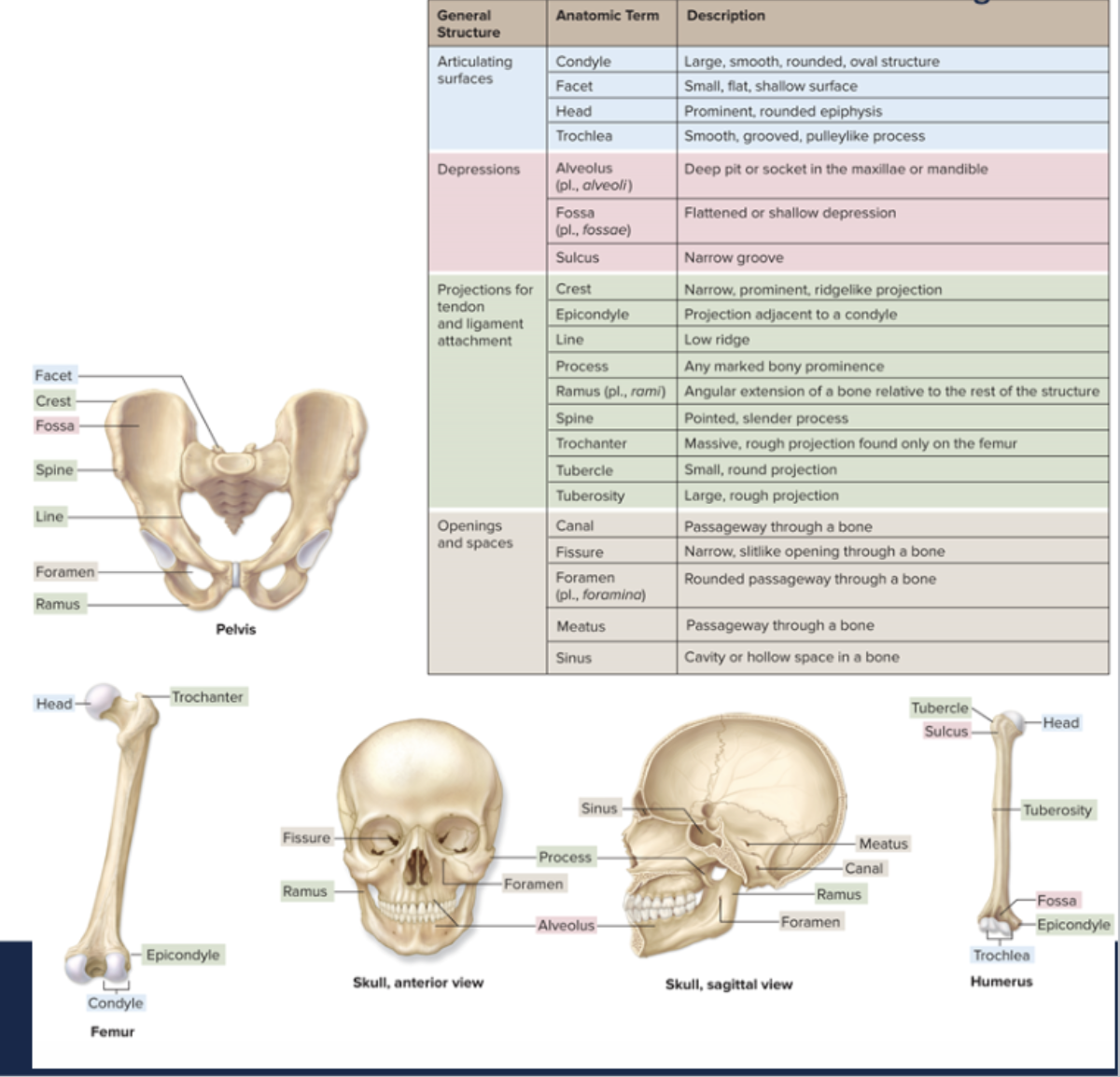
what are bone landmarks? what are they used for?
they are the bumps, grooves, and hole on bones
landmarks (markings) are used for muscle attachment, passageways for blood vessel and nerves, support, and movement (joints)
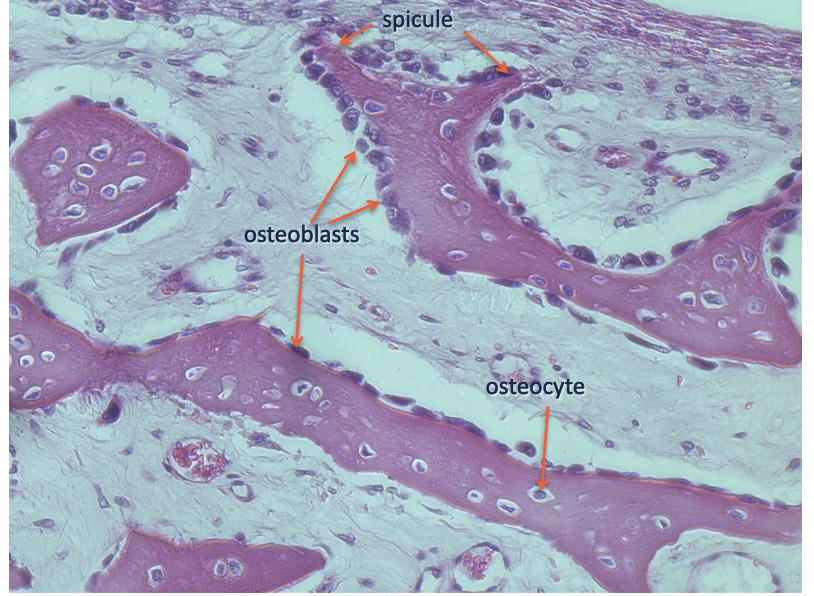
cells of bone
osteoprogenitor cells: stem cells that mature to become osteoblast
osteoblasts: immature bone cell that secretes osteoid, the organic component of bone matrix
osteocytes: mature bone cell; maintains the bone matrix
osteoclasts: multinucleate cell; secretes acids and enzymes to dissolve bone matrix
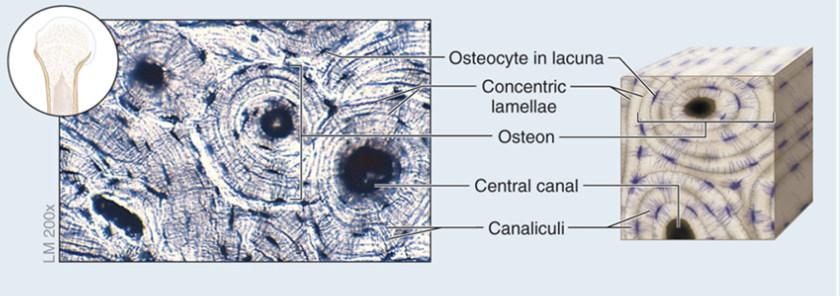
osteon components for compact bone
central canal
concentric lamellae
osteocytes
canaliculi
bone: supporting connective tissue
structure: calcified extracellular matrix containing osteocytes trapped in lacunae
compact bone: arranged in osteons (concentric lamellae arranged around a central canal)
spongy bone: a meshwork that has a different organization from compact bone
function: provides levers for body movement, supports soft structures, protects organs, stores calcium + phosphorus
spongy bone: contains hemopoietic tissue & is the site for hemopoietic
location: bones of the body
spongy (cancellous) bone
locations: flat bones of the skull, ribs, sternum, mandible
Head (epiphysis) of long bones
composed of: trabeculae, parallel lamellae
endochondral ossification: 6 steps
A hyaline cartilage model of bone forms
cartilage calcifies, a periosteal bone collar forms
Primary ossification center forms in diaphysis (shaft)
Secondary ossification centers form in epiphyses
Bone replaces cartilage, except articular cartilage & epiphyseal plates
Epiphyseal plates ossify & form epiphyseal lines
forms most bones in the body, long bones, & end bones of clavicle & replaces cartilage w/ bone
cartilage is formed first, then bone is laid down on it
replaces cartilage w/ bone
begins in embryological/ as a fetus
intramembranous ossification: 4 steps
ossification centers form within mesenchyme
osteoid undergoes calcification
woven bone & periosteum form
Lamellar bone replaces woven bone, compact & spongy bone form
forms the flat bones of skull, face, jaw, center of clavicle
incomplete ossification at birth can be seen as the “soft spot” on a baby’s head
bone is directly formed on meserichymal tissue]does not form an intermediate cartilage
bone growth in membrane; osteoblasts help synthesize the bones
developing membrane bone (developing spongy bone)
hemopoiesis
blood cell production
ossification
formation and development of bone
osteons
basic functional & structural unit of mature compact bone
trabeculae
highly porous form of bone tissue that is organized into a network of interconnected rods & plates which surround pores that are filled with bone marrow
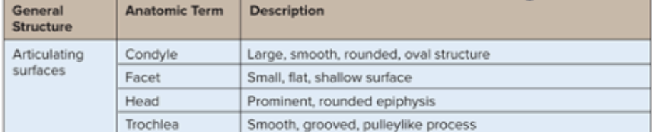
articulating surfaces
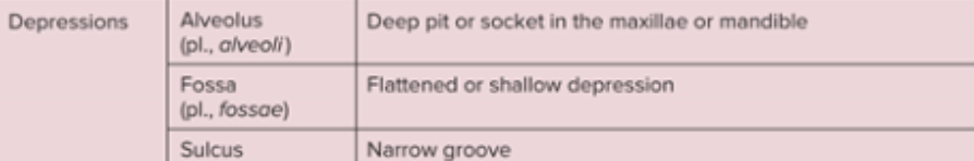
depressions
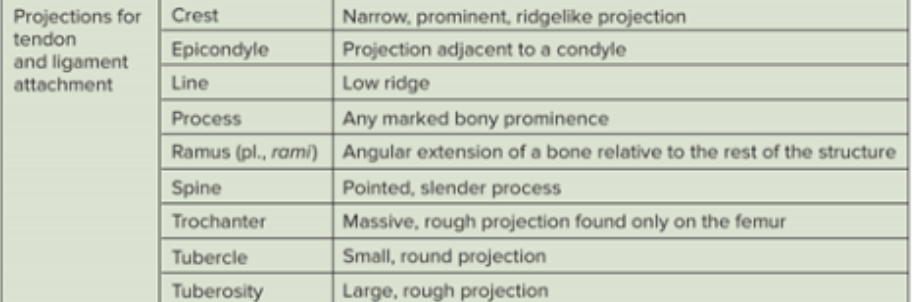
projections for tendon & ligament attachment
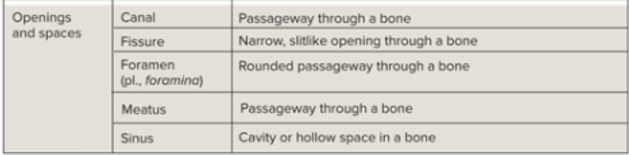
openings & spaces
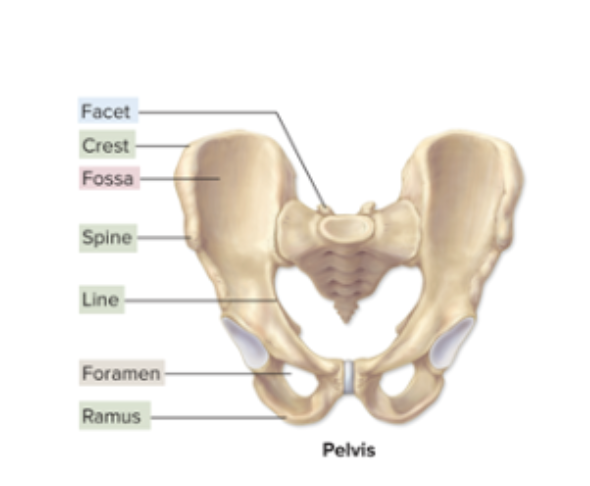
pelvis
femur
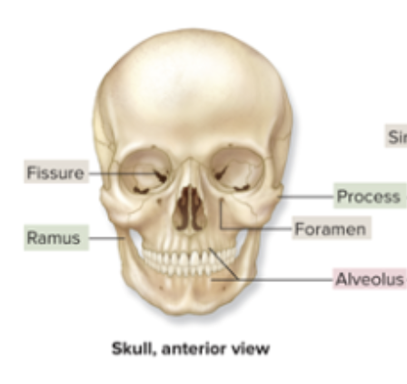
skull, anterior view
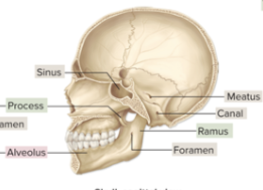
skull, sagittal view
humerus
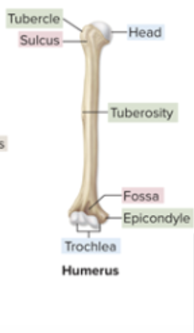
bone markings
bumps, holes, and ridges on bones where muscles, tendons, and ligaments are attached and where blood vessels and nerves pass through
articulation
a joint; where two bones meet
fontaneis
spaces between bones in an infant skull
canaliculi
tiny canals that radiate outward from the Haversian canal to lacunae to supply the bone cells with nutrients
diaphysis
shaft of a long bone; composed of compact bone
epiphysis
end of a long bone; composed mostly of spongy bone
epiphyseal line
marking left on the bone from growth at the epiphyseal plate
remodeling
process of breaking down and reforming bone that occurs throughout life to maintain proportion and strength and well as healthy calcium levels
fracture
broken bone
rickets
soft bones caused by lack of vitaminD, calcium, or phosphorus
cranial bones
fun fact
part of the axial skeleton (skull is a part of it), contains 8 cranial bones that form the brain case and 14 facial bones that protect and support the entrances of: the digestive and respiratory tracts
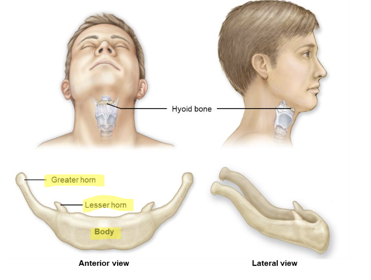
the hyoid bone is the only bone in humans that does not articulate with any other bone
whats the function of the axial skeleton
to support and protect the organs in body cavities and attach to the muscles of the head, neck, trunk, and muscles for respiration
function of the appendicular skeleton
to attach the limbs to the axial skeleton and functions the upper and lower limbs of the body
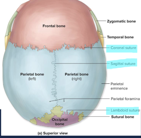
cranial sutures
coronal suture
sagittal suture
lambdoid suture
squamous suture
8 cranial bones
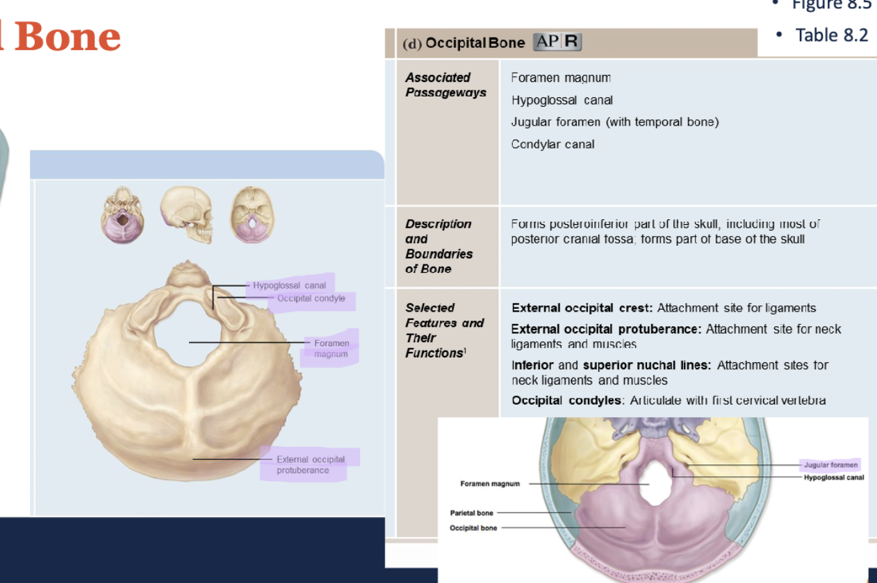
occipital bone (1)
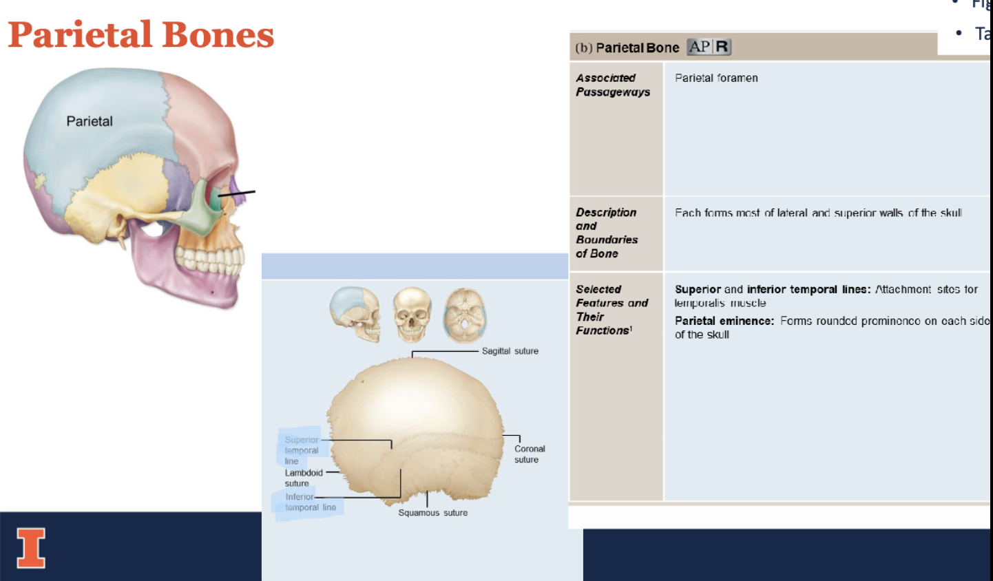
parietal bone (2)
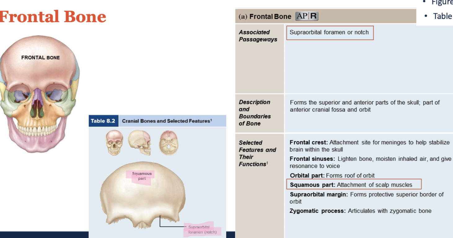
frontal bone (1)
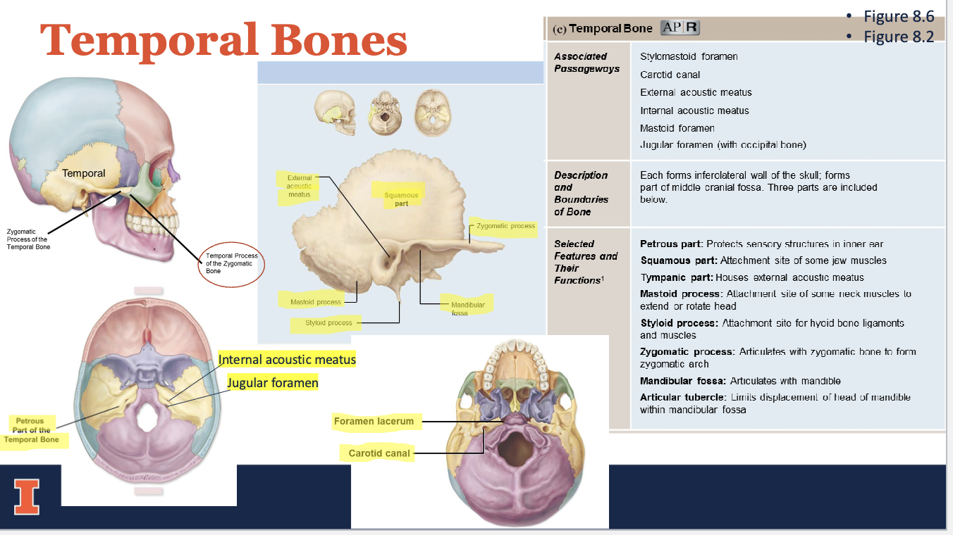
temporal bones (2)
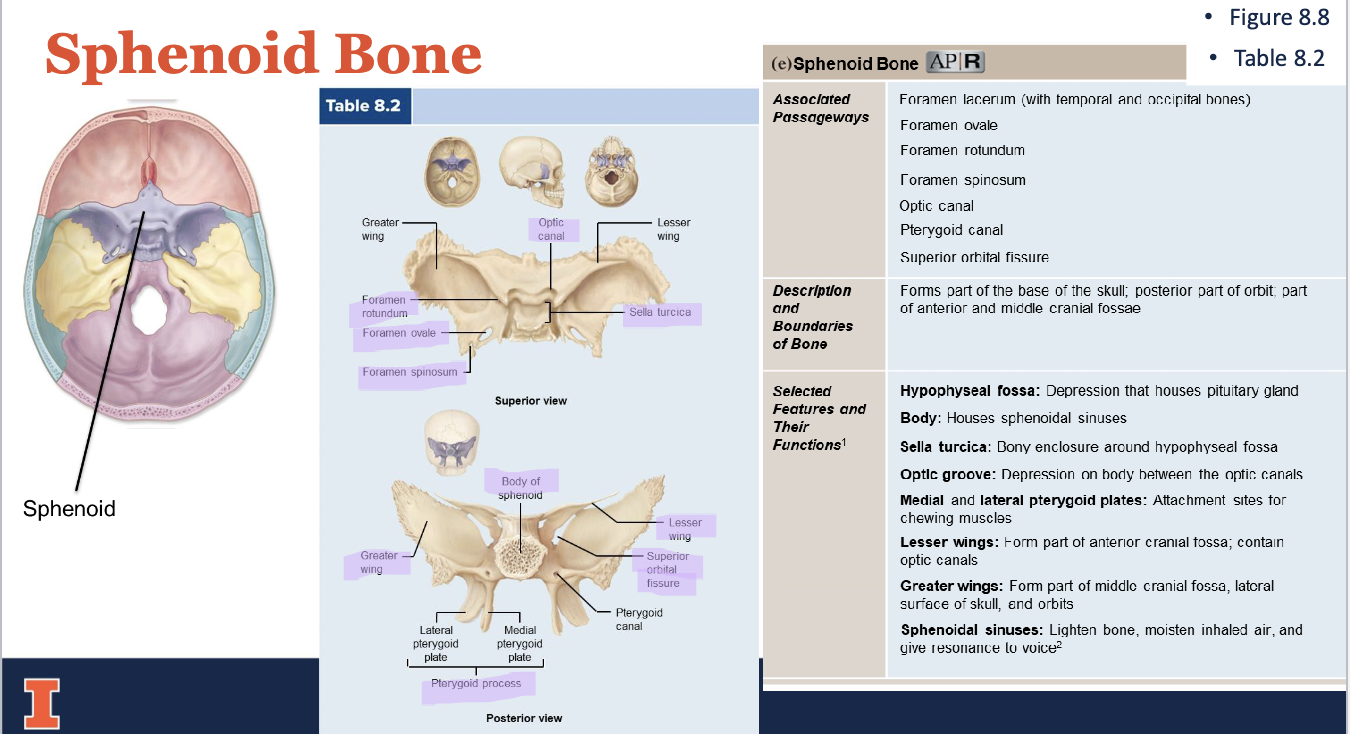
sphenoid (1)
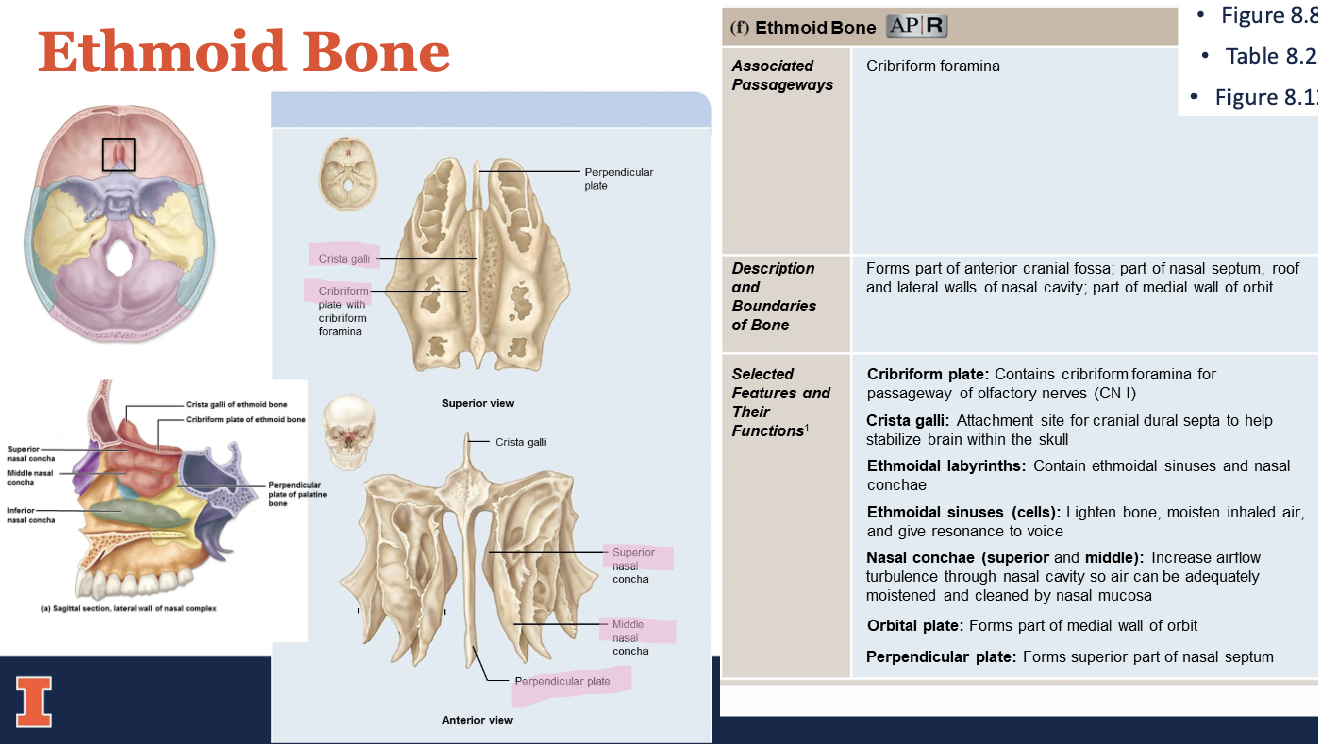
ethmoid (1)
occipital bone
parietal bones
frontal bone
temporal bones
sphenoid bone
ethmoid bone
facial bones
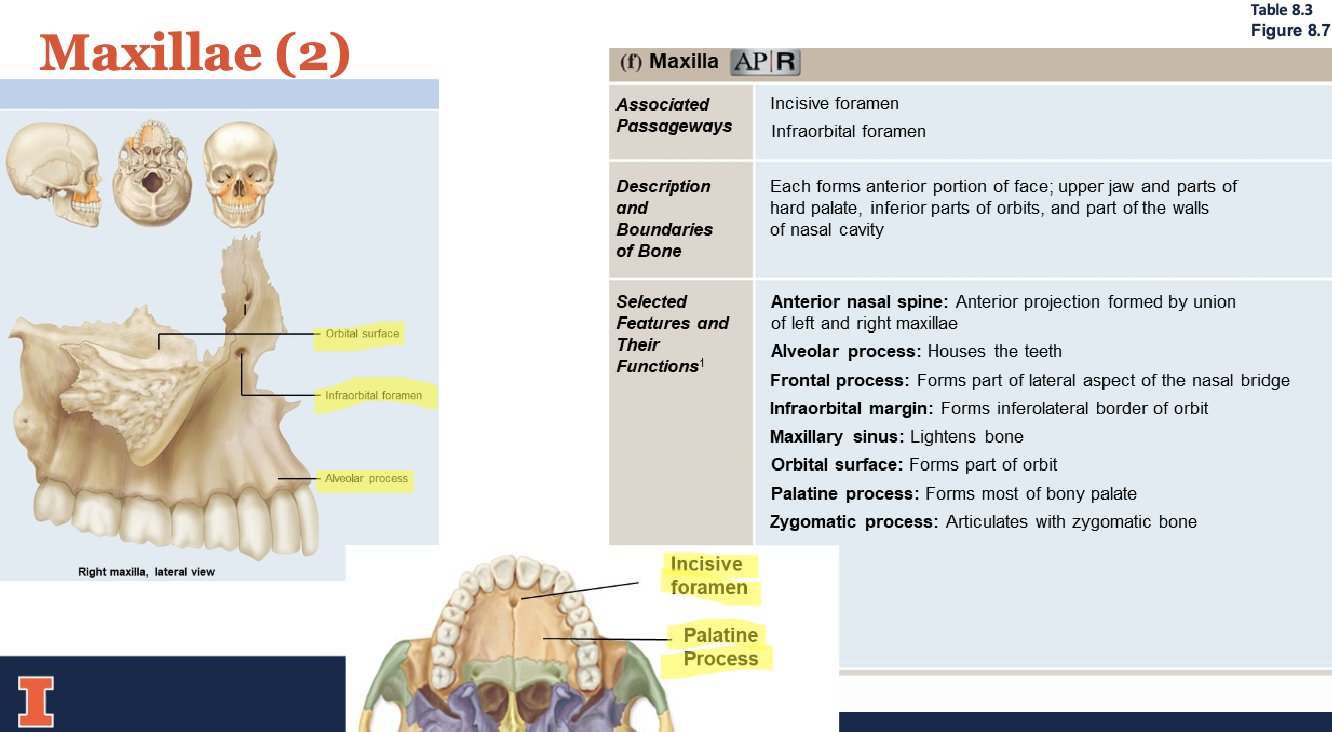
-Maxillae (2)
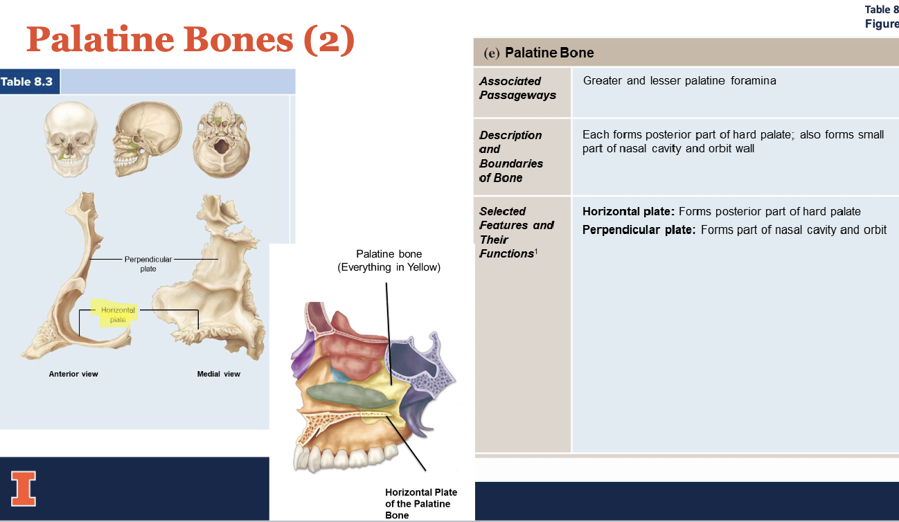
-Palatine Bones (2)
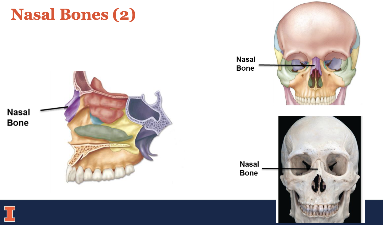
-Nasal Bones (2)
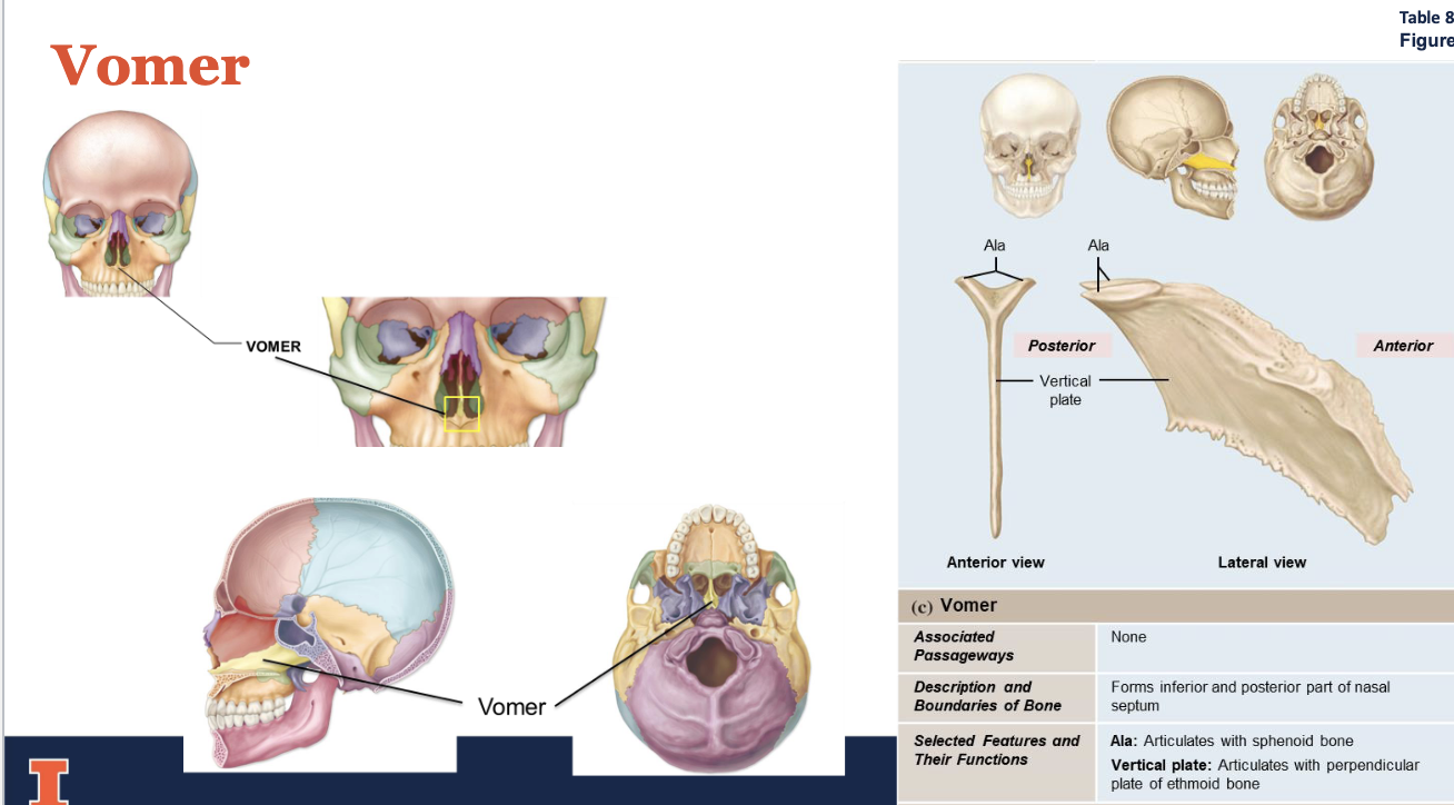
-Vomer
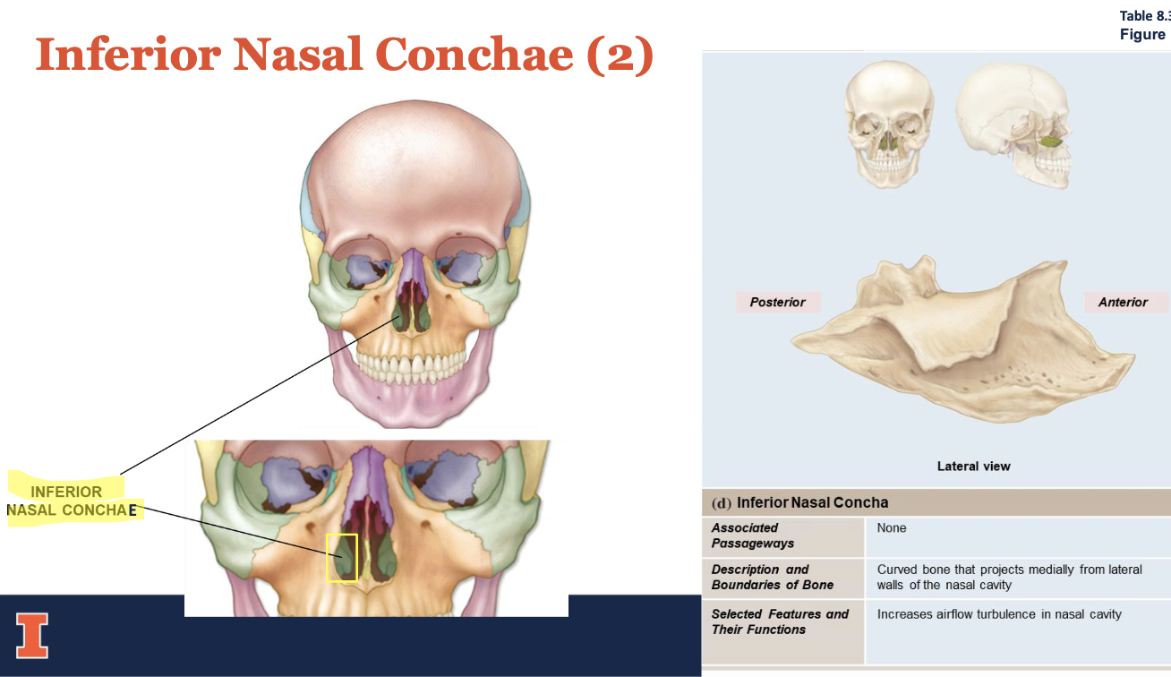
-Inferior Nasal Conchae (2)
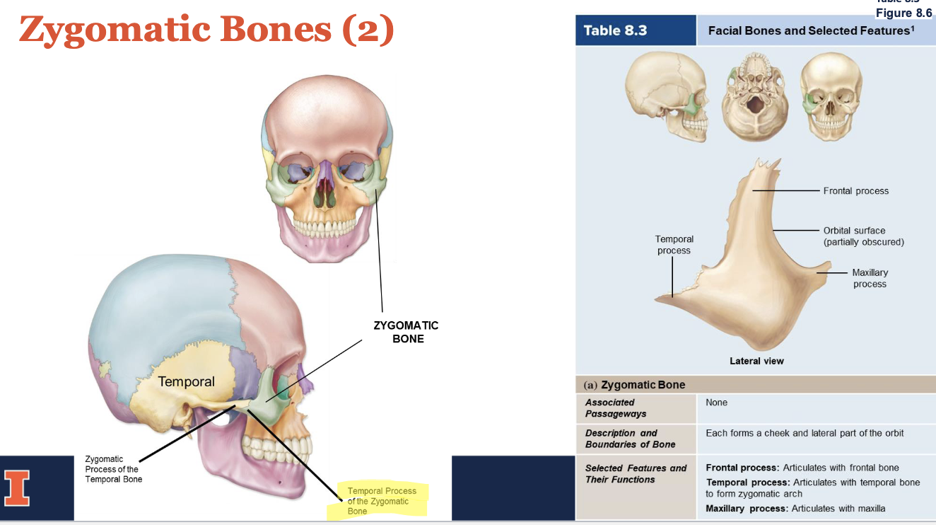
-Zygomatic Bones (2)
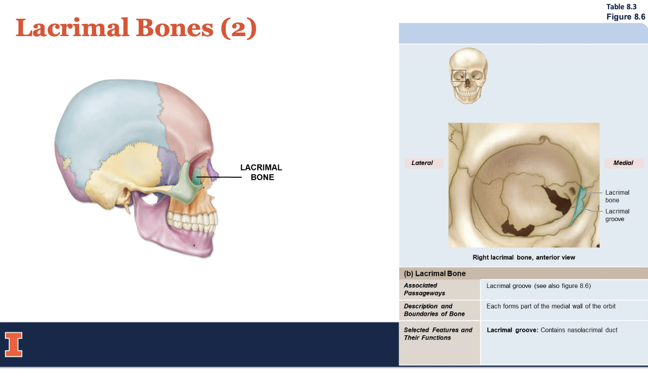
-Lacrimal Bones (2)
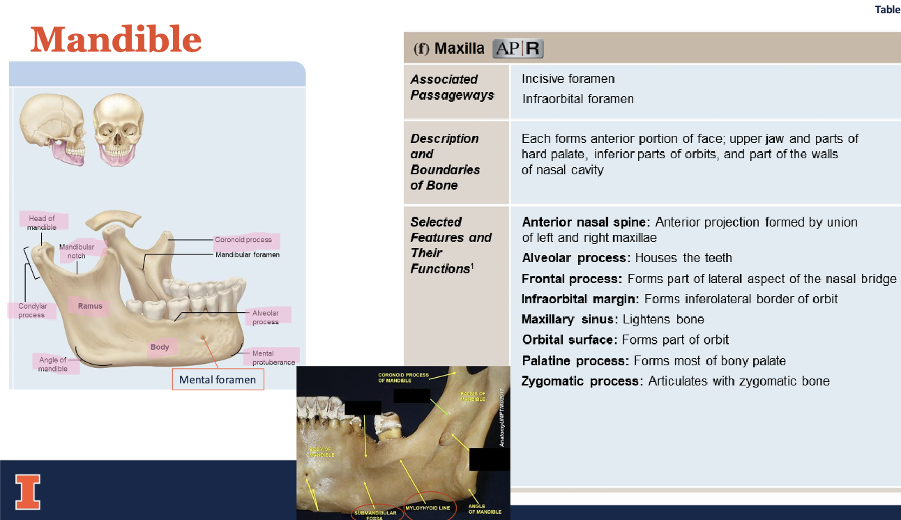
-Mandible
maxillae (2)
palatine bones (2)
nasal bones (2)
vomer
inferior nasal conchae (2)
zygomatic bones (2)
lacrimal bones (2)
mandible
hyoid bone
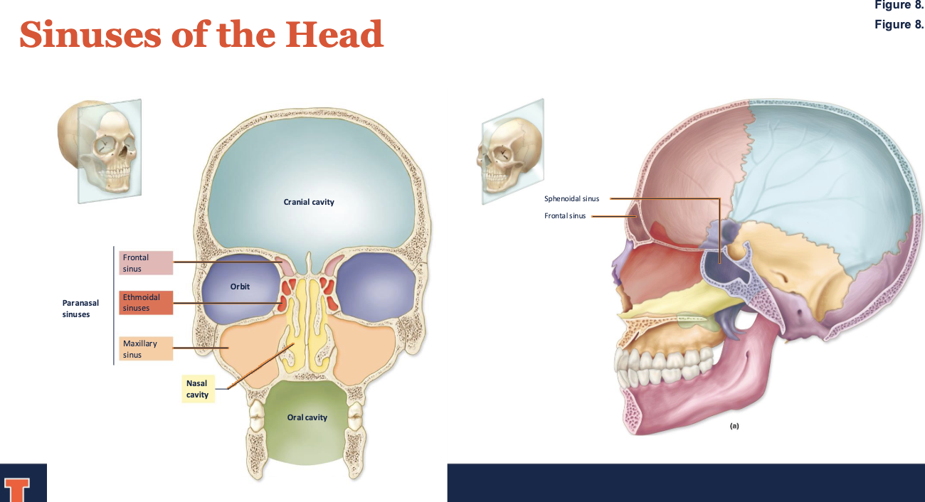
sinuses of the head
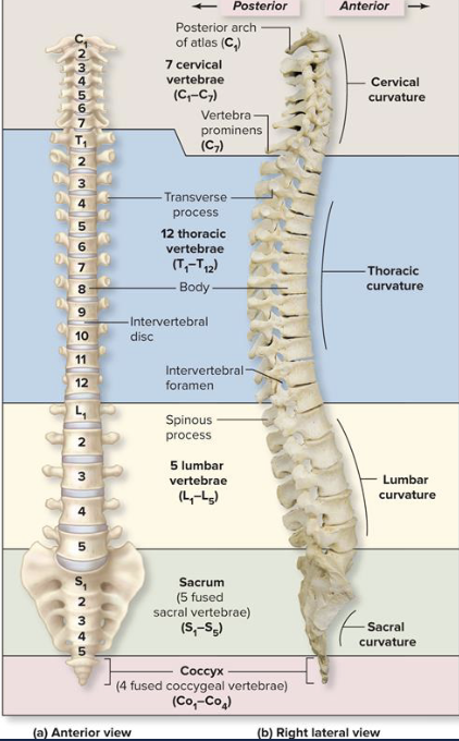
vertebral column
24 individual vertebrae and fused the sacrum and coccyx
Vertebral column divisions
• 5 divisions or regions
• Identified by capital letter for region followed by numerical subscript
- Indicates sequence from superior to inferior locations
• Cervical vertebrae
• 7 vertebrae, C1–C7
• Thoracic vertebrae
• 12 vertebrae, T1–T12
• Lumbar vertebrae
• 5 vertebrae, L1–L5
• Sacrum
• Formed from 5 sacral vertebrae, S1–S5
• Coccyx (tailbone)
• Formed from 4 coccygeal vertebrae: Co1–Co4
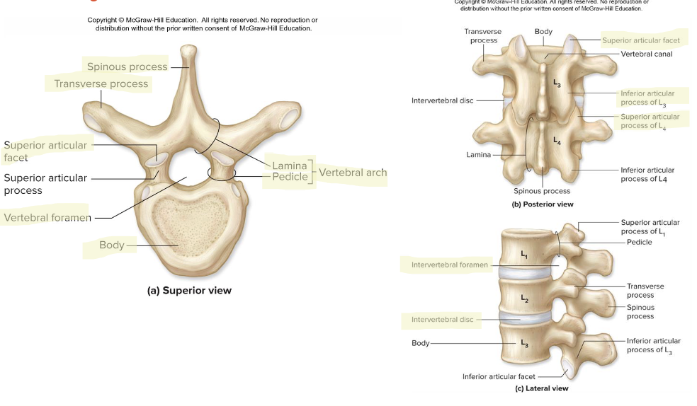
vertebral anatomy
Body
➢ Vertebral arch
➢ Vertebral foramen
➢ Vertebral canal
➢ Intervertebral foramina
➢ Pedicles
➢ Laminae
➢ Spinous process
➢ Transverse process
➢ Superior and Inferior articular processes
➢ Intervertebral discs
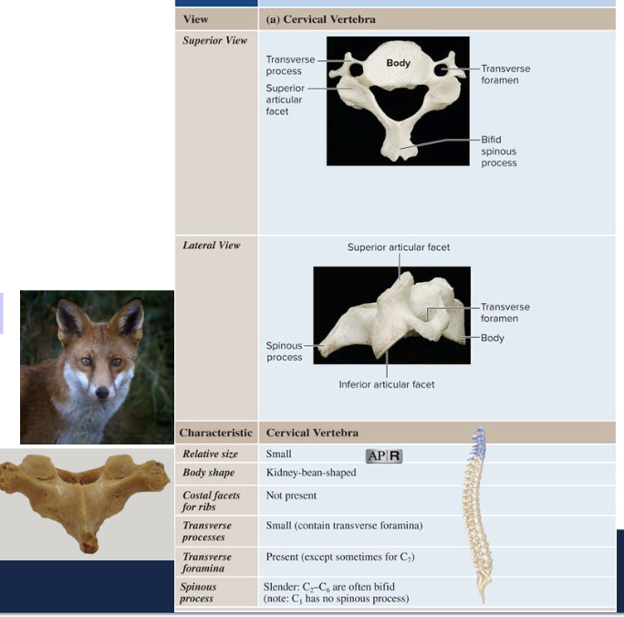
regions: cervical vertebrae
The Cervical Vertebrae (C1-C7)
• Transverse processes
➢ Are fused to costal processes
➢ Fusion creates transverse foramen (protect arteries and veins)
• Also contain long spinous processes with bifid tip
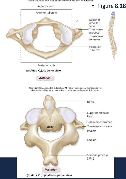
cervical vertebrae atlas (c1) and axis (c2)
Atlas
• Articulates with occipital condyles of skull at superior articular facet (or surface)
• Articulates with axis at the inferior articular facets
• Lacks body or spinous process
• Has a large, round foramen within anterior and posterior arches
• anterior tubercle serves as site of attachment for cervical ligaments and muscles
Axis
• Supports the atlas at the Superior Articular Facet
• Has heavy spinous process
• To attach muscles of head and Neck
Axis and atlas bodies fuse during development to form the dens
- dens disassociates from atlas to allow rotation
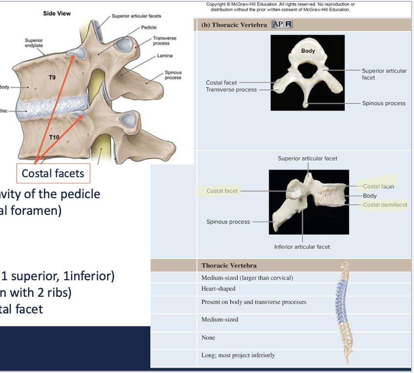
regions: (T1-T12) thoracic vertebrae
Have heart-shaped bodies
Larger bodies than in C1–C7
Smaller vertebral foramen than in C1–C7
Long, slender spinous processes
Inferior vertebral notch is the lower concavity of the pedicle
(forms ½ of the border of the intervertebral foramen)
Have costal facets:
• articulate with heads of ribs
• T1 - T8 have 2 sets of costal facets (1 superior, 1inferior)
on each side (allows for articulation with 2 ribs)
• T9 – T10 have a single superior costal facet

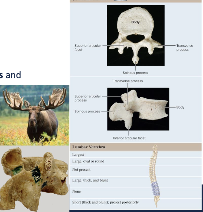
regions (L1-L5) lumbar vertebrae
Thickened vertebral body, pedicles and lamina
Have narrow vertebral foramen
Contain superior and inferior articular processes and
facets for articulation with adjacent vertebrae
Transverse processes
• Slender
• Project dorsolaterally
Spinous process
• Short, heavy
• For attachment of lower back muscles
sacrum
Anteriorly curved, triangular bone: forms posterior wall of pelvic cavity
5 fused vertebrae, fused by age 20-30
Apex pointed bone projecting inferiorly
Base broad superior surface
Articulates with L5 superiorly via pair of superior articular processes
Sacral canal Continuation of vertebral canal
• Continues through sacrum on posterior side, allows for passage of sacral roots of spinal nerves
• Terminates in inferior opening, sacral hiatus
Median sacral crest Dorsal ridge formed by fusion of spinous processes: Four pairs of sacral foramina open to either side
Lateral sacral crest fused transverse processes
Ala broad lateral extensions of the sacrum
Auricular articular surface Site of articulation with os coxae of pelvic girdle: Forms strong sacroiliac joint
Sacral foramina permit passage of nerves to pelvic organs and gluteals
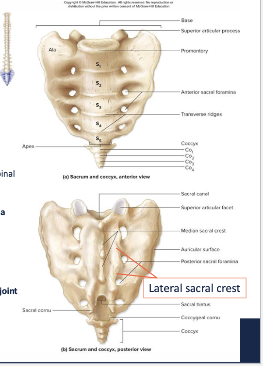
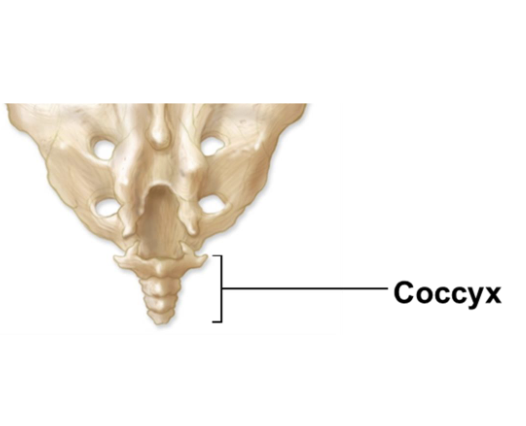
coccyx
Fusion of 4 coccygeal vertebrae
- Begins about age 25Attachment site for several ligaments and muscles
In elderly, may ultimately fuse with the sacrum
thoracic cage
Bony framework for the chest
Consists of
• Thoracic vertebrae posteriorly
• Ribs laterally
• Sternum anteriorly
Protective enclosure around thoracic organs
Attachment sites for many muscles
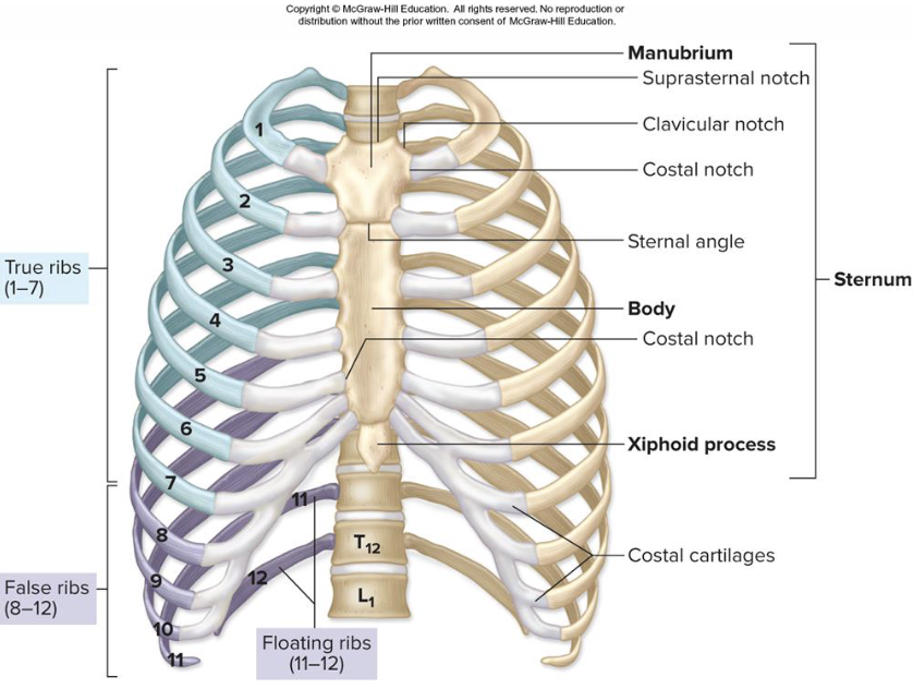
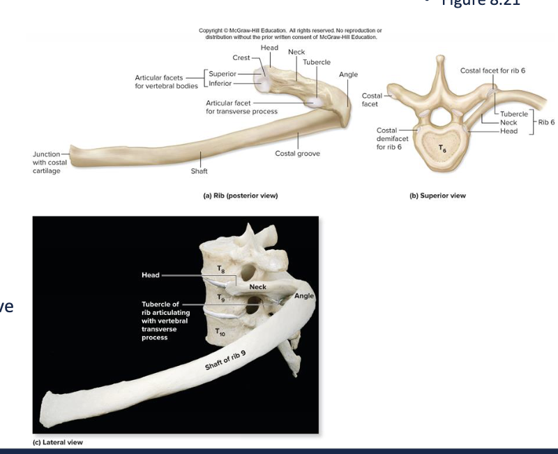
ribs
Elongated, curved, flattened bones
Originate on thoracic vertebrae
End in anterior wall of thorax
12 pairs of ribs
True ribs (ribs 1–7)
• Connect individually to the sternum by costal cartilages
• Smallest true rib is the first
False ribs (ribs 8–12)
• Costal cartilages not attached directly to the sternum
• Ribs of 11–12 without a connection to sternum called floating ribs
what consists of the ribs
Head at the vertebral end of the rib
• contains articular facets: articulate with superior costal facet of vertebrae
Neck lies between head and tubercle
Tubercle articulates with transverse costal facet of transverse process of vertebrae
Angle site where tubular shaft begins to curve
• Shaft attaches muscles of the pectoral girdle, trunk and intercostal muscles that move the ribs
Costal groove marks the path of nerves and blood
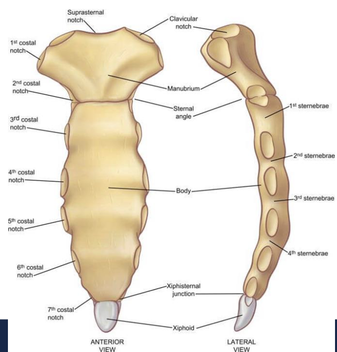
sternum
Breastbone: Flat bone forming anterior midline of thoracic wall
Manubrium widest most superior portion
• clavicular notches articulate sternum with clavicles
• suprasternal notch between the clavicular notches
• costal notches articulations for first ribs’ costal cartilages
Body longest part
• attaches to costal cartilages from ribs 2-7
• articulates with manubrium at sternal angle
Xiphoid process smallest part, at tip of sternum
• attaches to sternal body, doesn’t ossify until age 40
vertebral body
thick anterior weight-bearing structure
vertebral arch
posterior to body
vertebral foramen
opening enclosed by body with vertebral arch
vertebral canal
formed by stacked vertebral foramina, contains the spinal cord
pedicles
originate from posterolateral margins of body
laminae
extend posteromedially from posterior edge of pedicle
spinous process
projects posteriorly from laminae junctions
transverse process
lateral projections on both sides of vertebral arch
superior and inferior articular processes
originate at junction between pedicles and laminae, have smooth surface articular facet-articulate with vertebra either above or below
intervertebral discs
pads of fibrocartilage separating vertebral bodies, shock absorbers, allows vertebral column to blend
true ribs
ribs 1-7, connect individually to the sternum by costal cartilages
false ribs
ribs 8-12, costal cartilages not attached directly to the sternum