LAB9:Pelvis&hipbones,muscles
1/67
There's no tags or description
Looks like no tags are added yet.
Name | Mastery | Learn | Test | Matching | Spaced | Call with Kai |
|---|
No study sessions yet.
68 Terms
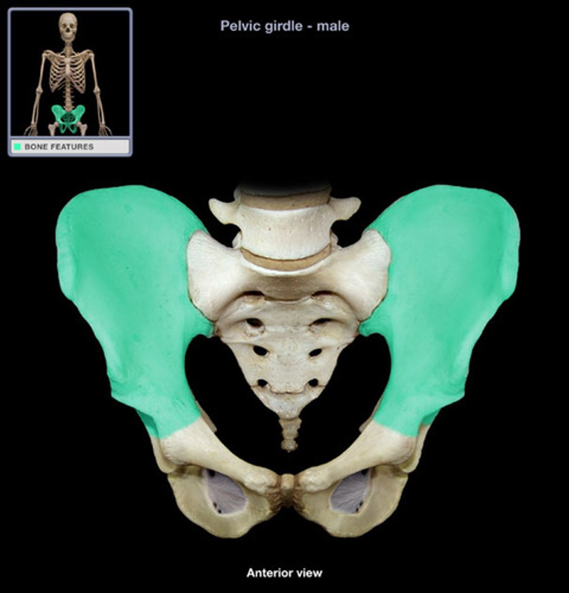
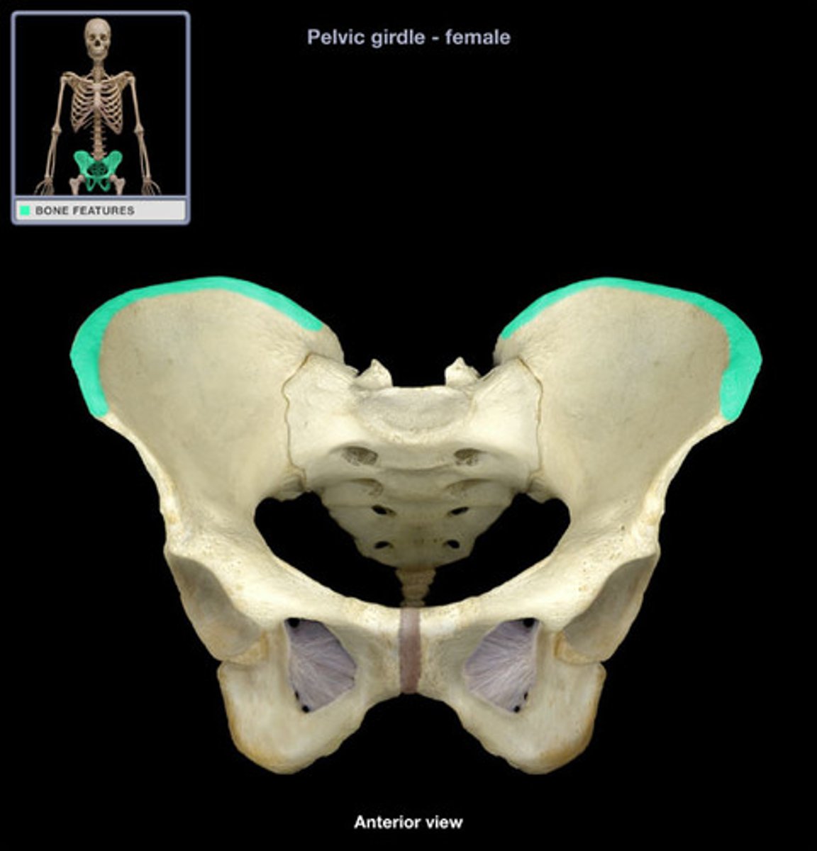
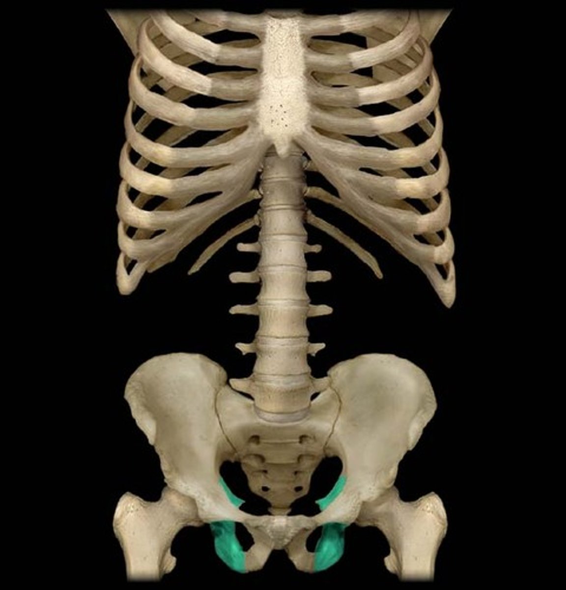
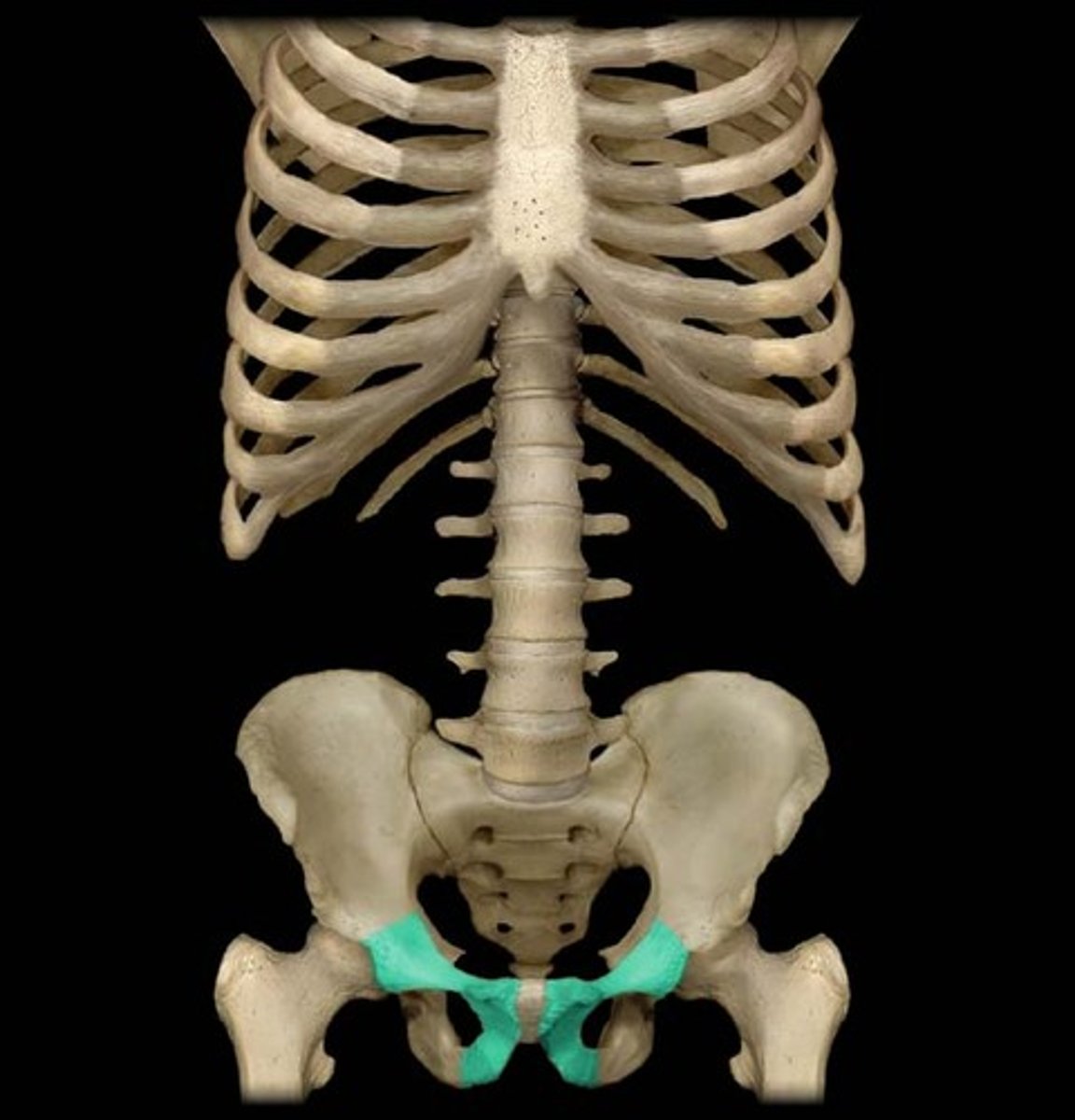
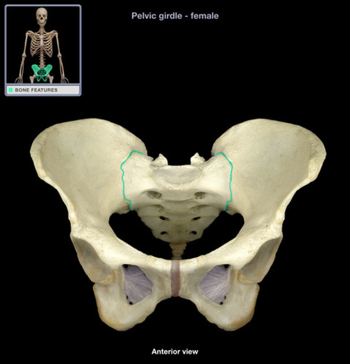
Pelvic Girdle
Bony structure connecting lower extremity to axial skeleton.
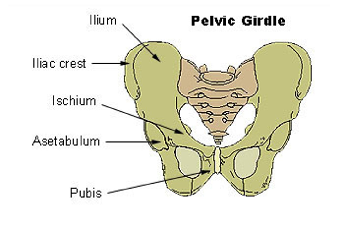
Coxal Bones
hip bones made up of ilium, ischium and pubis
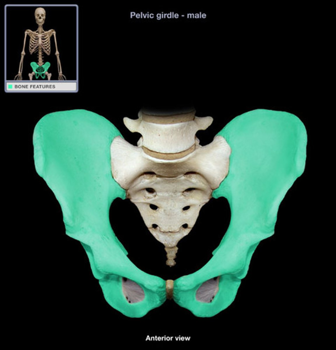
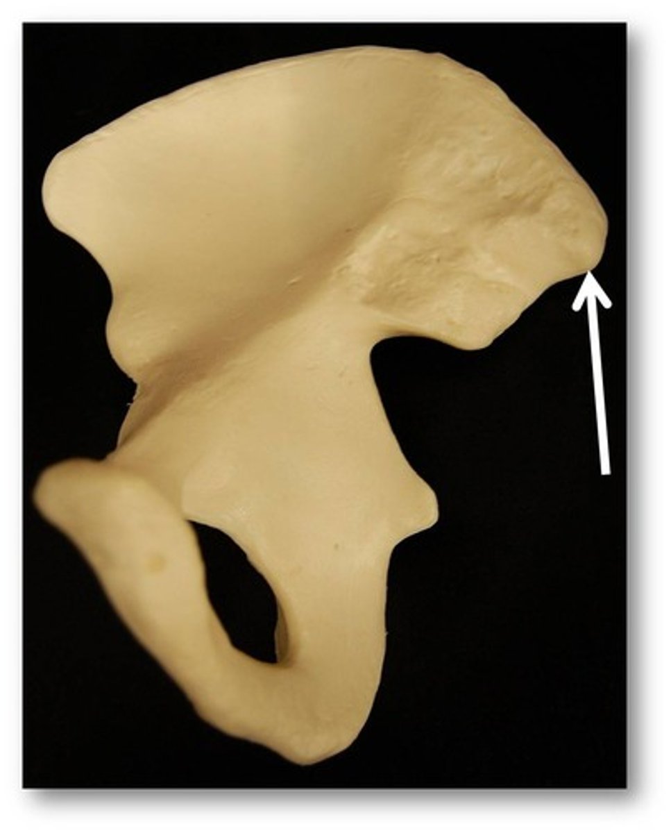
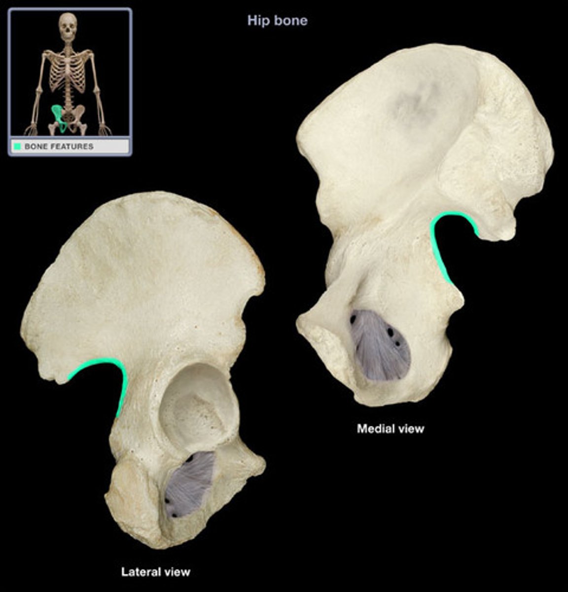
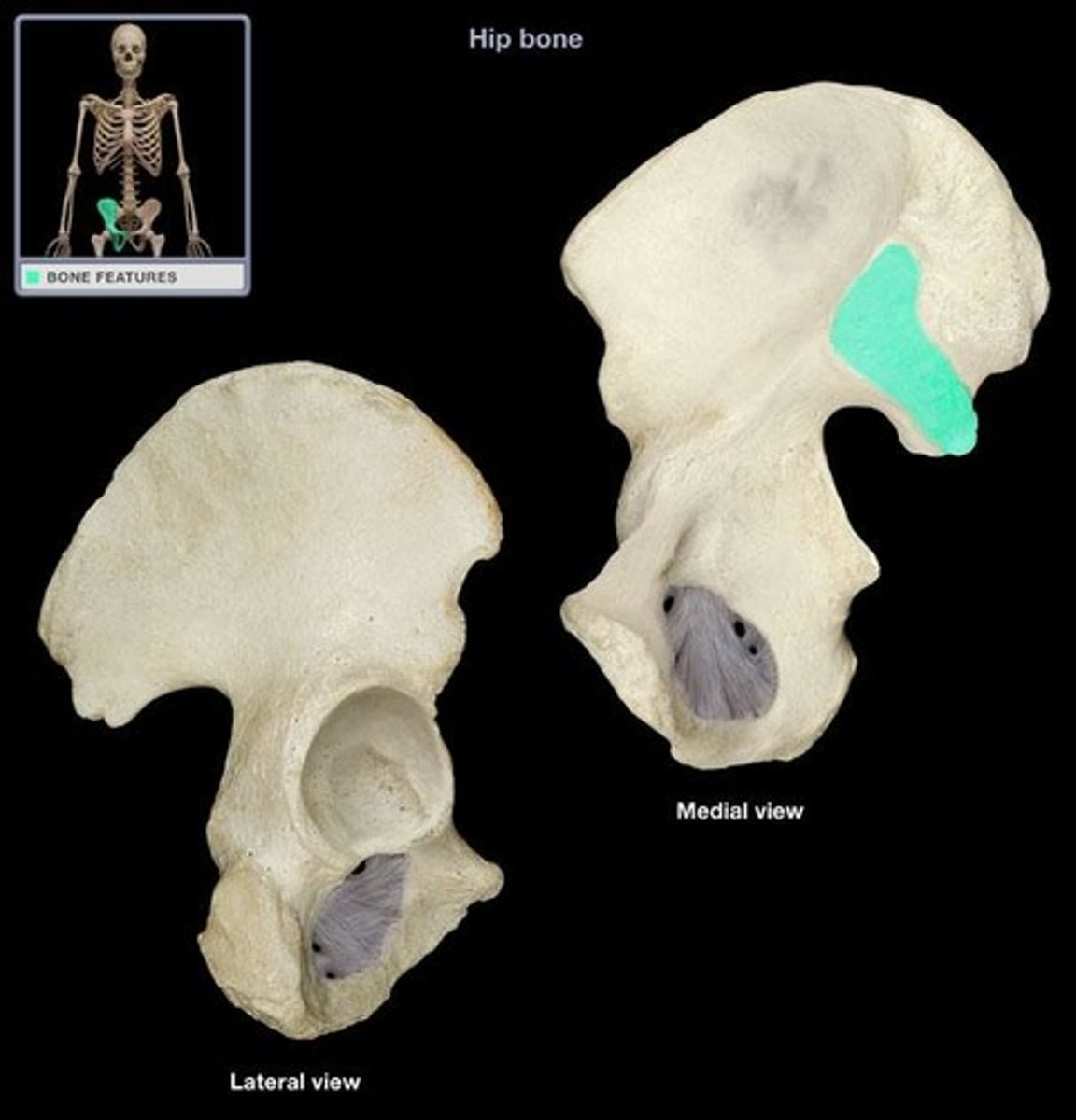
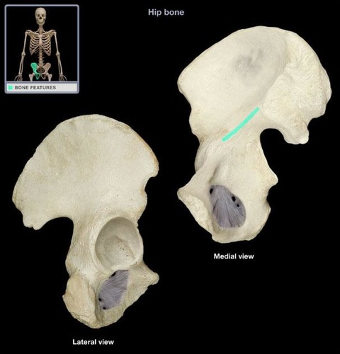
Ilium
Superior part of the hip bone.
Ilium diagram
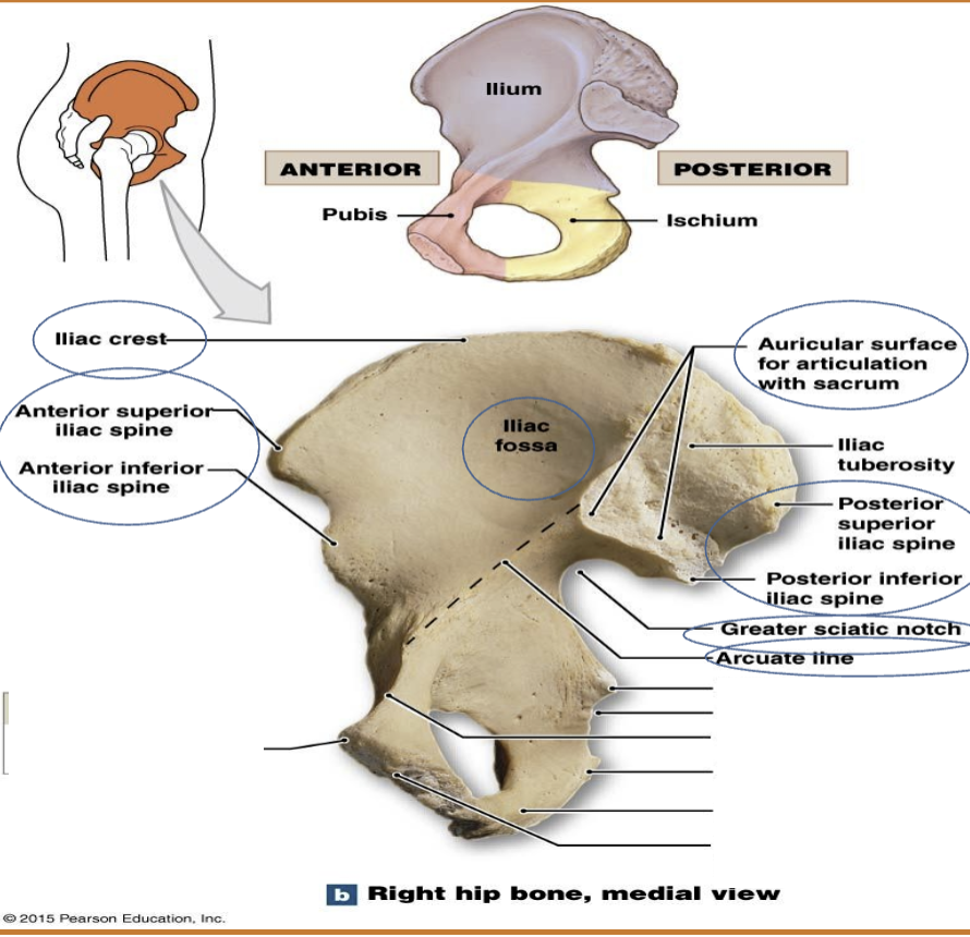
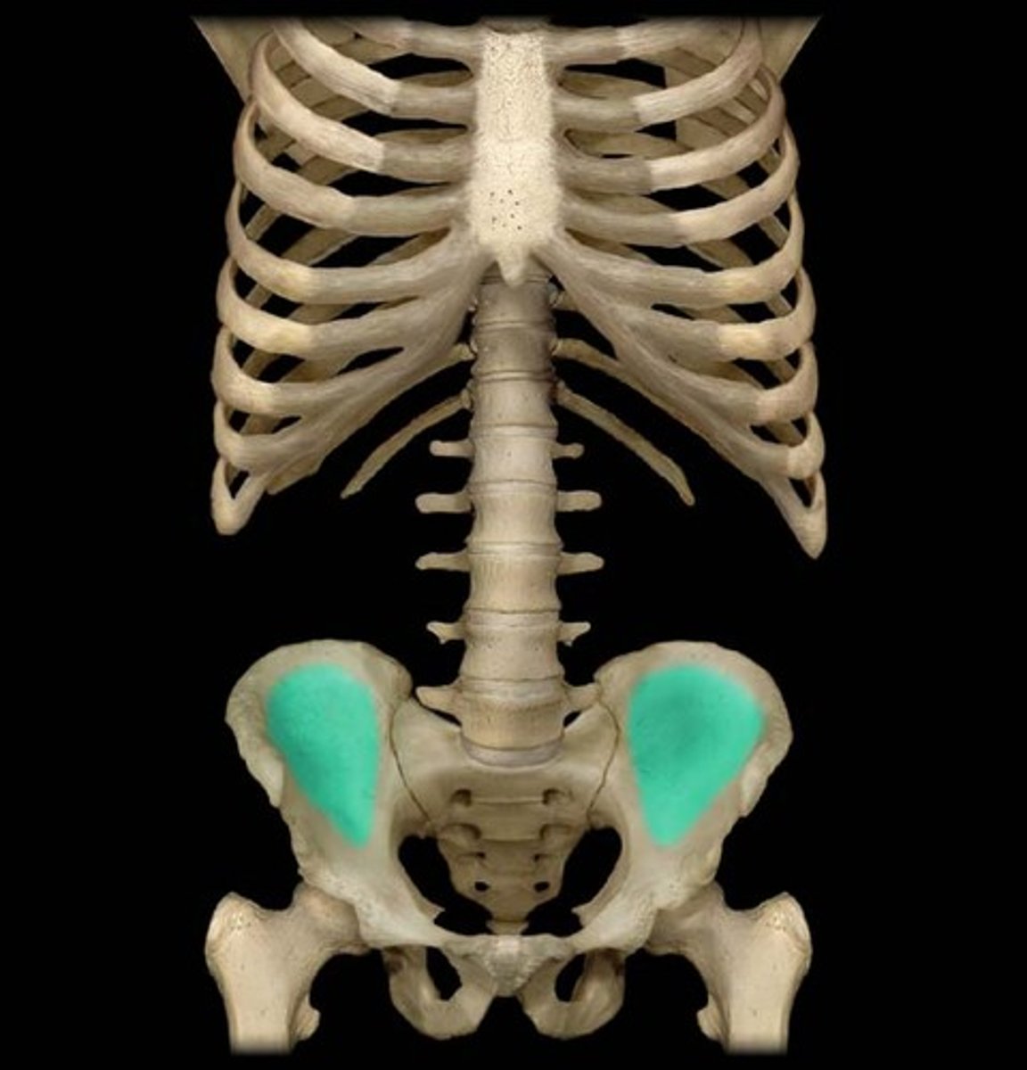
Iliac crest
Iliac fossa
Anterior superior & Anterior inferior iliac spine
Posterior superior & Posterior inferior iliac spine
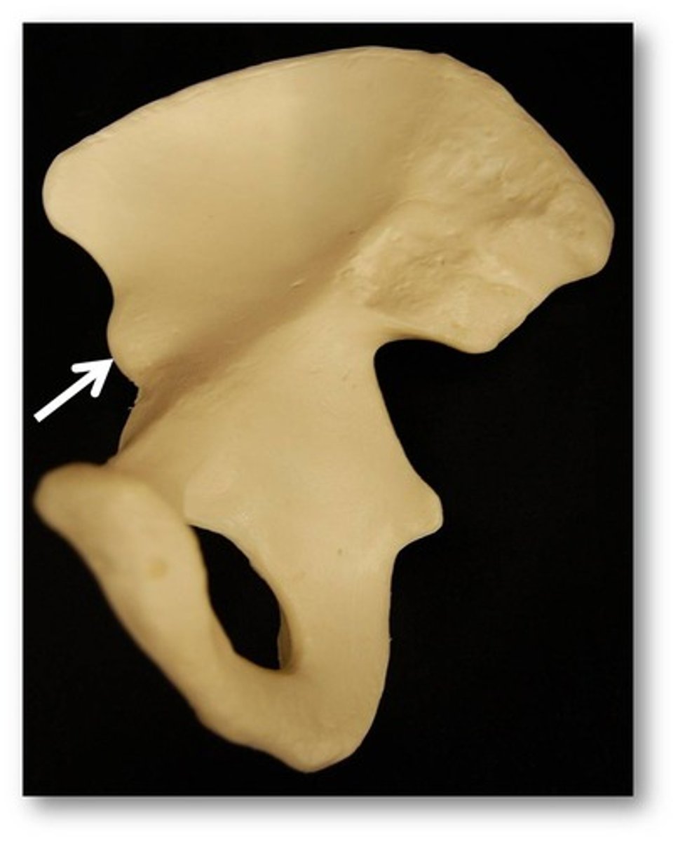
Greater sciatic notch of ilium
Auricular surface of ilium
Arcuate line of ilium
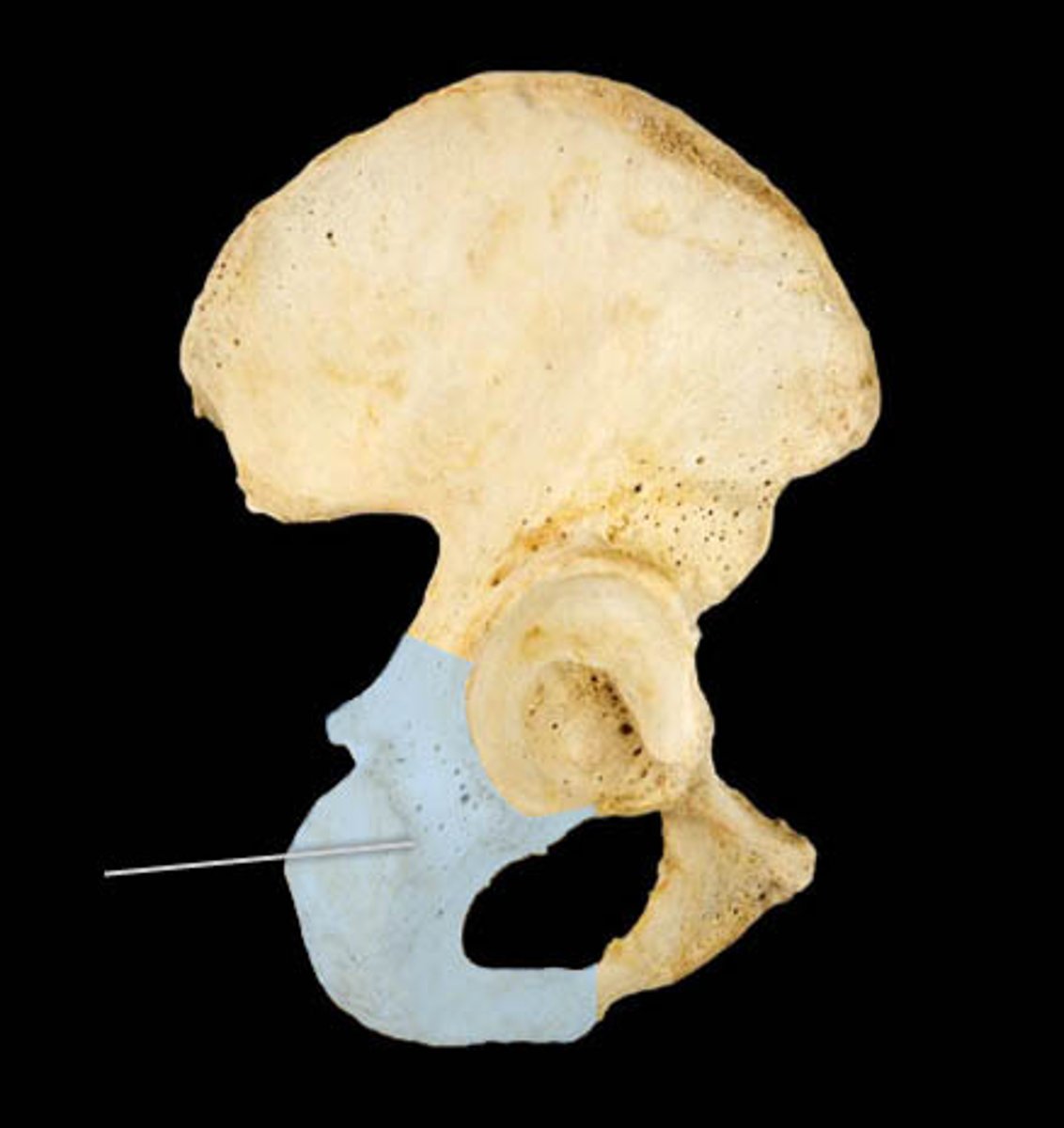
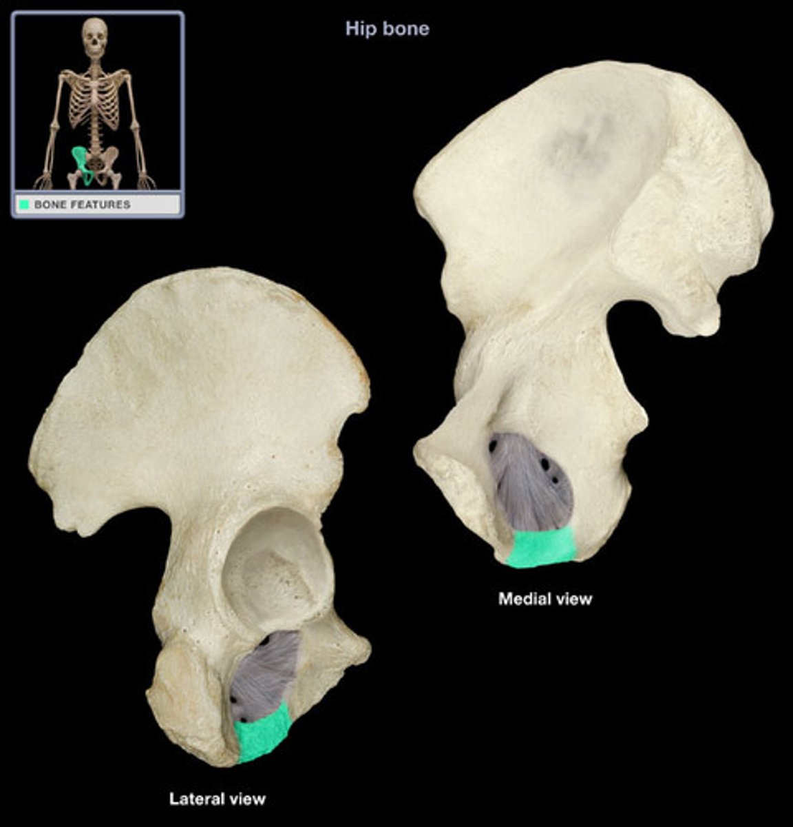
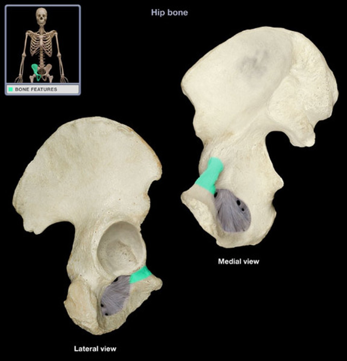
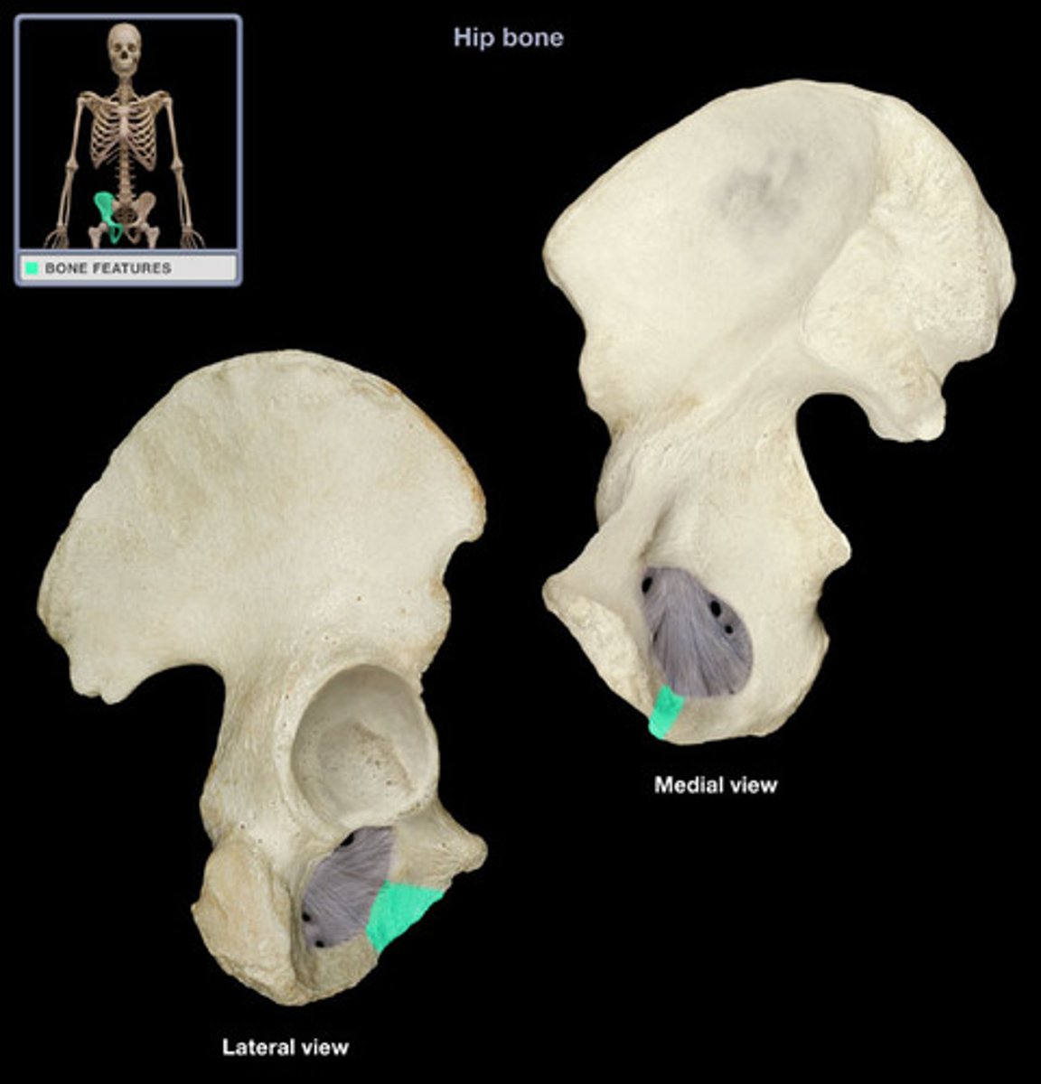
Ischium
Inferior, posterior part of the hip bone.
Ischium diagram
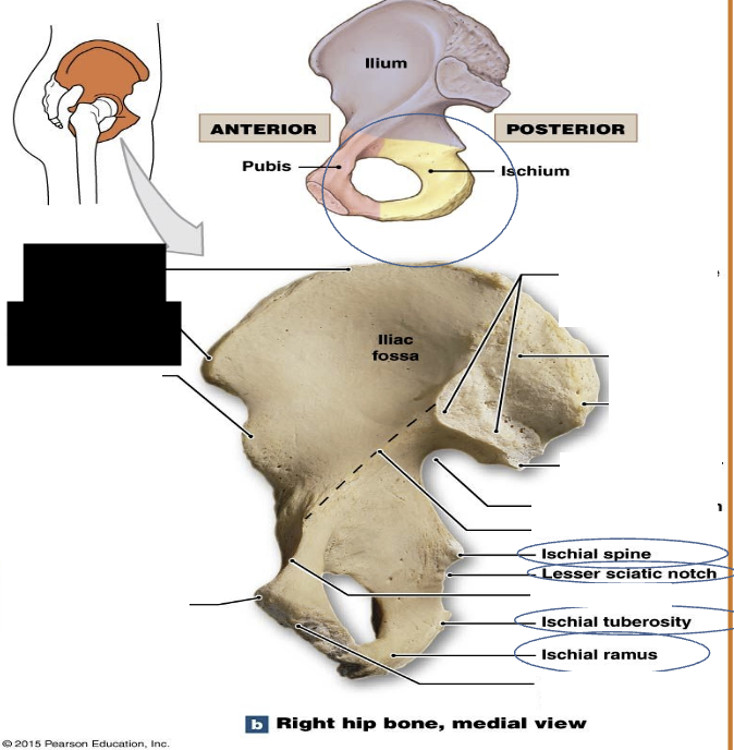
Body of ischium
Ramus of ischium
Ischial spine

Lesser sciatic notch of ischium

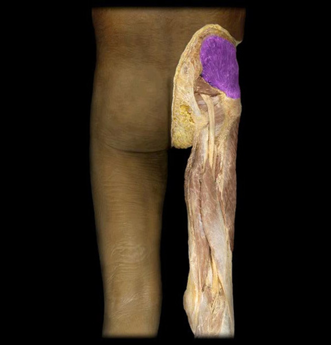
Ischial tuberosity (Sits bone)
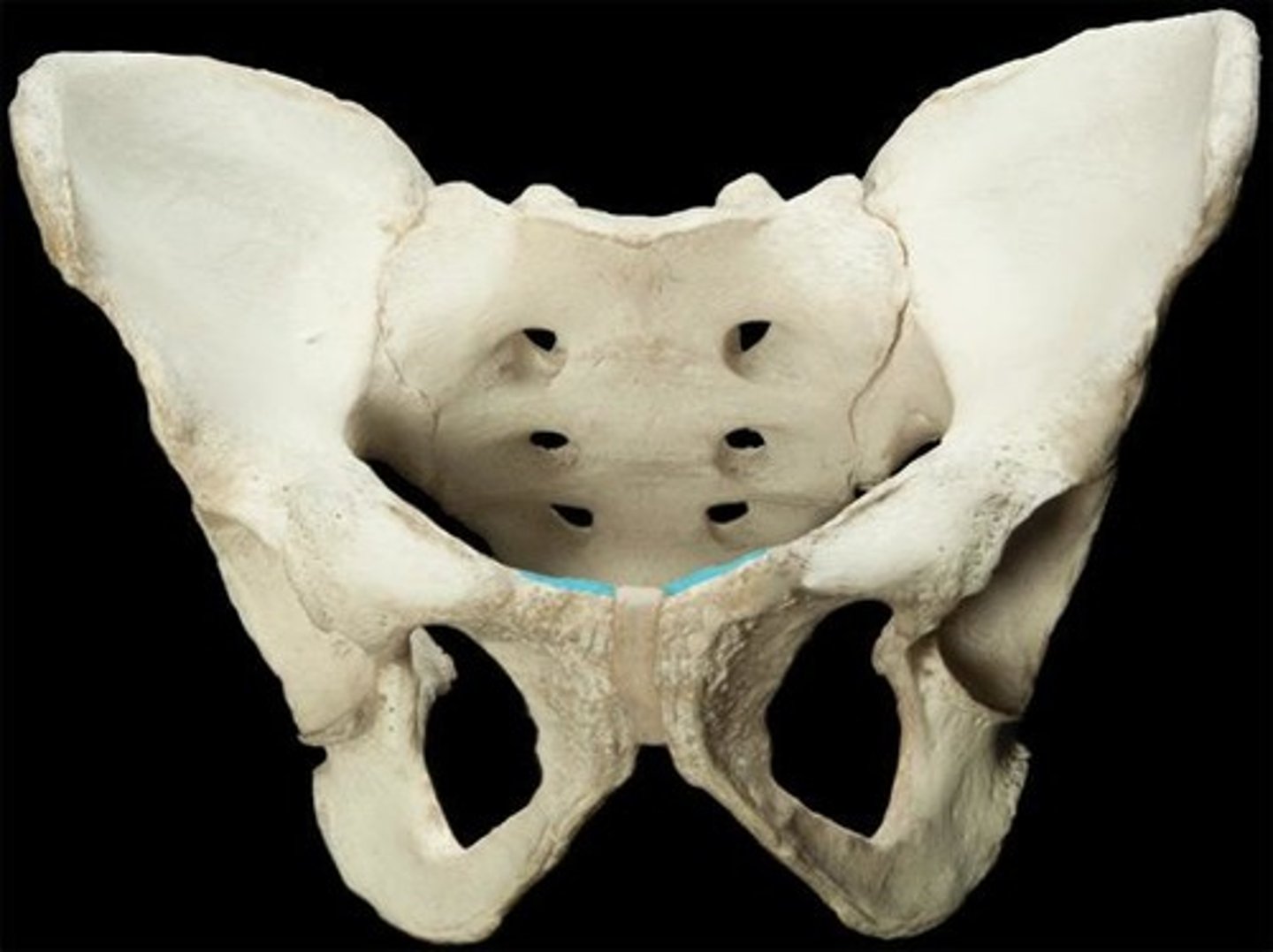
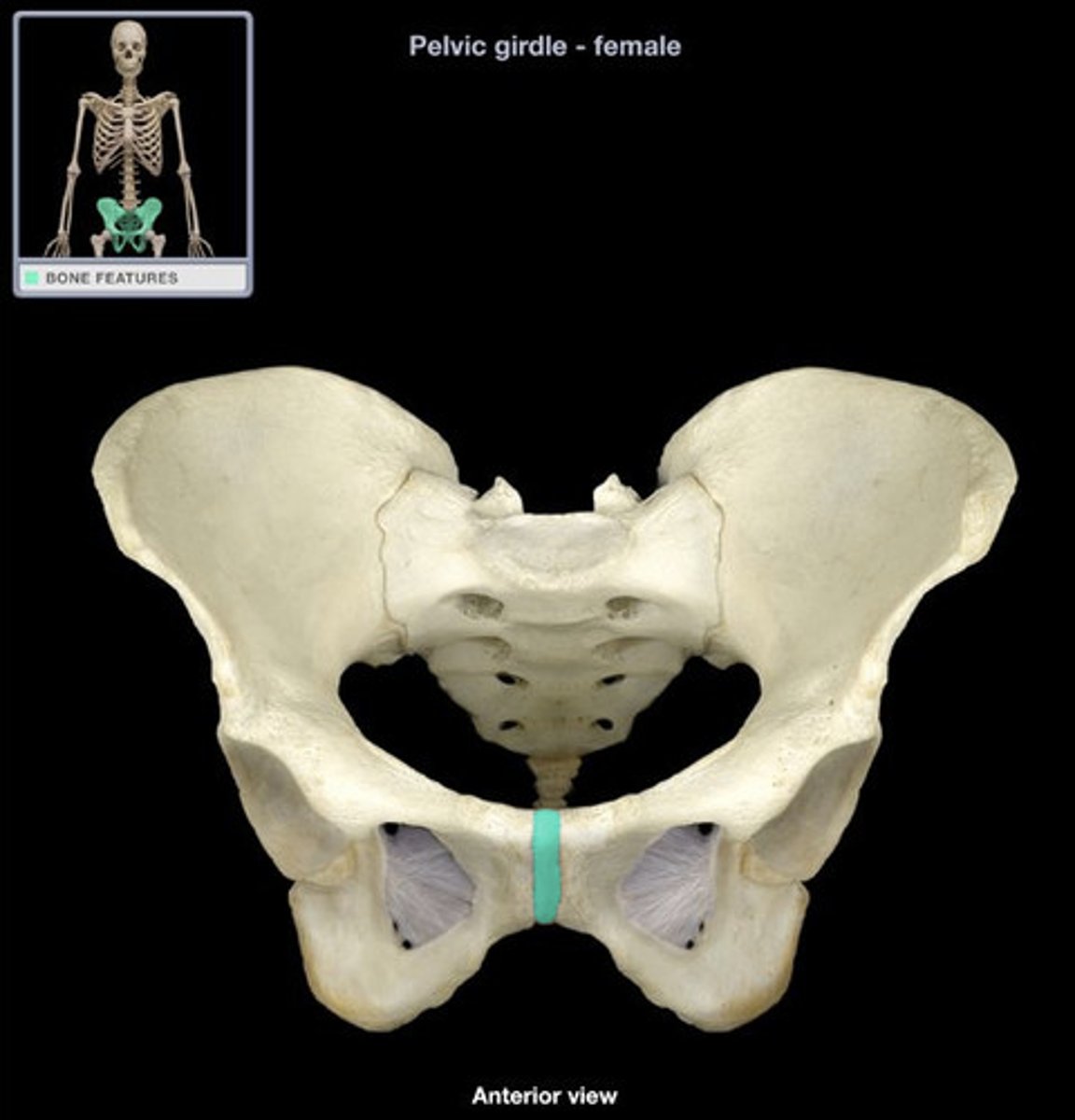
Pubis
Inferior, anterior part of the hip bone.

Pubis diagram
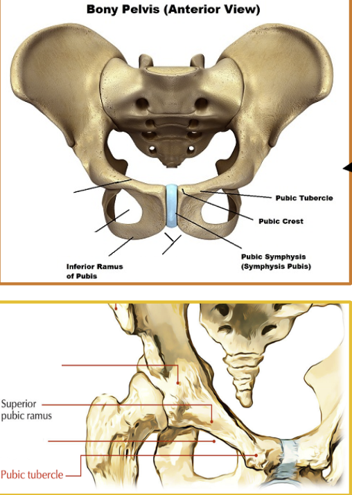
Superior ramus of pubis
Inferior ramus of pubis
Body of pubis
Pubic crest
Pubic tubercle
Pubic symphysis
Amphiarthrodial Cartilaginous joint at which two pubic bones fuse together
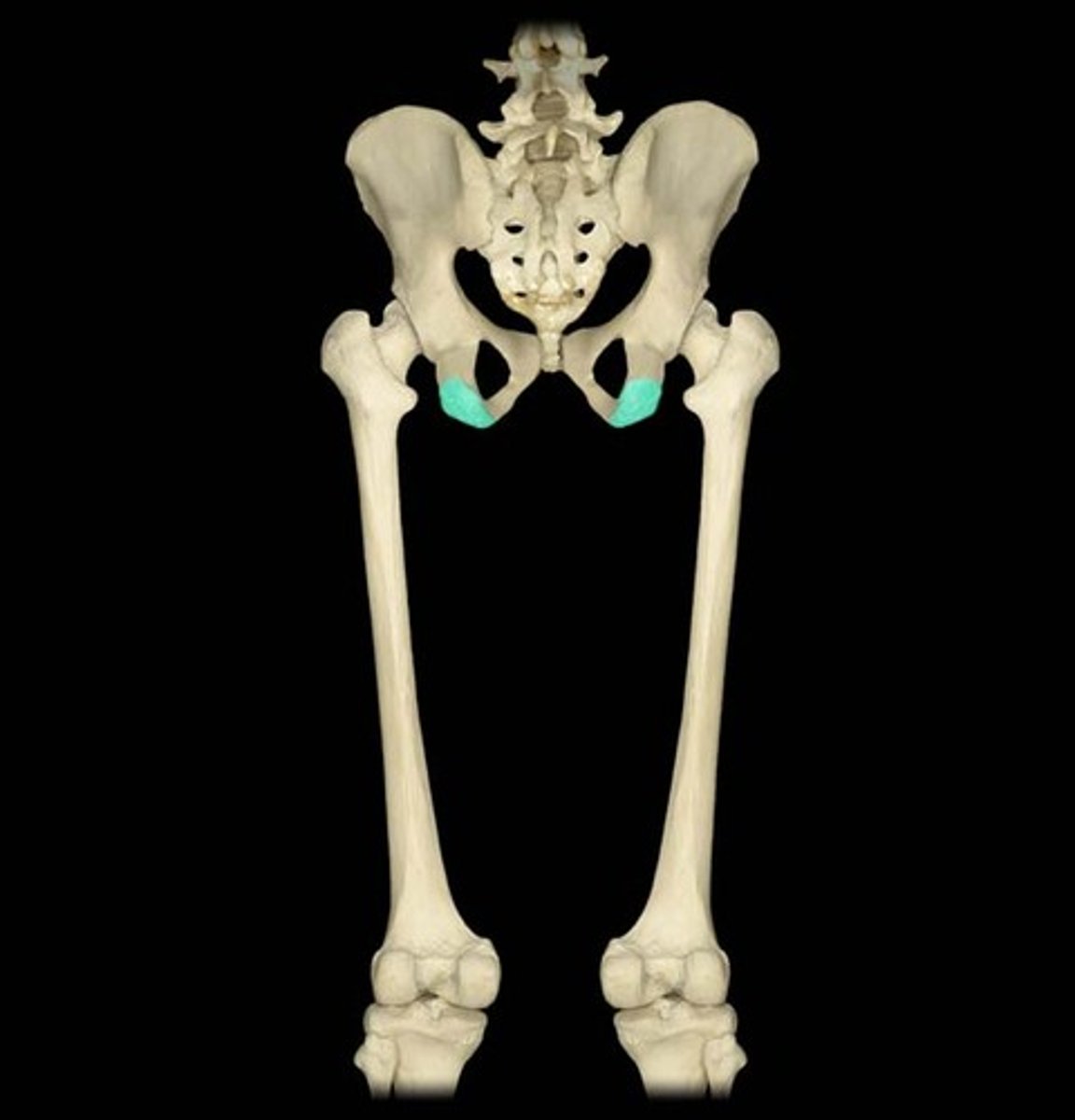
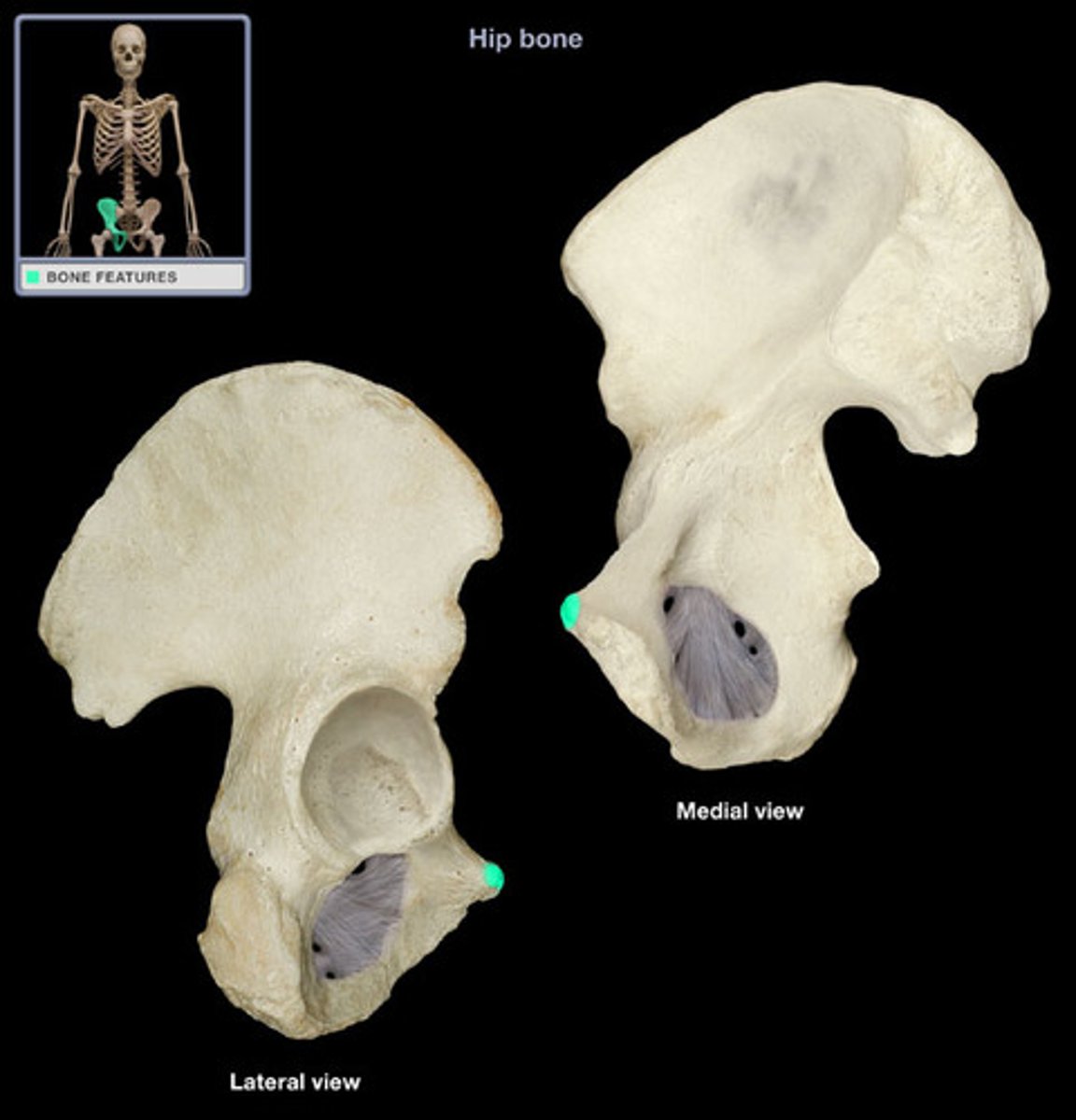
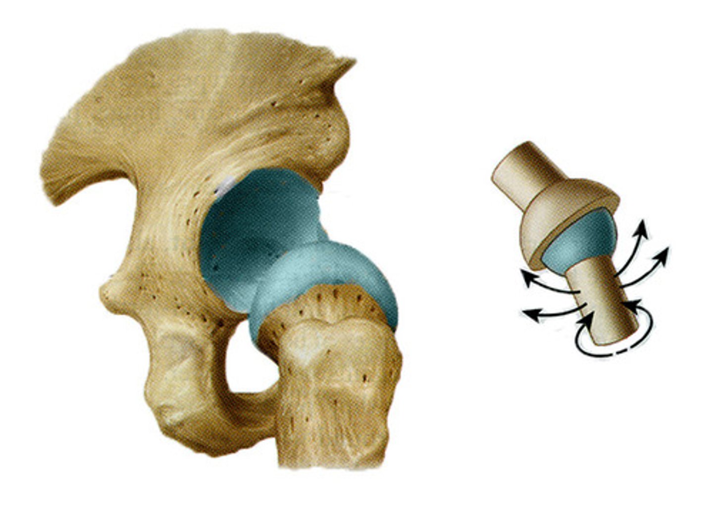
Acetabulum
Socket for the femur in the hip joint.
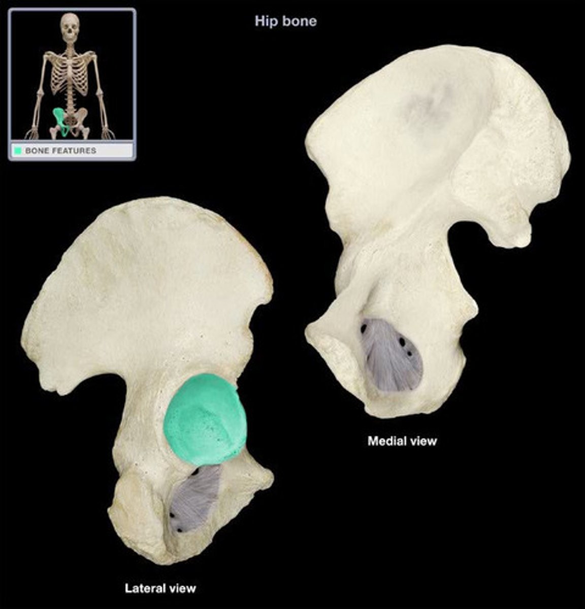
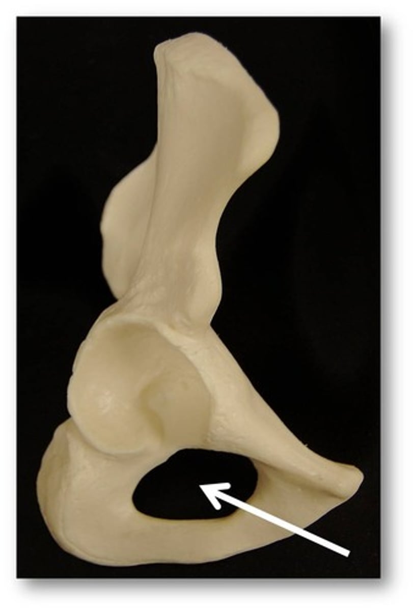
Obturator Foramen
Large opening in the pelvic girdle.
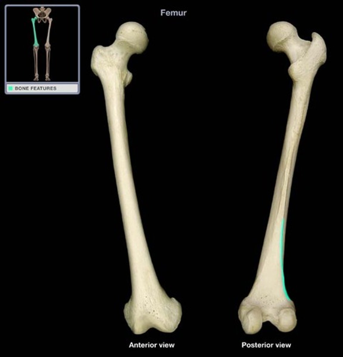
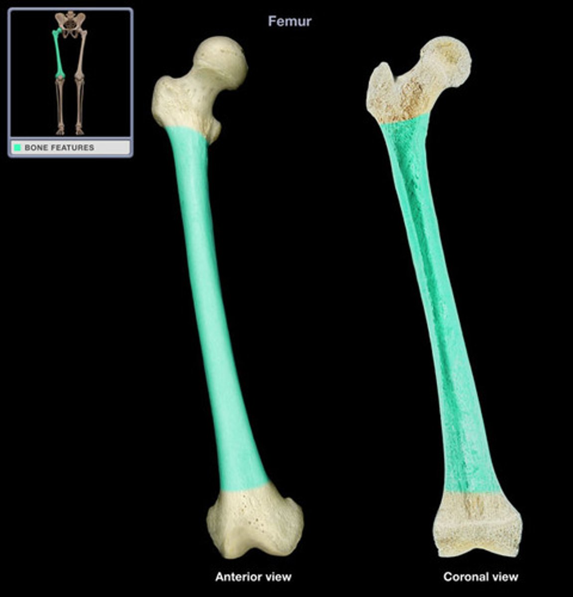
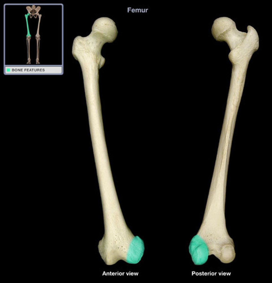
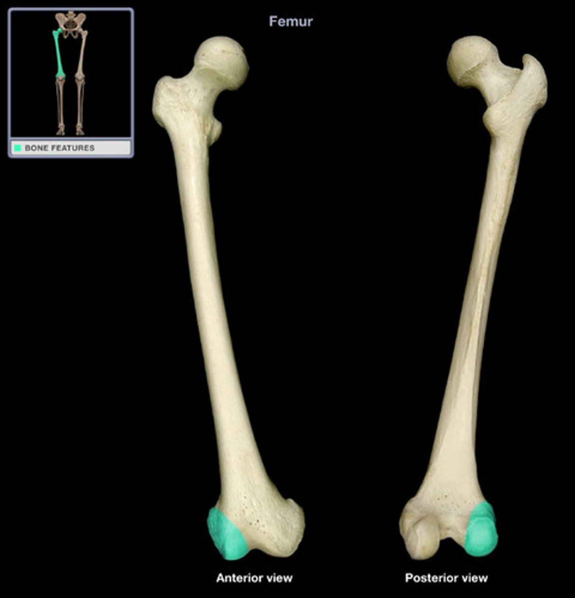
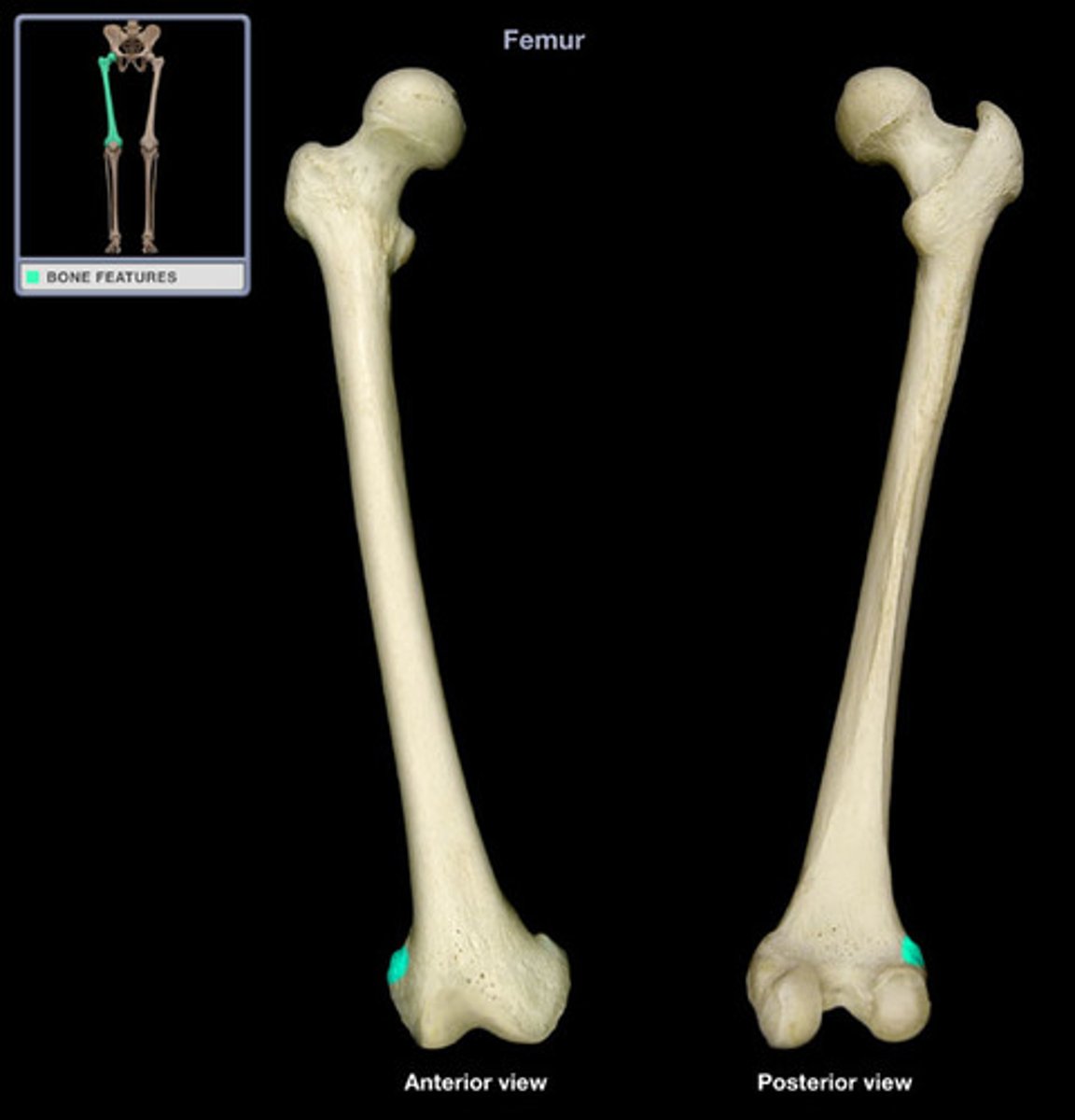
Femur
thigh bone of the leg; the longest and strongest bone in the body
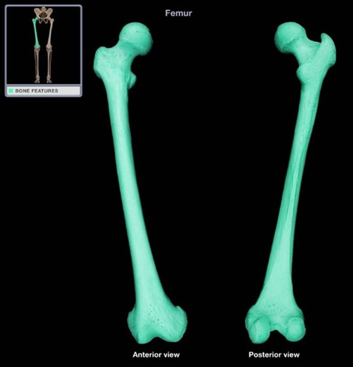
Femur diagram

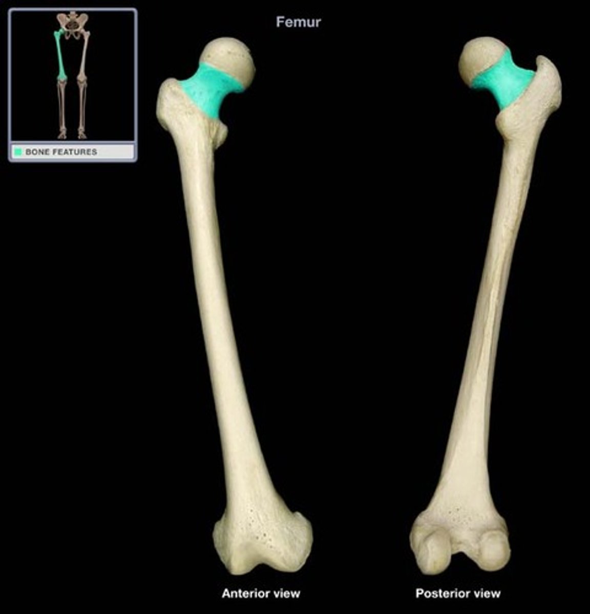
Head of femur
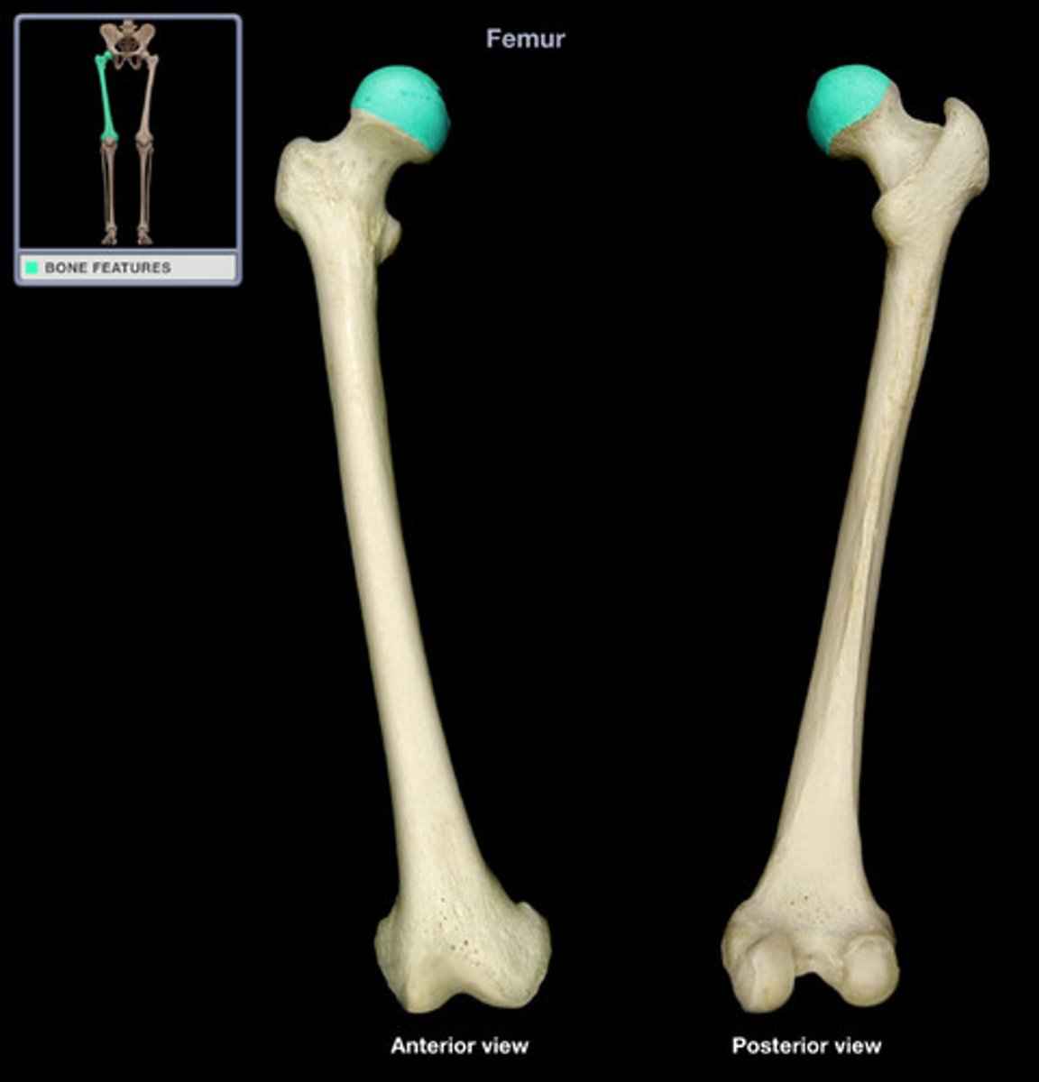
Neck of femur
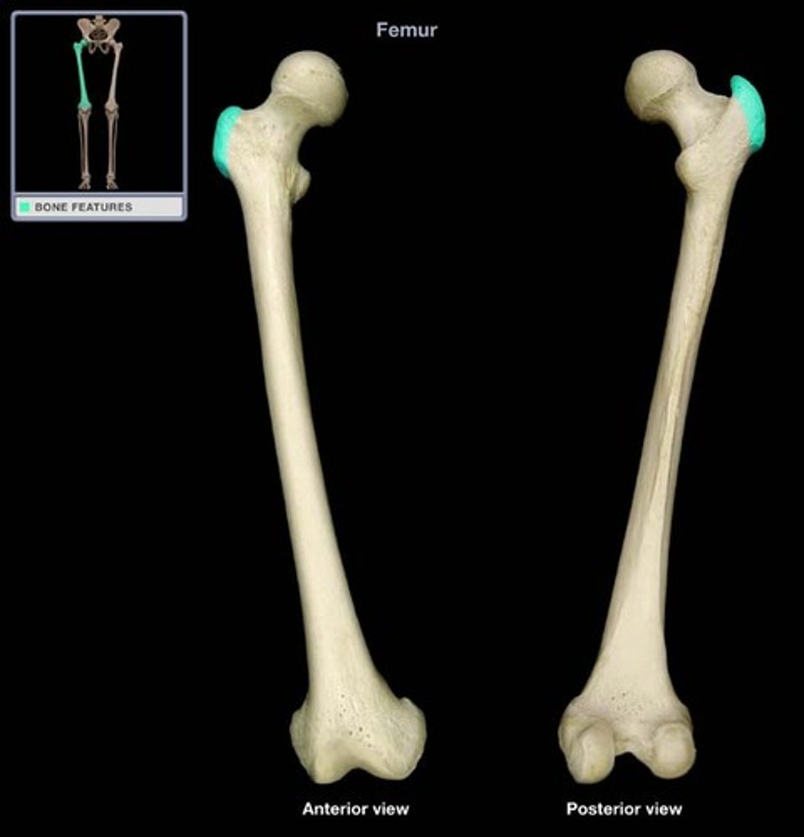
Greater trochanter of femur
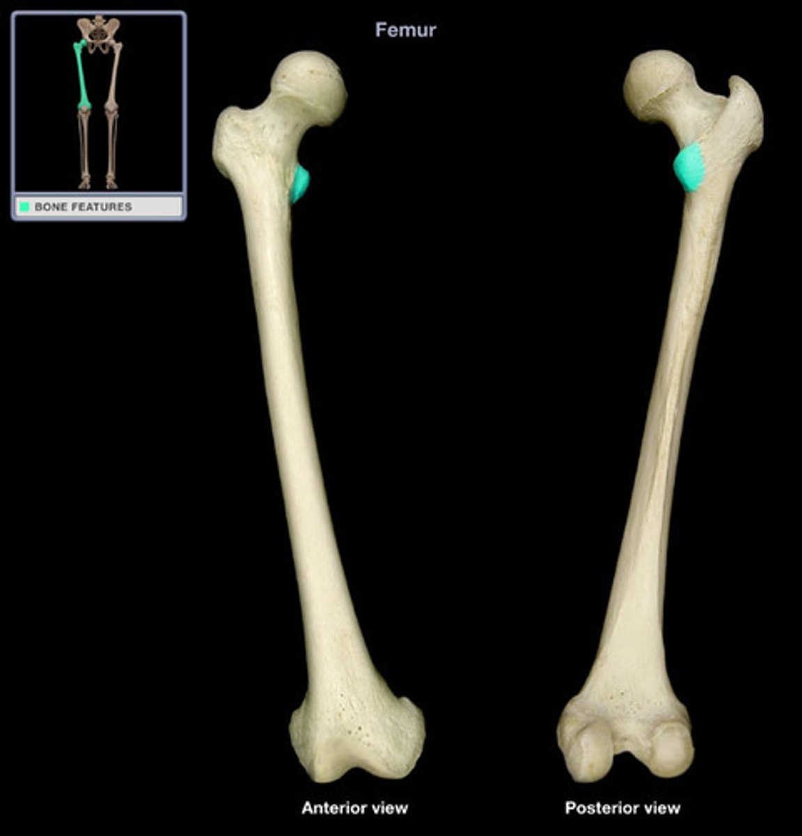
Lesser trochanter of femur
Intertrochanteric line of femur

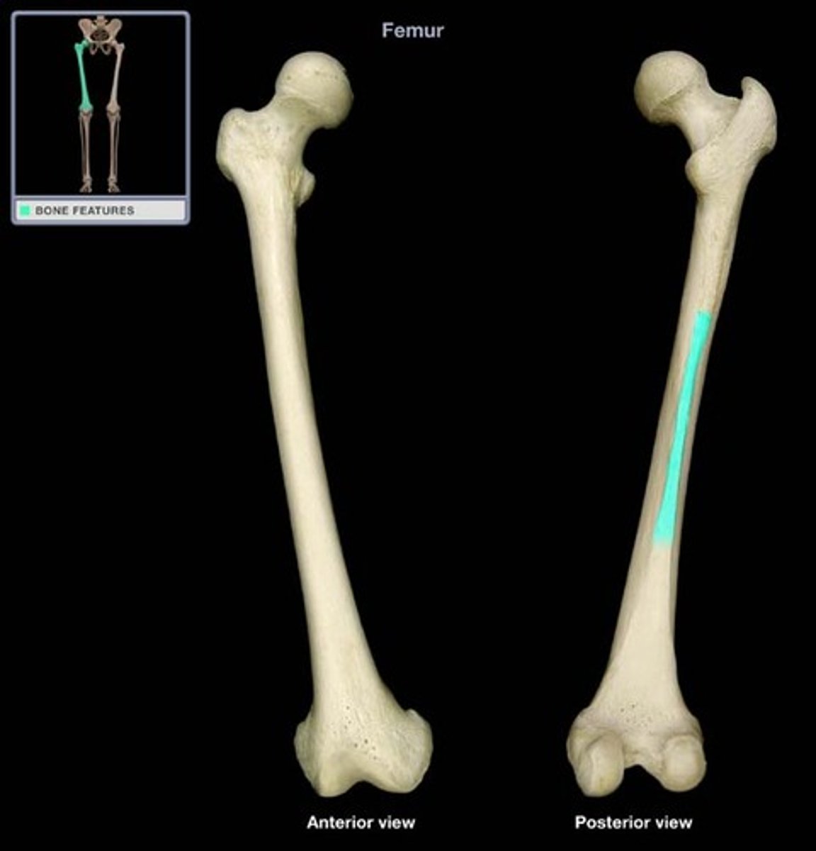
Linea aspera of femur
Medial and lateral supracondylar lines of femur
Femoral shaft
Gluteal tuberosity of femur

Medial condyle of femur
Lateral condyle of femur
Medial epicondyle of femur
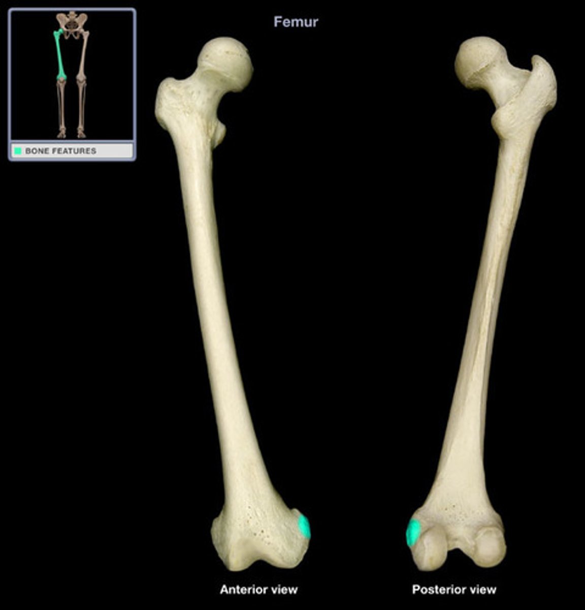
Lateral epicondyle of femur
Sacroiliac Joint
Diarthrodial Synovial gliding joint between sacrum and ilium.
Femoral Acetabular Joint
Diarthrodial Synovial ball and socket joint of hip.
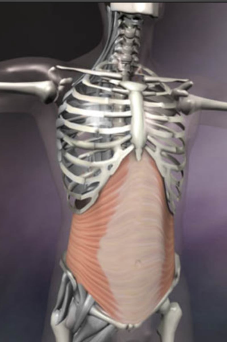
Transverse Abdominis
Origin - inguinal ligament, lumbar fascia, cartilages of last 6 ribs; iliac crest
Insertion - Linea alba, pubic crest
Action - compresses abdominal contents
Description - deepest; fibers run horizontally
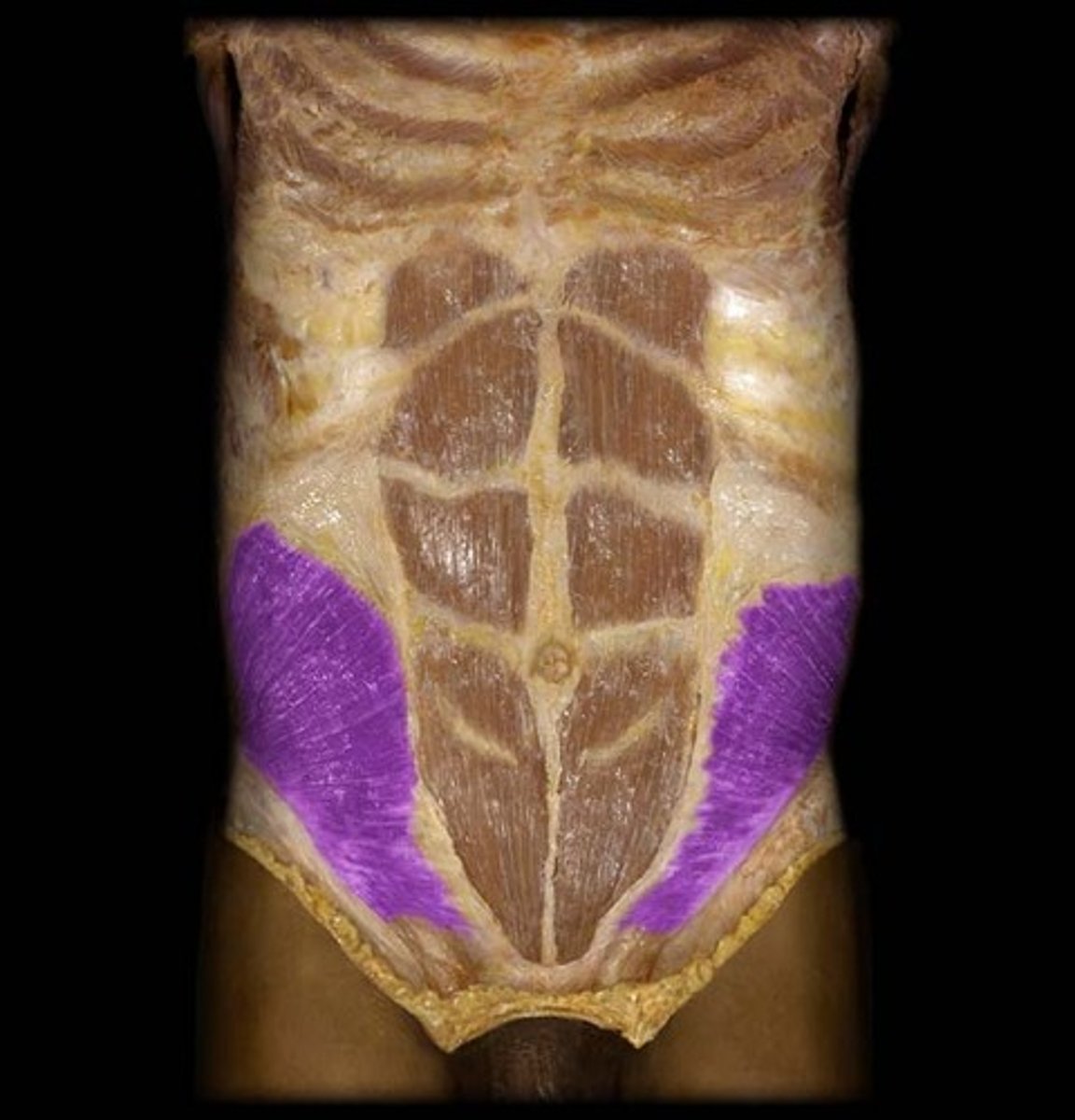
Internal Oblique
Origin - lumbar fascia; iliac crest and inguinal ligament
Insertion - Linea alba, pubic crest, last 3-4 ribs and costal margin
Action - Flex, Rotate and Laterally Bend the Spine
Description - fibers run upward and medially; BUTFANNED
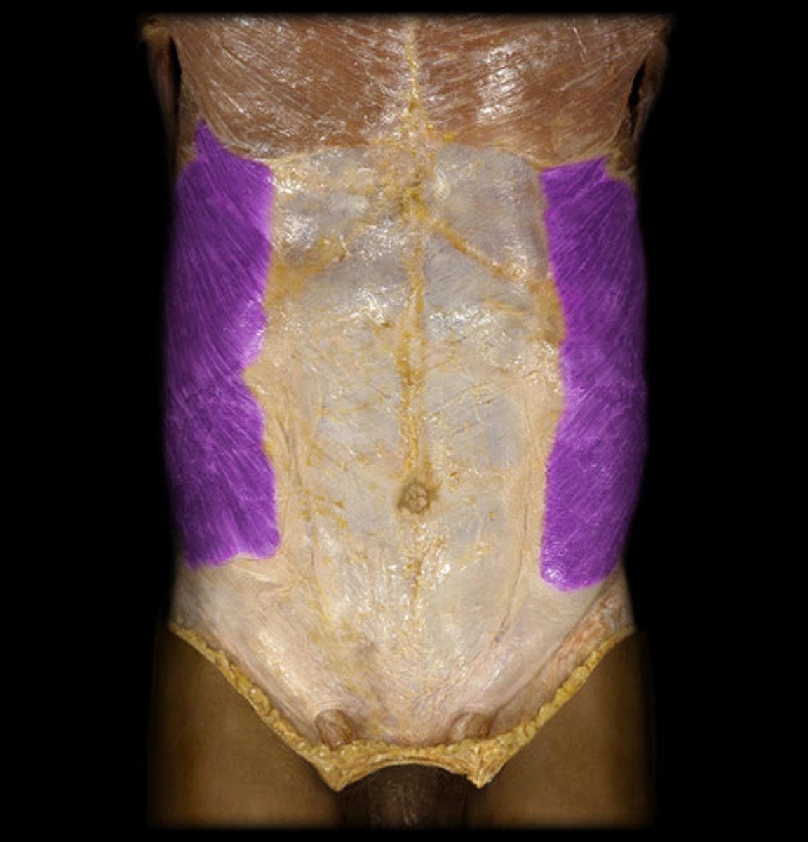
External Oblique
Origin - by fleshy strips from outer surfaces of lower 8 ribs
Insertion - Linea alba; pubic crest, pubic tubercle and iliac crest
Action - flex vertebral column/compress abdominal wall (BOTH)Rotate and lateral flexion (single)
Description - largest most superficial of 3 lateral muscles; fibers run downward and medially
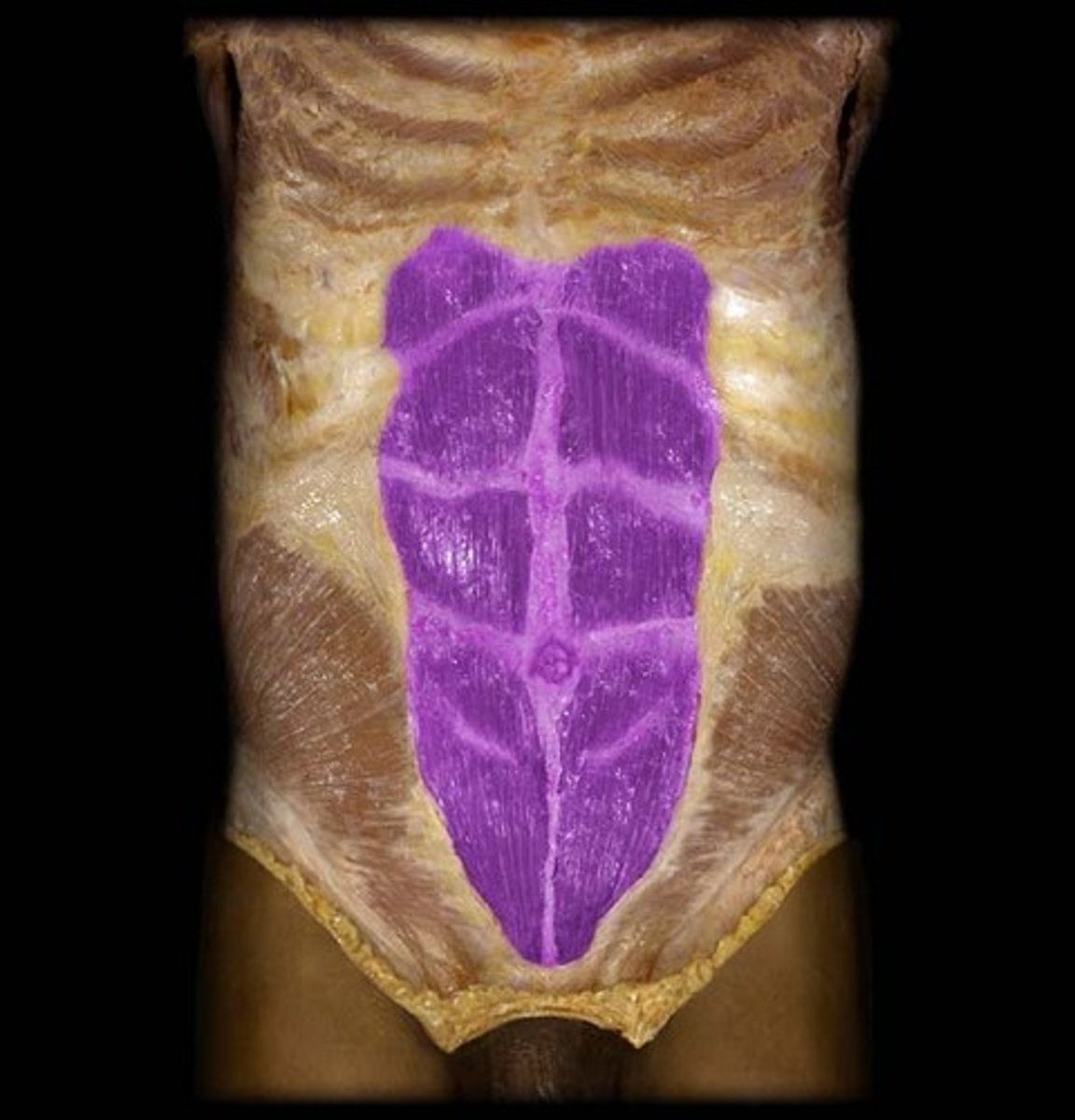
Rectus Abdominis
Origin - pubic crest
Insertion - xiphoid process and costal cartilages ribs 5-7
Action - flex and rotate lumbar region of vertebral column
Description - medial superficial muscle
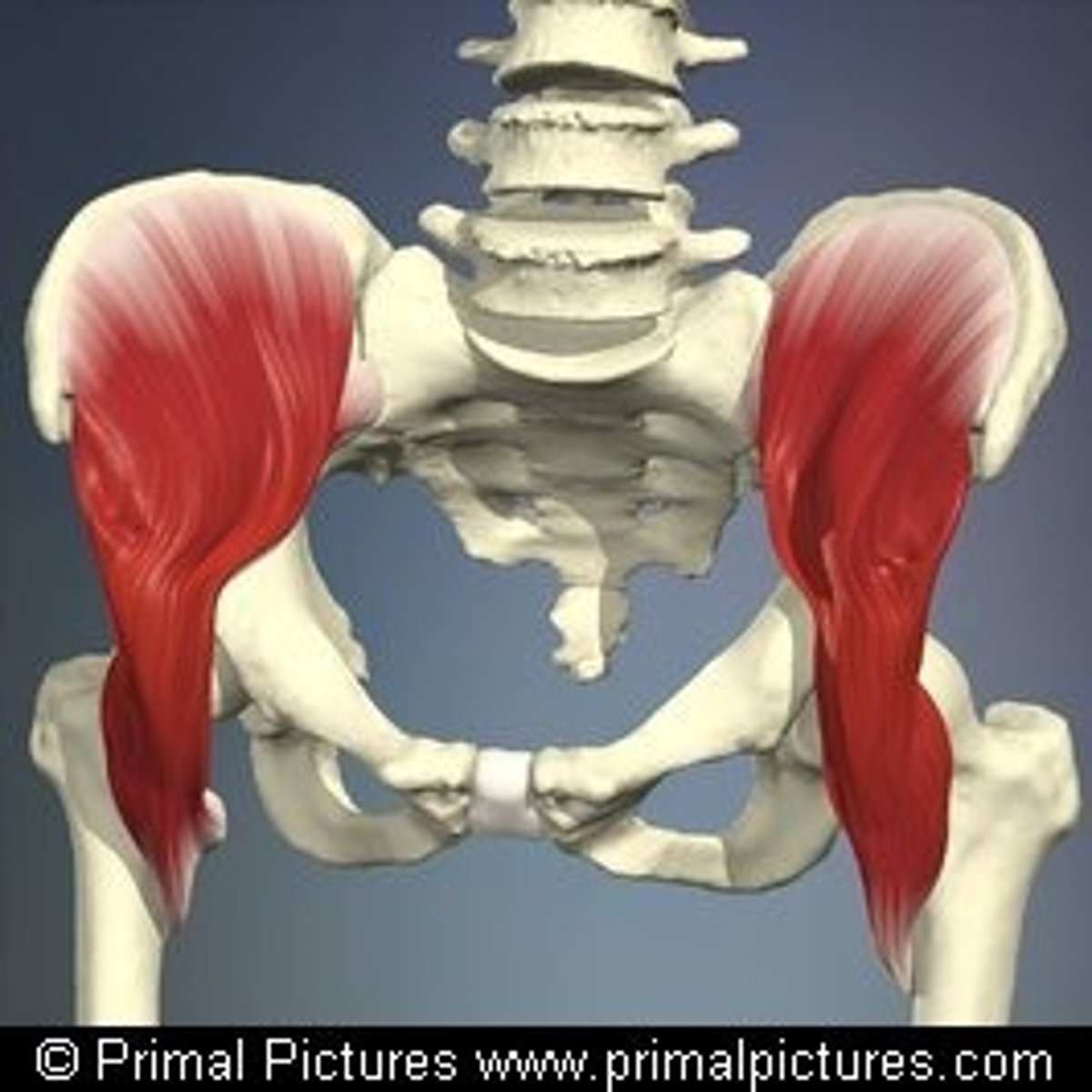
Iliopsoas Group
Includes iliacus and psoas major muscles.
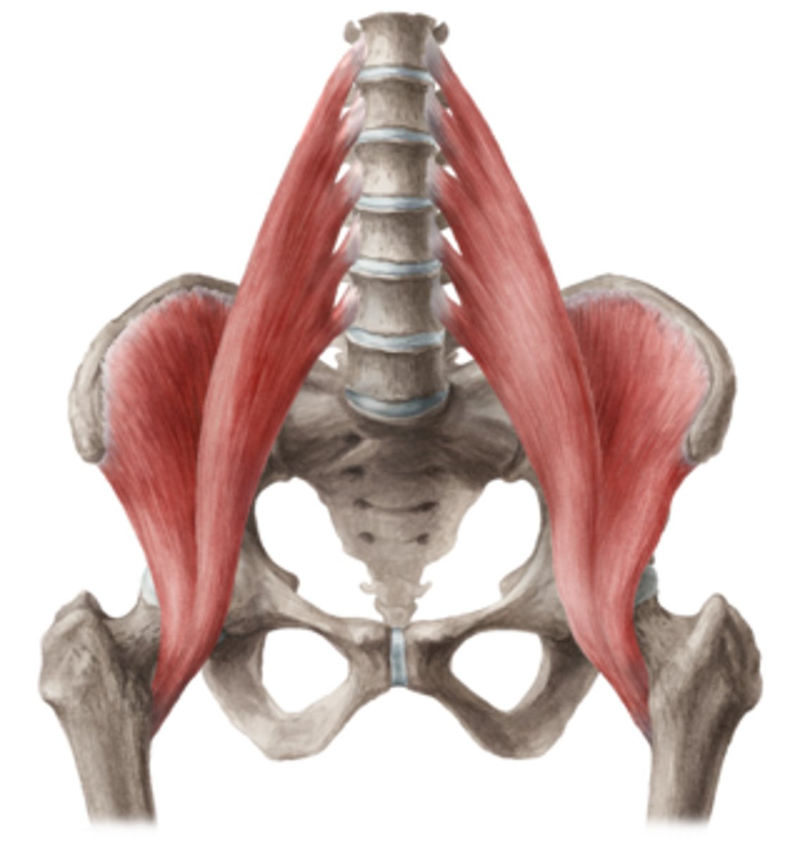
Iliacus
Origin - iliac fossa, crest and ala of sacrum
Insertion - lesser trochanter of femur
Action - thigh flexion; or flexing trunk on thigh
Description - large, fan-shaped lateral muscle
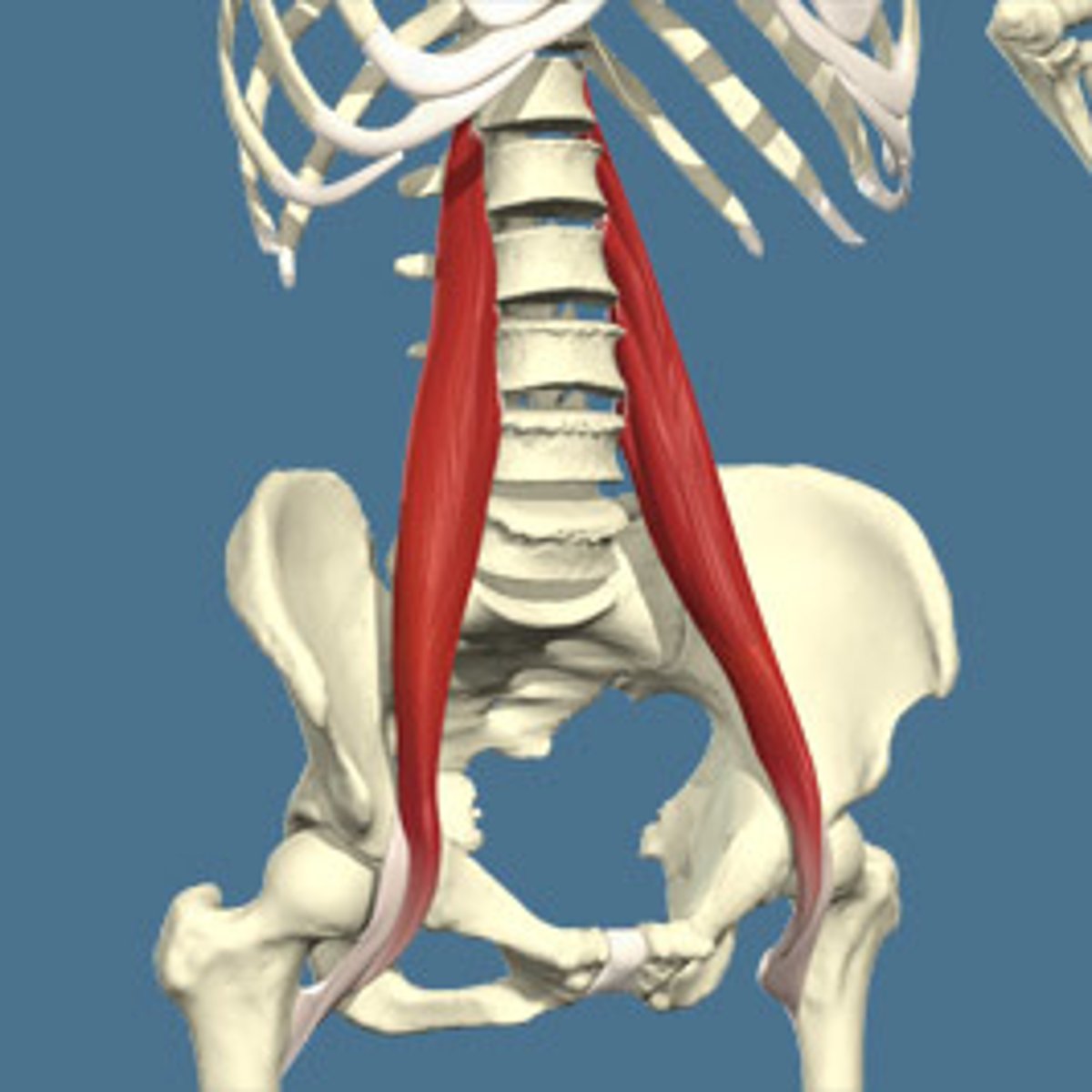
Psoas major
Origin - transverse processes/bodies/disc of lumbar vertebrae and T12
Insertion - lesser trochanter of femur
Action - thigh flexion; or flexing trunk on thigh
Description - long, thick medial portion
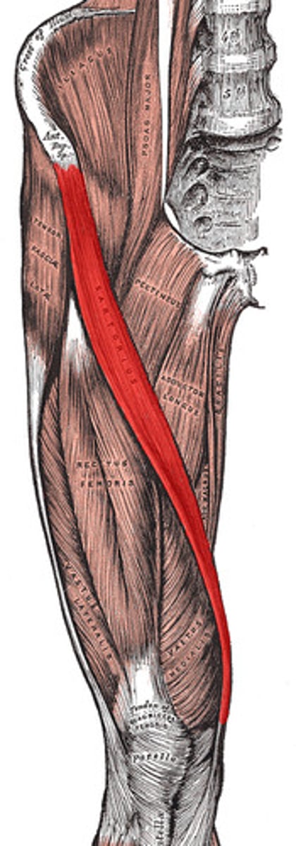
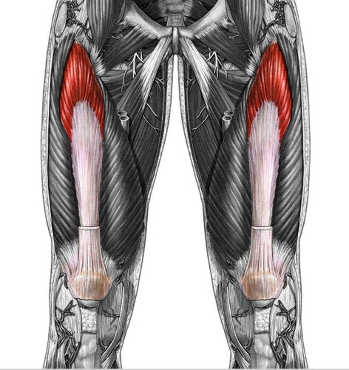
Sartorius
Origin - anterior superior iliac spine
Insertion - winds around medial aspect of the knee into medial aspect of the proximal tibia
Action - flexes, abducts, and laterally rotates thigh
Description - "straplike" superficial muscle running obliquely across anterior surface of thigh to knee; crosses both hip and knee joints
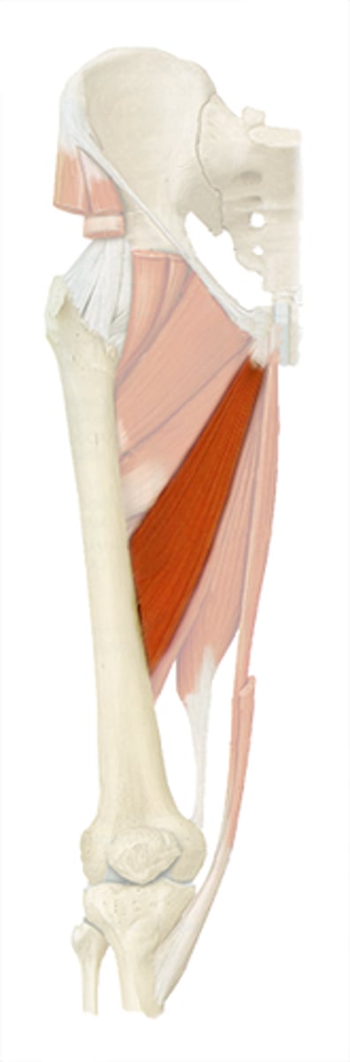
Adductor Longus
Origin - pubis
Insertion - Linea aspera
Action - adducts, flexes, and medially rotates thigh
Description -most anterior of adductor muscles
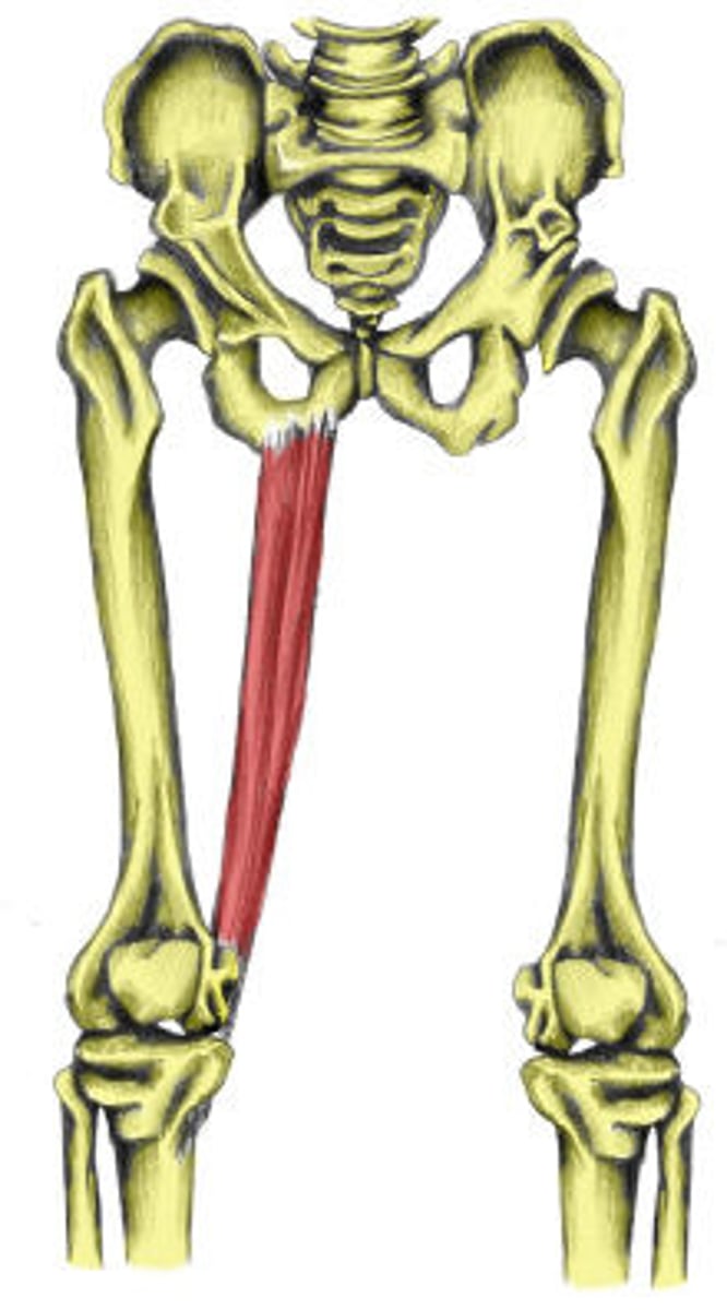
Gracilis
Origin - anterior medial edge of descending ramus of pubis
Insertion - anterior medial surface of the tibia just below the medial condyle
Action - flexes, abducts, and internal rotation
Description - "straplike" medial muscle -weak assist to knee flexion
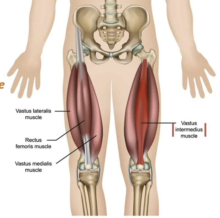
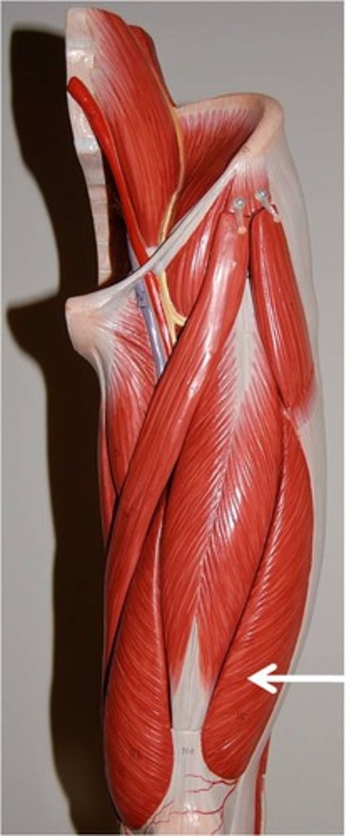
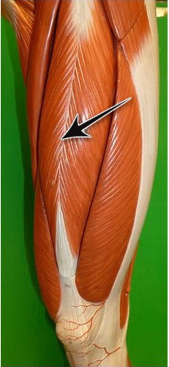
Quadriceps Group
The group of muscles consisting of vastus medialis + vastus medialis oblique (VMO), vastus lateralis, vastus intermedius, and rectus femoris. Extends the leg.
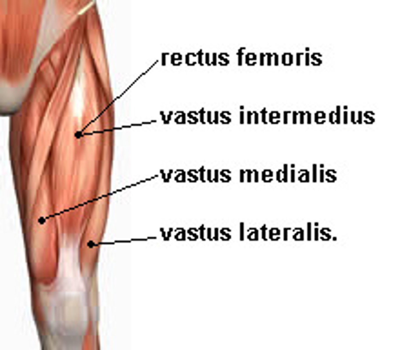
Quadriceps diagram
Vastus Medialis
Origin - Linea aspera, intertrochanteric and medial supracondylar lines
Insertion - patella and tibial tuberosity via patellar ligament
Action - extends knee and stabilizes patella (VMO)
Description - Inferomedial aspect of thigh
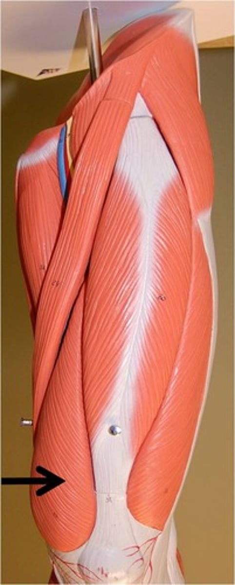
Vastus Lateralis
Origin - greater trochanter, intertrochanteric line, Linea aspera
Insertion - patella and tibial tuberosity via patellar ligament
Action - extends and stabilizes knee
Description - lateral aspect of thigh
Vastus Intermedius
Origin - anterior and lateral surfaces of proximal femur shaft
Insertion - patella and tibial tuberosity via patellar ligament
Action - extends knee
Description - between lateralis and medialis under rectus femoris
Rectus Femoris
Origin - anterior inferior iliac spine and superior margin of acetabulum
Insertion - patella and tibial tuberosity via patellar ligament
Action - extends leg and flexes thigh at hip
Description - superficial muscle of anterior thigh
Tensor Fascia Latae
Origin - anterior aspect of iliac crest and anterior superior iliac spine
Insertion - iliotibial tract
Action - stabilizes leg and trunk on thigh by making IT band taut
Description - anterolateral aspect of thigh
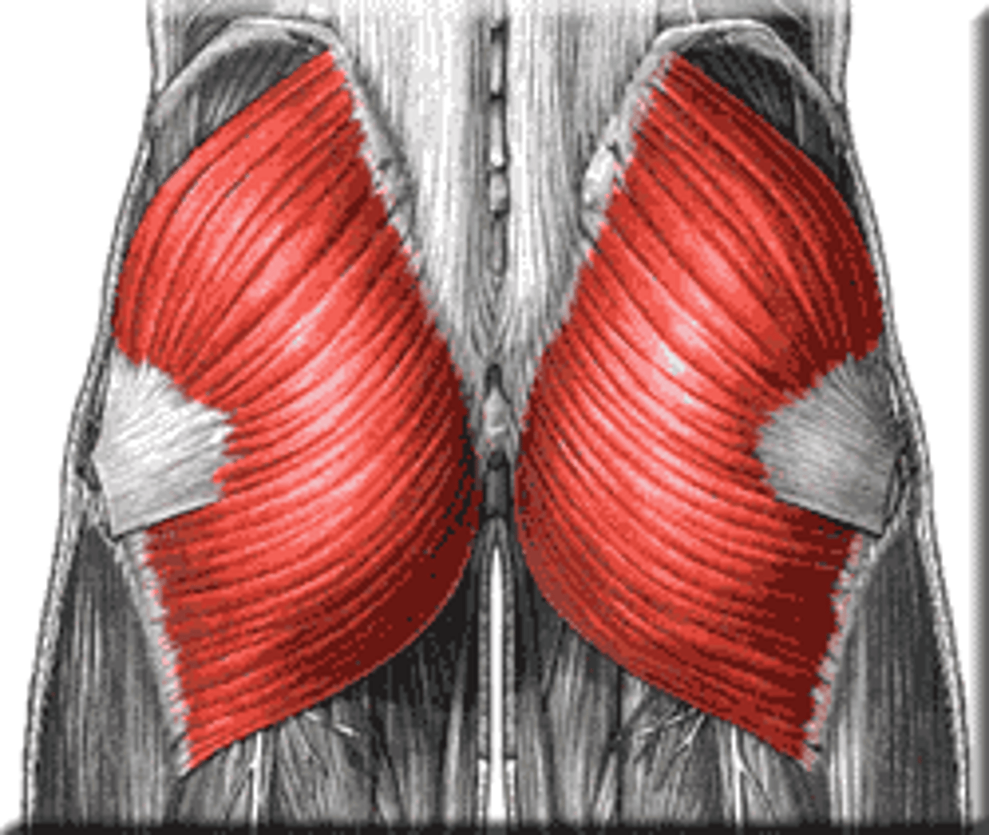
Gluteus Maximus
Origin - dorsal ilium, sacrum and coccyx
Insertion - gluteal tuberosity of femur/IT tract
Action - extensor of thigh
Description - largest most superficial gluteus muscle
Gluteus Medius
Origin - anterior and posterior gluteal lines on lateral surface of ilium
Insertion - by short tendon into lateral aspect of greater trochanter of femur
Action - abducts and medially rotates thigh
Description - thick muscle covered by maximus
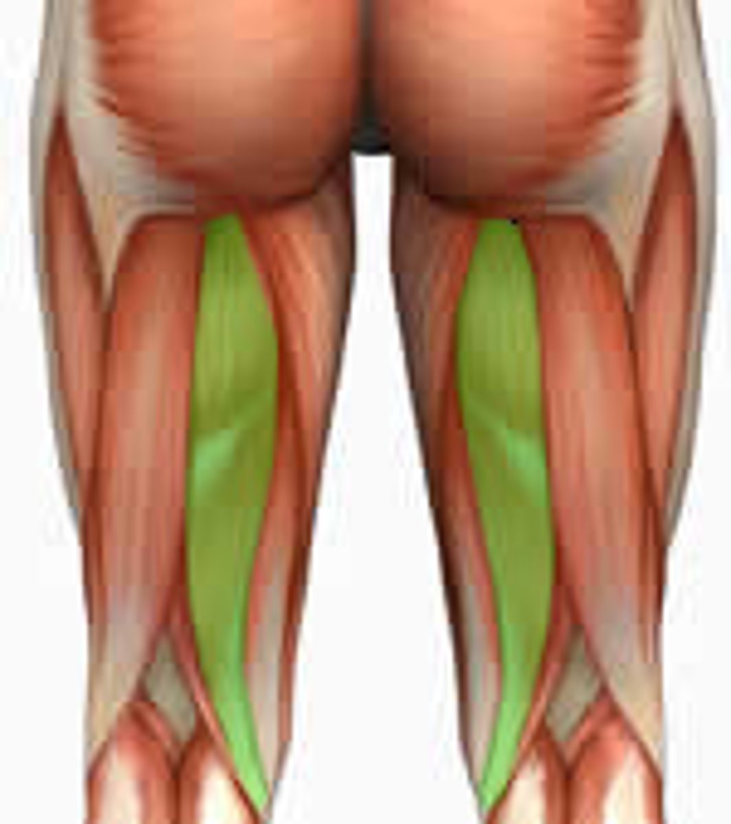
Hamstring Group
Group of muscles consisting of biceps femoris, semimembranosus, semitendinosus
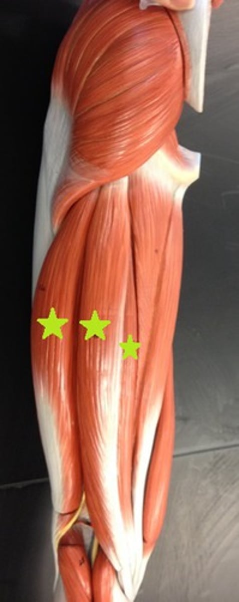
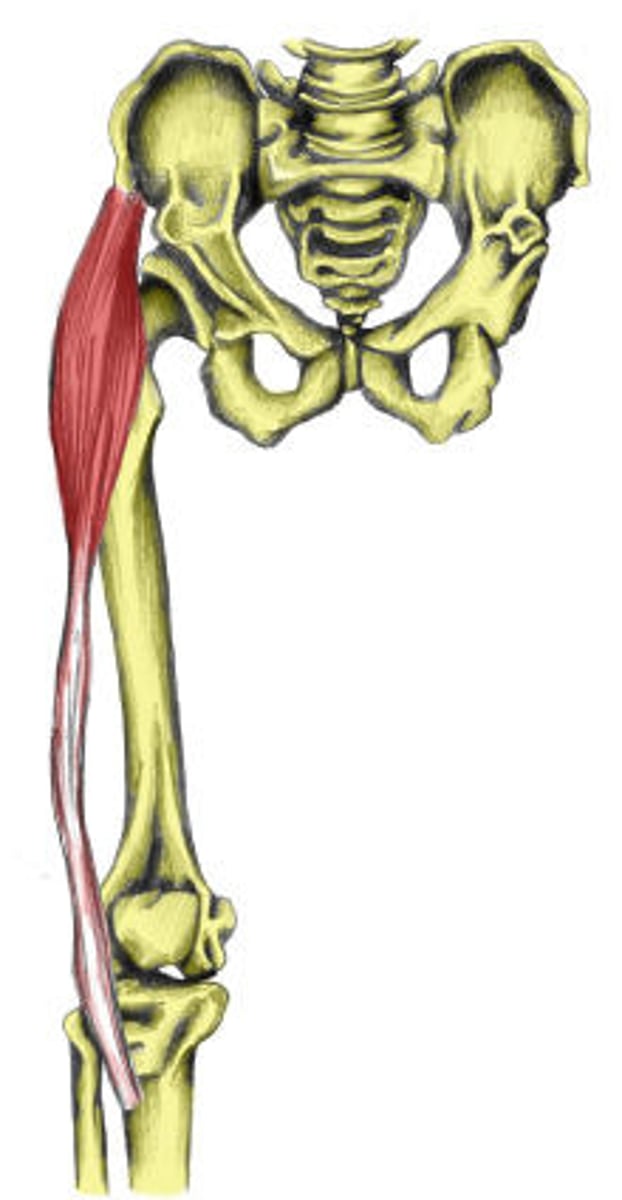
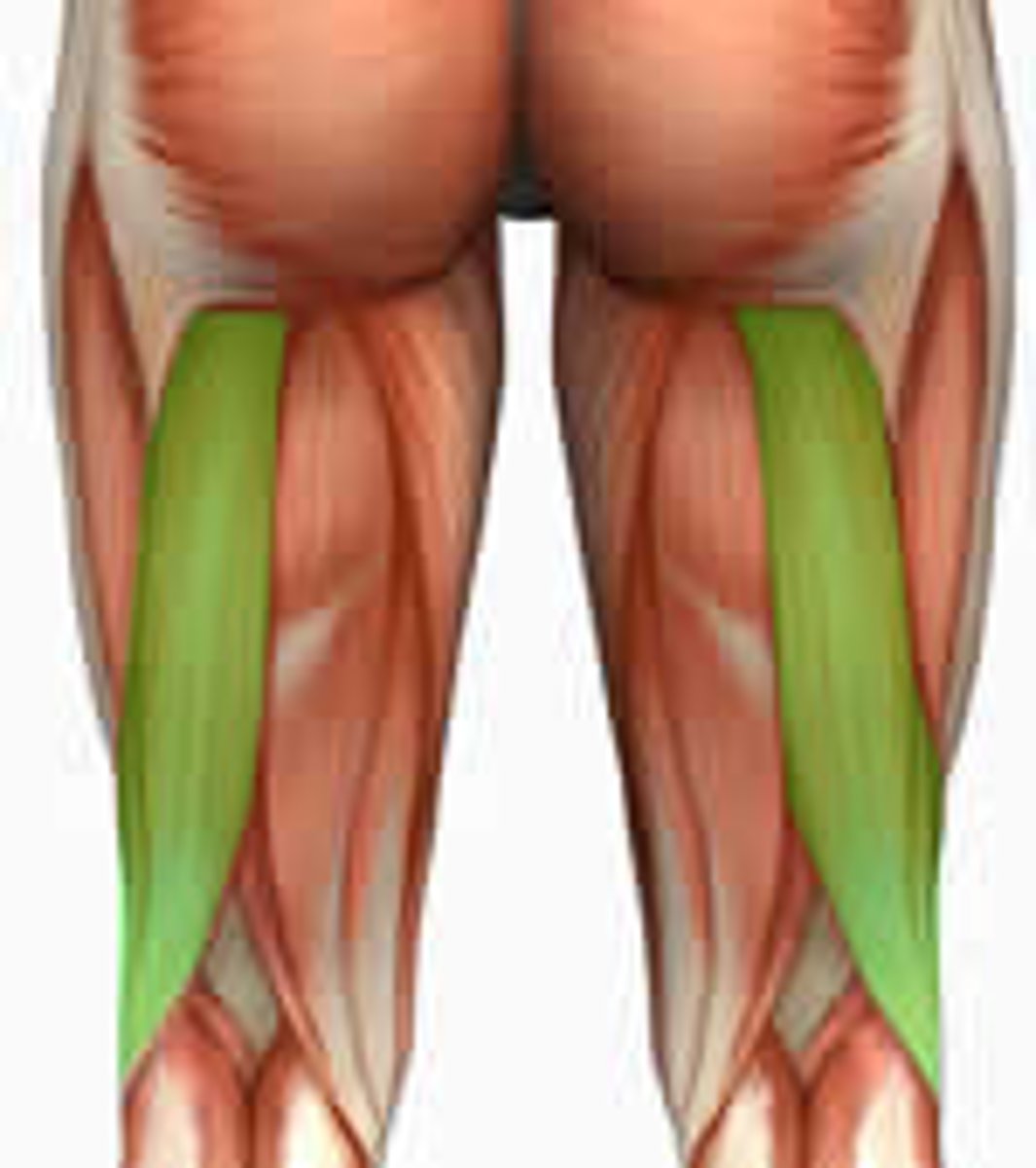
Biceps Femoris
Origin - ischial tuberosity (long head); linea aspera, lateral supracondylar line, and distal femur (short head)
Insertion - head of fibula and lateral condyle of tibia
Action - extends thigh and flexes leg
Description - most lateral muscle of group; 2 heads
Semimembranosus
Origin - ischial tuberosity
Insertion - medial condyle of tibia
Action - extends thigh and flexes knee
Description - deep to semitendinosus
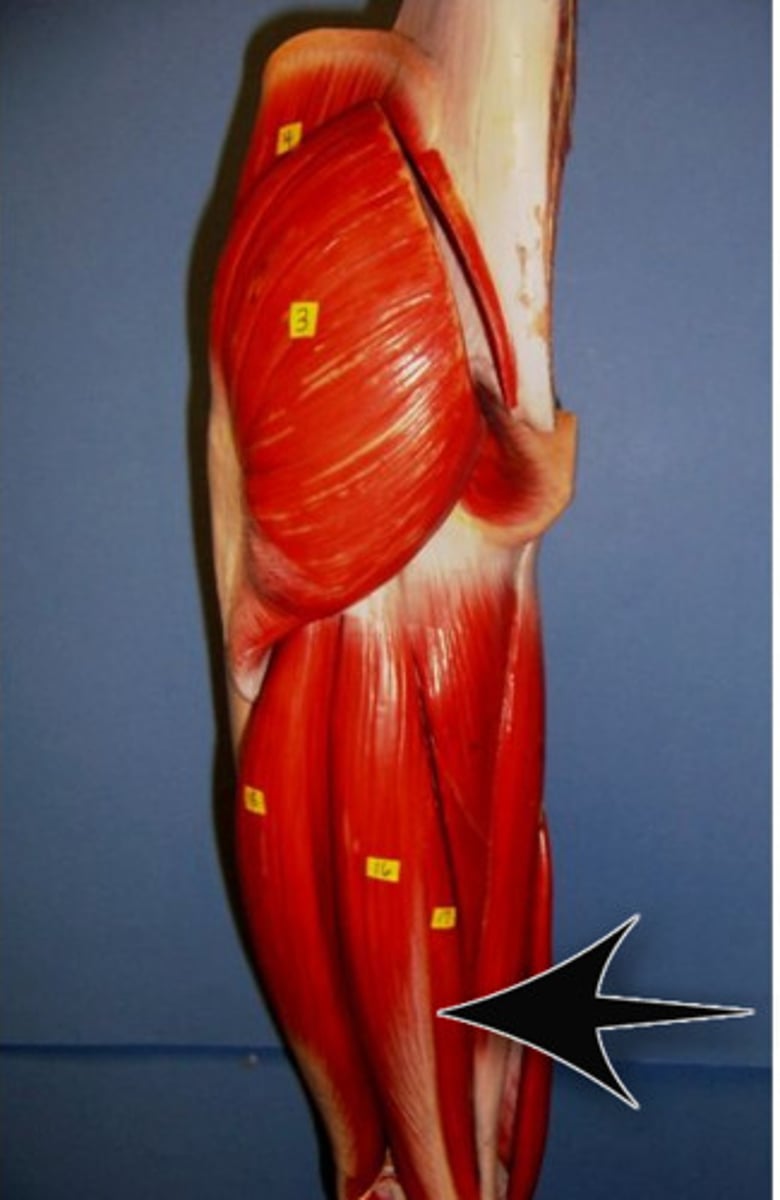
Semitendinosus
Origin - ischial tuberosity
Insertion - medial aspect upper tibial shaft
Action - extends thigh and flexes knee
Description - medial to biceps femoris; strap-like superficial muscle