SS - ORAL CAVITY & TONGUE
1/32
There's no tags or description
Looks like no tags are added yet.
Name | Mastery | Learn | Test | Matching | Spaced | Call with Kai |
|---|
No analytics yet
Send a link to your students to track their progress
33 Terms
ORAL CAVITY STRUCTURE & FUNCTION
STUCTURE
The oral cavity has two parts; oral vestibule and oral cavity proper
The teeth and gums separate the two.
FUNCTION
Ingestion
Mastication (Chewing)
Digestion
Pressurise air (EX: whistling)
Vocal tract
obstruct/partially obstruct sound
Modify resonance of sound by changing shape
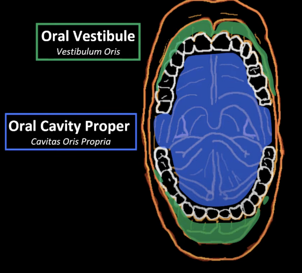
BOUNDARIES OF ORAL CAVITY PROPER
Anterior Boundary → Teeth
Lateral Boundary → Teeth (in alveolar ridges)
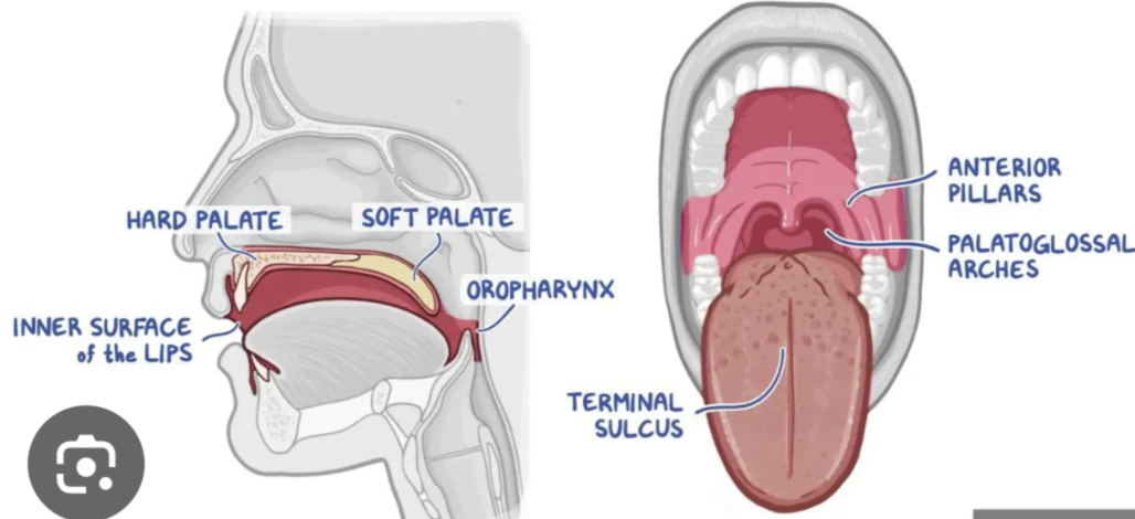
Posteriorly → Palatoglossal arch (anterior faucial pillar)
Superior → Soft & Hard Palates
Inferior → Tongue
FUNCTION:
Intake and digestion of food and water
Formation of speech
Respiration
BOUNDARIES OF ORAL VESTIBULE
Vestibule of mouth is basically sides of mouth inside the cheek (the space in between)
Space between the lips and cheeks, and the teeth/gingiva
FUNCTION:
Containing food during mastication, aided by muscles such as the buccinator which helps keep food between the teeth.
Houses the opening of the parotid duct opposite the upper second molar, which secretes saliva into this space.
LIPS: STRUCTURE & FUNCTION
|
 |
|---|

CHEEKS: STRUCTURE & FUNCTION
 |
 |
|---|
GUMS: STRUCTURE & FUNCTION
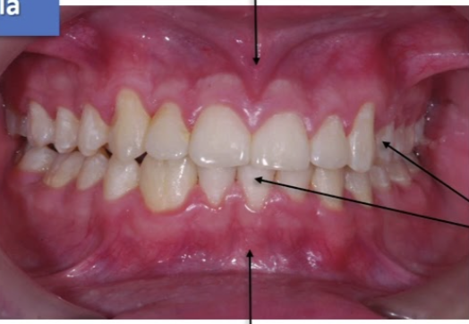
Gums is a mucous membrane covering the alveolar process of the maxilla and mandible
They form a place for the teeth to be rooted in
Gums are composed of dense fibrous tissue
Gingival papillae = projections you see between the neck of the teeth

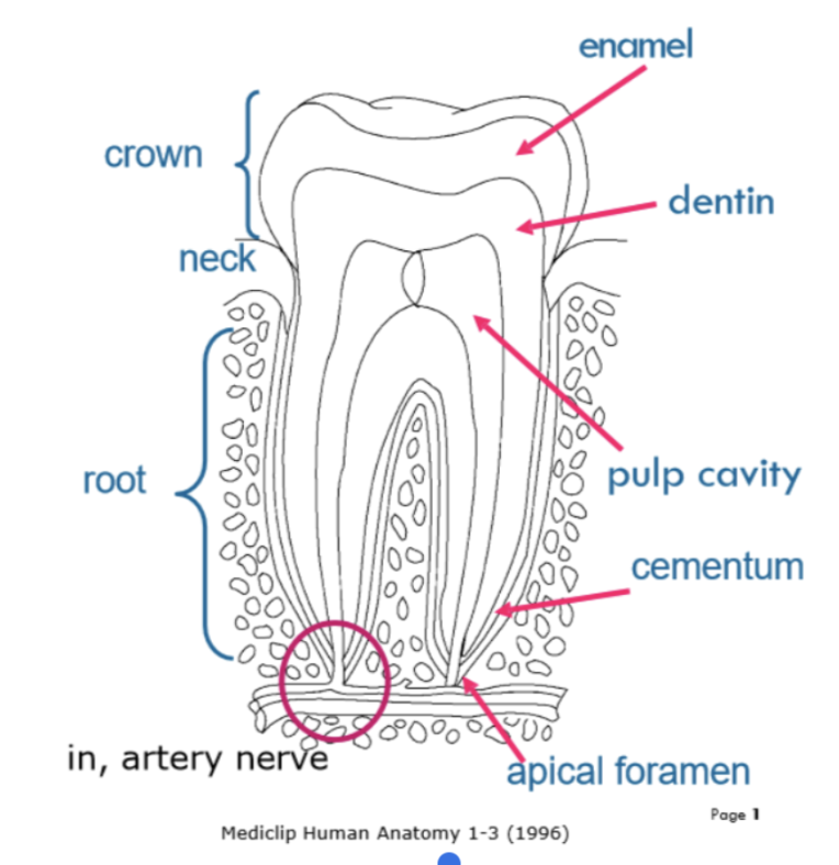
TEETH: STRUCTURE & FUNCTION
|
|
|---|
TEETH; LIFE CYCLE
PRENATAL GROWTH
Deciduous & permanent teeth developed in utero (in uterus in the womb)
Deciduous teeth begin developing at 5-6 weeks
Perm. teeth develop above deciduous teeth at about 10 weeks
Development follows development of the whole skeleton
Teeth do not “erupt” (emerge from gums) until approx 6 months after birth
ERRUPTION
Calcification (when mineralisation occurs): dentin & enamel harden
Interosseus eruption: some bone of the mandible & maxillae above the teeth resorb (breaks down) to make way for the tooth’s movement toward the gum
Clinical eruption: teeth emerge through gums
TEETH; TOOTH WEAR
Attrition = physiological wearing away of enamel as a result of tooth to tooth contact (EX: grinding)
Abrasion = physical wear via abnormal mechanical process (EX: violent brushing)
Erosion = loss of tooth surface via chemical process (not bacterial (EX: diet related acids, like coke)

TEETH STRUCTURE
Deciduous teeth = 5 in each quadrant = 20 total
Perm teeth = 7 in each quadrant = 28 total
Third molars = wisdom teeth = thus 32 total
Children have two molars, but when they get perm. teeth they get 2 premolars and then 2 molars, then wisdom teeth

TEETH; DENTAL SUCCESSION DECIDUOUS TEETH
Erupts from 6 mo. - 2yrs
Sheds from 6 yrs - 15 yrs
TOOTH APPEAR | SHED (full eruption) |
Central Incisors (6-8 mo.) | 6-8 yrs |
Lateral Incisors (8-20 mo.) | 6.5 - 8.5 years |
1st Molar & Canines (15-20 mo.) | 9.5 - 12 yrs |
2nd Molar (20-24 mo.) | 10 - 11.5 yrs |
TEETH; DENTAL SUCCESSION PERMANENT TEETH
TOOTH | SHED (full eruption) |
Central & Lateral Incisors, 1st Molars | 6-9 yrs |
1st & 2nd Premolars & Canines | 9 - 12 years |
2nd Molars | 11 - 13 yrs |
3rd Molars | 17 - 25 yrs |
GENERAL STRUCTURE & FUNCTION OF SALIVARY GLANDS
All exocrine glands have ducts.
FUNCTION:
Initiate digestion
Soften bolus
Allows for frictionless movement for speech
STRUCTURE:
Saliva is made up of two components
Serous Component = contains enzymes that helps break down & digest food we eat
Mucous component - lubricates the inner surfaces of our mouth & lubricates the food we eat so it passes easily (Mainly produces mucin that absorbs water to create a lubricator called mucus)
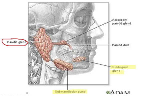
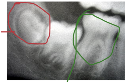
PAROTID GLANDS
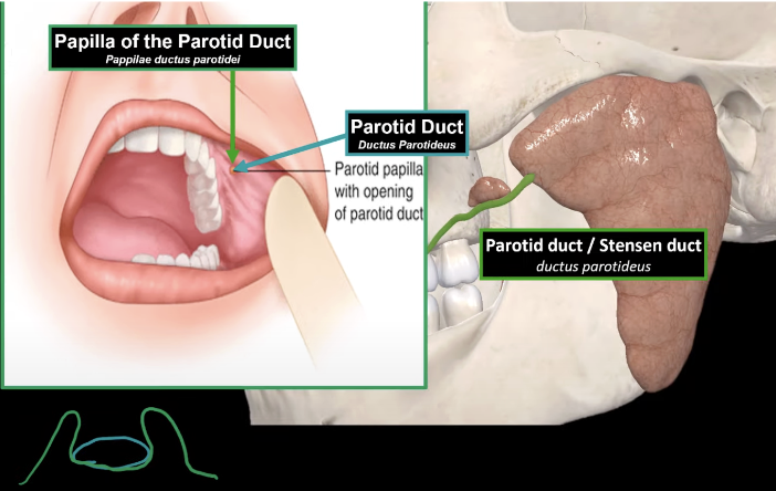
Opens into the buccal cavity above the upper second molar tooth (via the parotid duct)
Largest salivary gland humans have
Purely serous gland (helps break down food)
Function = produce watery secretion containing enzymes
Located on the lateral surface of the head, anterior to the auricle
When the parotid gland produces its serous secretion, it's going to send them through a duct called the parotid duct
This opens up at the upper side of the cheeks. The opening of the parotid duct has a papilla round it, called the papilla of the parotid duct.
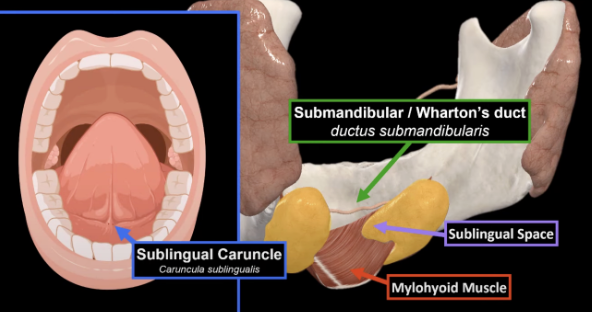
SUBMANDIBULAR GLANDS
OPENS IN THE FLOOR OF THE MOUTH
Contains seromucous glands = produce digestive enzymes & mucin that goes together with water to produce mucous (function)
Lies in submandibular space
Lies mainly under mylohyoid muscle
Submandibular duct = opens up in the middle of the floor of the oral cavity in an elevation called the sublingual caruncle
SUBLINGUAL GLANDS
OPENS UNDER THE TONGUE
Located in the sublingual space
The anterior part of the gland is seromucous
The posterior part of gland is purely mucous
Sublingual gland has two types of ducts
1) Major sublingual duct function = joins with submandibular duct to open into sublingual caruncle
2) Minor sublingual ducts function = open as small holes within the sublingual folds

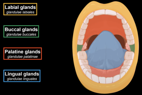
LABIAL GLANDS (LIPS) & MINOR GLANDS
Between the orbicularis oris & the mucous membrane of the lips
Saliva is secreted by salivary glands which is situated around the oral cavity
TONGUE: BRIEF STRUCTURE & FUNCTION
|
|
|---|
TONGUE STRUCTURE - GENERAL
Tongue is a muscle organ, made up by the apex, body & root
Body
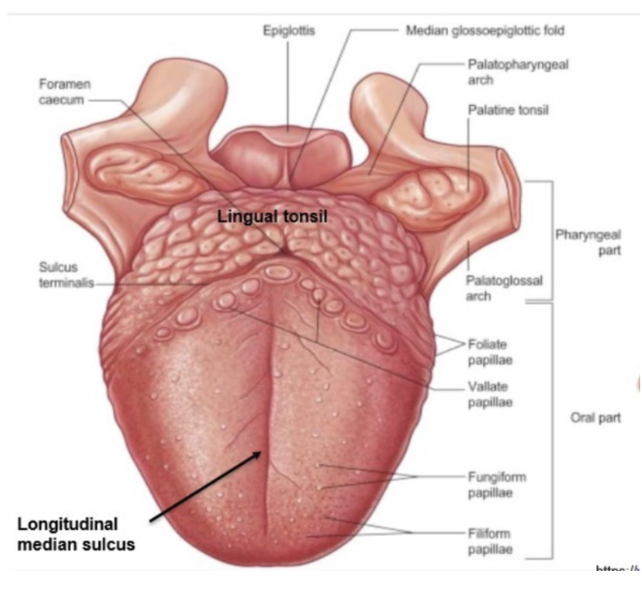
Median sulcus: A line running down the middle of the tongue.
Sulcus Terminals: A V-shaped groove separating the body from the root.
Foramen caecum: A small pit at the tip of the terminal sulcus, a remnant of the thyroglossal duct from fetal development.
Lingual tonsil: Lymphatic tissue at the root of the tongue, forming bumps on its surface.
Glossoepiglottic folds: Two folds of tissue connecting the tongue to the epiglottis, keeping it in place.
Frenulum: A thin band attaching the tongue to the floor of the mouth.
Sublingual fold: A ridge on the lower border of the tongue with openings for sublingual gland ducts.
TONGUE STRUCTURE - PAPILLAE
You have something called lingual papillae. These are rough sensory structures that contains taste buds to sense taste
1) Filiform papilla - gives the tongue its roughness as it contains a thick layer of epithelial tissue = touch
2) Fungiform papilla = small elevation that carries taste receptor buds
3) Vallate papilla = contains taste buds & located near terminal sulcus
4) Foliate papilla = contains taste buds & located on lateral regions of tongue
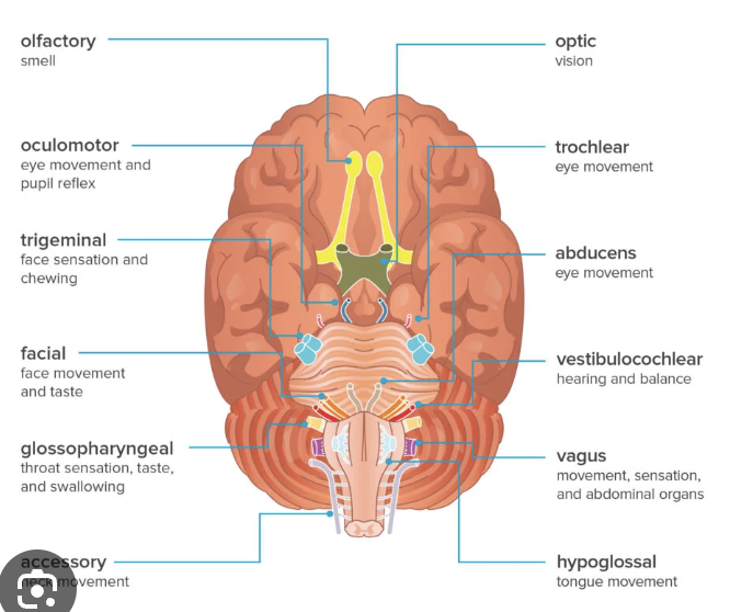
TONGUE STRUCTURE - SENSORY INNERVATION (TASTE & GENERAL)
Taste sensation for anterior 2/3 of tongue are innervated by VII (facial nerve)
General sensation for anterior 2/3 of tongue are innervated by V (Trigeminal)
Taste sensation for posterior 1/3 of tongue are innervated by IX (Glossopharyngeal)
General sensation for the posterior 1/3 of tongue are innervated by IX (Glossopharyngeal)
TONGUE STRUCTURE - EXTRINSIC MUSCLE - GENIOGLOSSUS
Genioglossus 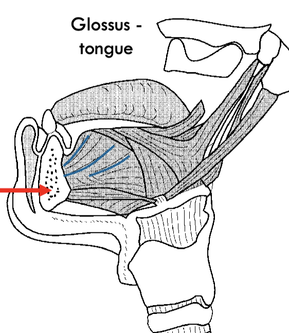 |
|
|
|
TONGUE STRUCTURE - EXTRINSIC MUSCLE - STYLOGLOSSUS
Styloglossus  |
|
|
|
TONGUE STRUCTURE - EXTRINSIC MUSCLE - HYOGLOSSUS
Hyoglossus  |
|
|
|
TONGUE STRUCTURE - EXTRINSIC MUSCLE - PALATOGLOSSUS
Palatoglossus  |
|
|
|
TONGUE STRUCTURE - INTRINSIC MUSCLE - SUPER LONGITUDINAL
Super Longitudinal  |
|
|
|
TONGUE STRUCTURE - INTRINSIC MUSCLE - INFERIOR LONGITUDINAL
Inferior Longitudinal  |
|
|
|
TONGUE STRUCTURE - INTRINSIC MUSCLE - TRANSVERSE
Transverse  |
|
|
|
TONGUE STRUCTURE - INTRINSIC MUSCLE - VERTICAL
Vertical  |
|
|
|
TONGUE DIAGRAMS


FLOOR OF ORAL CAVITY - MYLOHYOID
Floor of the oral cavity consists of 3 main muscles that primarily go from the inner surface of the mandible, to the hyoid bone.
MYLOHYOID MUSCLE (under the geniohyoid) Innervated by Vmand |
|  |
FLOOR OF ORAL CAVITY - ANTERIOR BELLY OF DIGASTRIC
ANTERIOR BELLY OF DIGASTRIC MUSCLE Innervated by Vmand |
|  |
*****SUBLINGUAL GLAND IS ABOVE THESE MUSCLES
****SUBMANDIBULAR GLAND IS BELOW THESE MUSCLES
FLOOR OF ORAL CAVITY - GENIOHYOID
GENIOHYOID MUSCLE (above mylohyoid) Innervated by hypoglossal |
|  |