BSCI201 Opoku exam 2 articulations, skeletal, integumentary
1/154
There's no tags or description
Looks like no tags are added yet.
Name | Mastery | Learn | Test | Matching | Spaced | Call with Kai |
|---|
No analytics yet
Send a link to your students to track their progress
155 Terms
Bursae
tiny sacs of fluid that are located near joints and help reduce friction
tendon sheath
elongated bursa that wraps around a tendon. DIRECTLY ATTACHED
Osteoarthritis
Wearing down of the cartilage tissue (protective tissue). Bursae leak fluid causing bones to directly touch
rheumatoid arthritis
causes joint pain and will damage joints on both sides of the body; autoimmune disease. Bursae inflamed
synathrotic joints
immovable
amphiarthrotic joints
slightly movable
diarthrotic joints
freely movable
cartilaginous joints
cartilage (hyaline synarthrotic and fibrocartilage amphiarthrotic); no joint cavity
synovial joints
joint cavity present *all synovial joints are diarthrotic
pivot joint
rotation of the atlanto-axial joints; head goes NO
pivot joint f/s
F: diarthrotic
S: synovial
condyloid joint
atlanto-occipital extension; head goes NO
condyloid joint f/s
F: diarthrotic
S: synovial
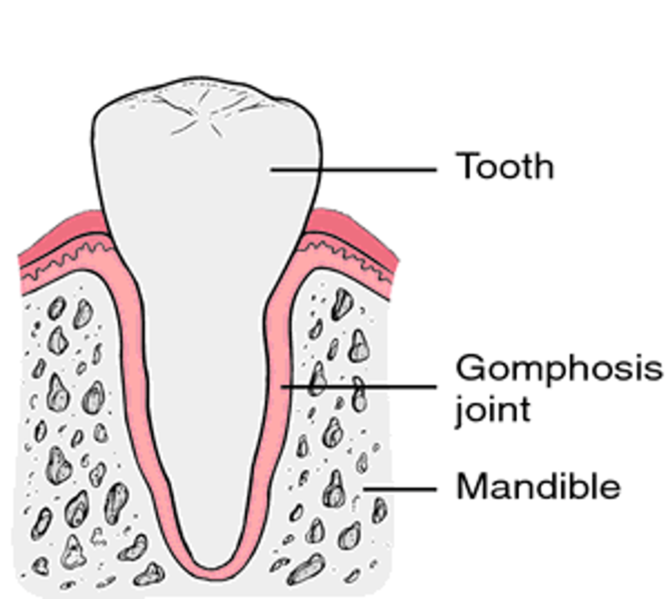
gomphoses joint (ADULT)
fibrous joint that attaches the tooth to its socket
gomphoses joint f/s
f: synarthrotic
s: fibrous
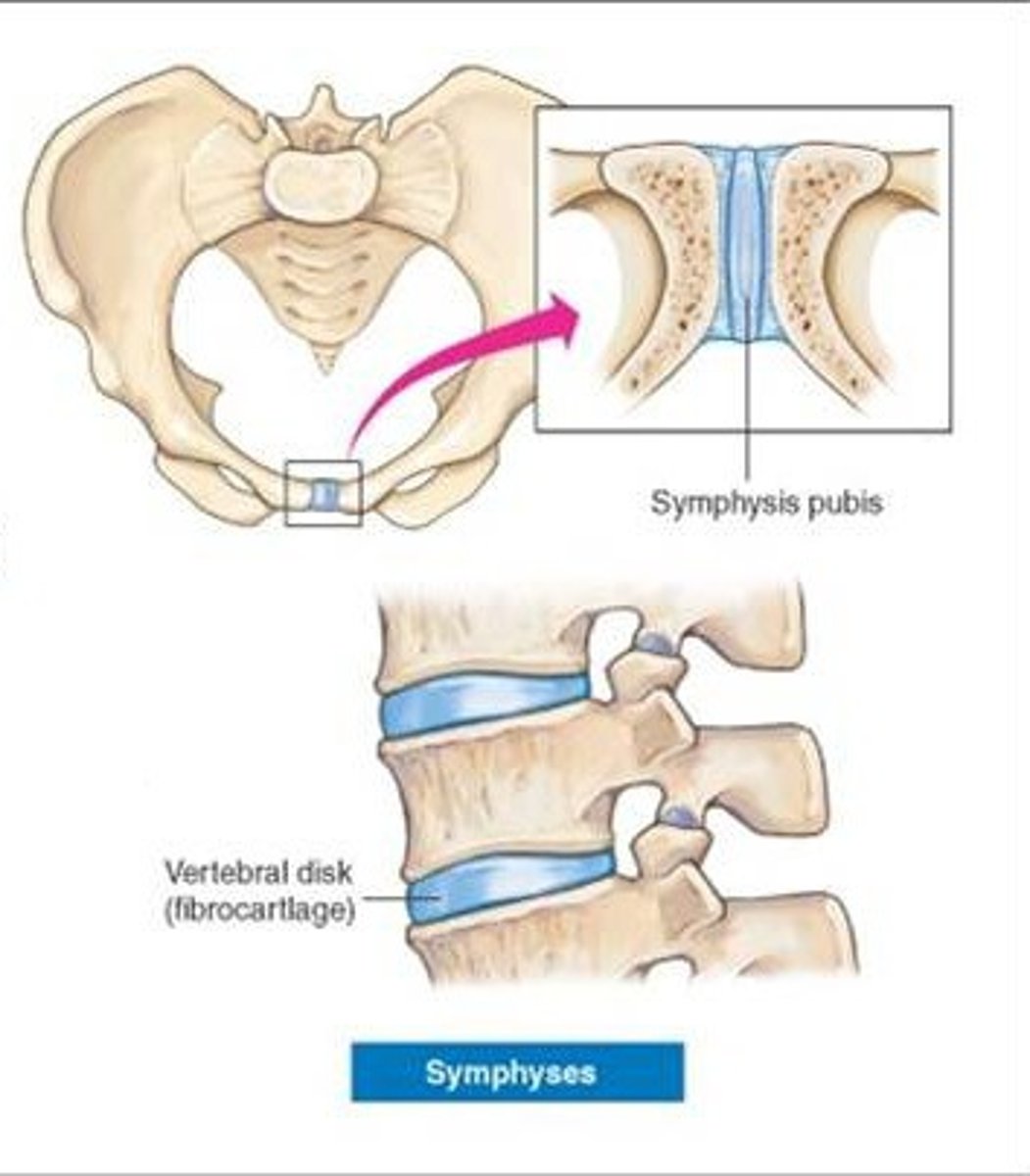
intervertebral discs f/s
f: amphiarthrotic
s: cartilaginous
symphysis joint
f: ampiarthrotic
s: cartilaginous
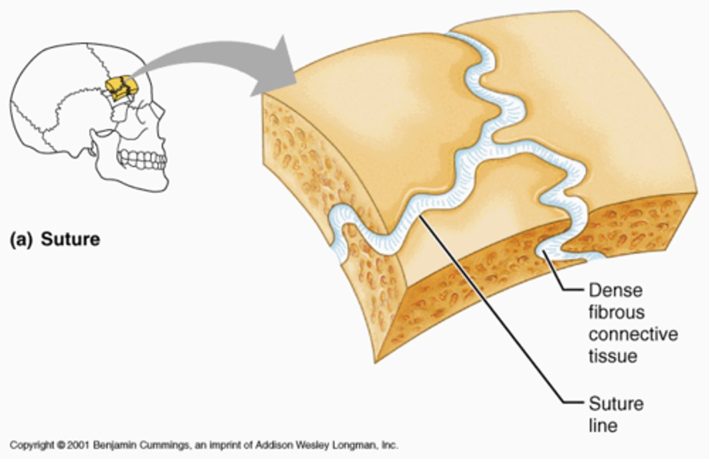
sutures joint (CHILD)
f: amphiarthrotic
s: cartilaginous
glinging
slipping movements
rotation
movement of a bone around its longitudinal axis
circumduction
proxial end of a limb remains stationary and the distal end moves in a circle
adducton
movement of a limb towards the midline
abduction
movement of a limb away from the midline
extension
increases angle of a joint an increases distance between two bones
flexion
decreases the angle of a joint and reduces distance between two bones
plane joint example
intercarpal and intertarsal joints
hinge joint example
elbow and knee
pivot joint example
atlas and axis
condyloid joint example
atlanto occipital joint and knuckles
saddle joint example
only thumb
ball and socket joint example
shoulder and hip
plane joint movement and articulating surface
gliding, flat and flat
hinge joint movement and articulating surface
flexion/extension, trough and cylindrical projection
pivot joint movement and articulating surface
rotation, rounded protrusion, and ring/sleeve
condyloid joint movement and articulating surface
everything, oval protrusion/depression
saddle joint movement and articulating surface
everything, concave/convex and reverse
ball/socket joint
all but rotation, spherical head, cup-like socket
fibrous joints
consists of inflexible layers of dense connective tissue, holds the bones tightly together
cartilagenous joints
joints filled with cartilage, typically are immovable or slightly movable
Gomphosis
A type of fibrous joint such as a tooth into the alveolus
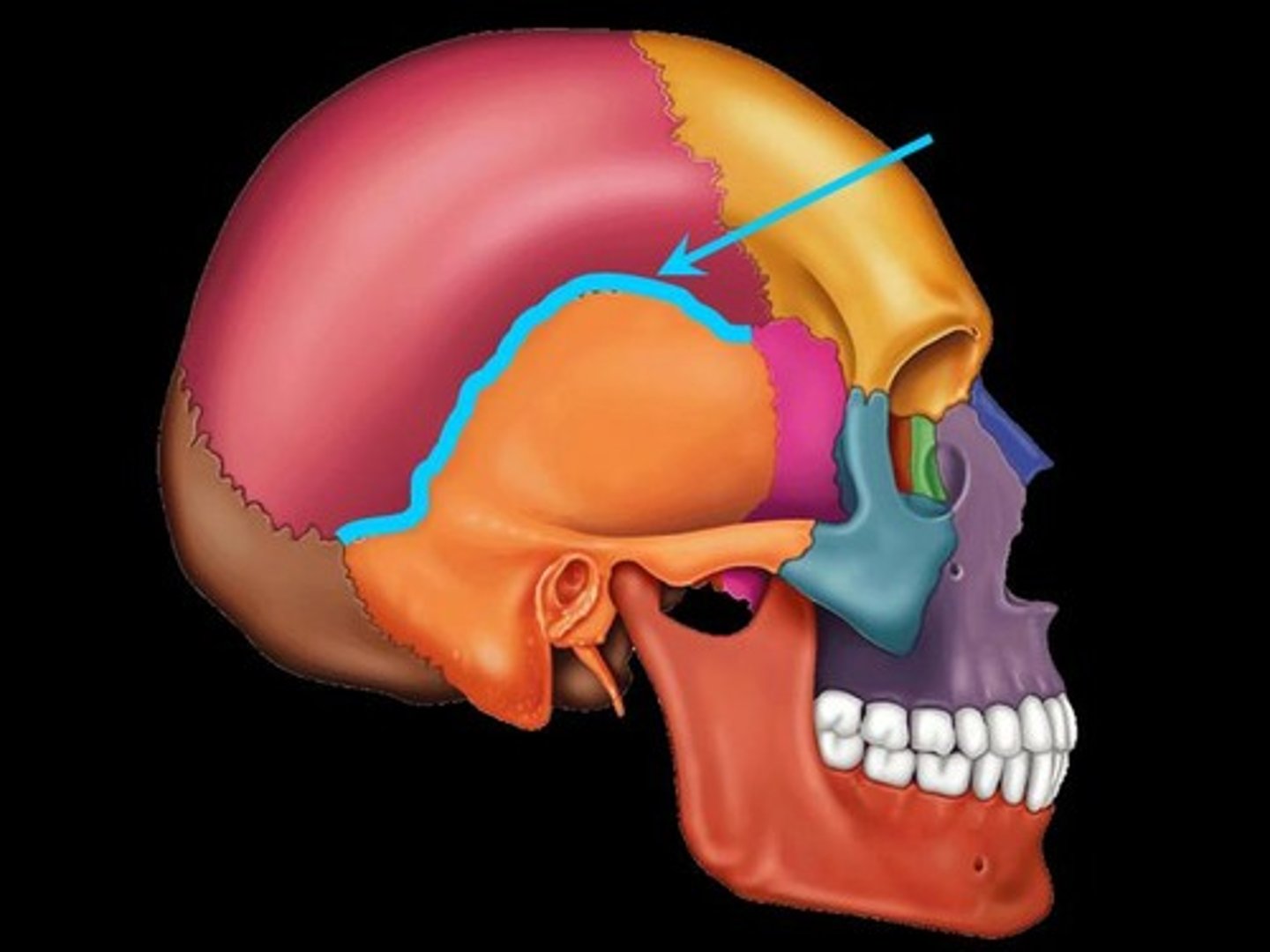
suture
a fibrous joint between flat bones of the skull
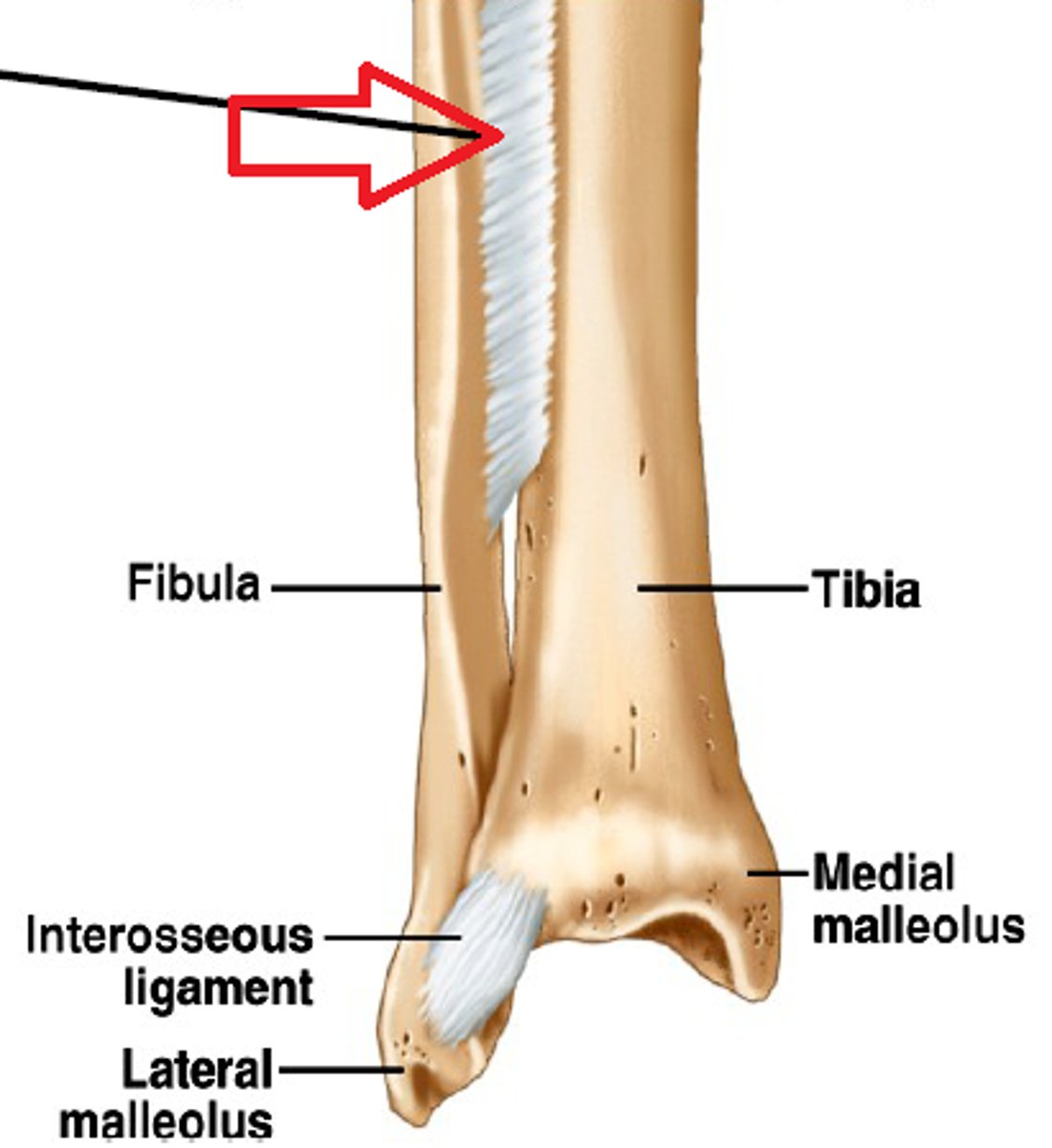
Syndesmosis
a fibrous joint at which two bones are bound by long collagen fibers
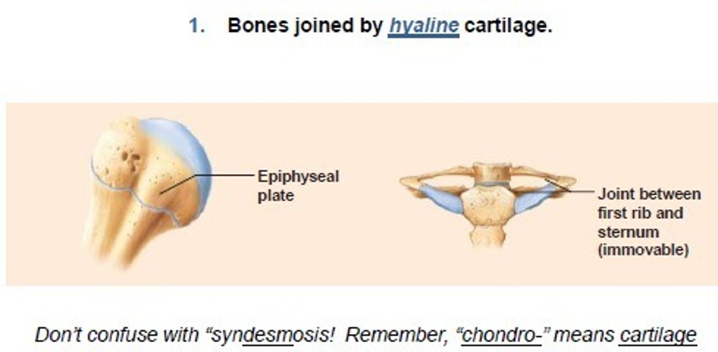
type of cartilaginous joint: synchondroses
bones join by hyaline cartilage--in the epiphyseal plates of chilren
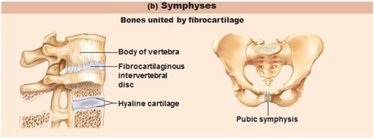
type of cartilaginous joint: symphyses
bones join by fibrocartilage
Where is a saddle joint found?
base of the thumb
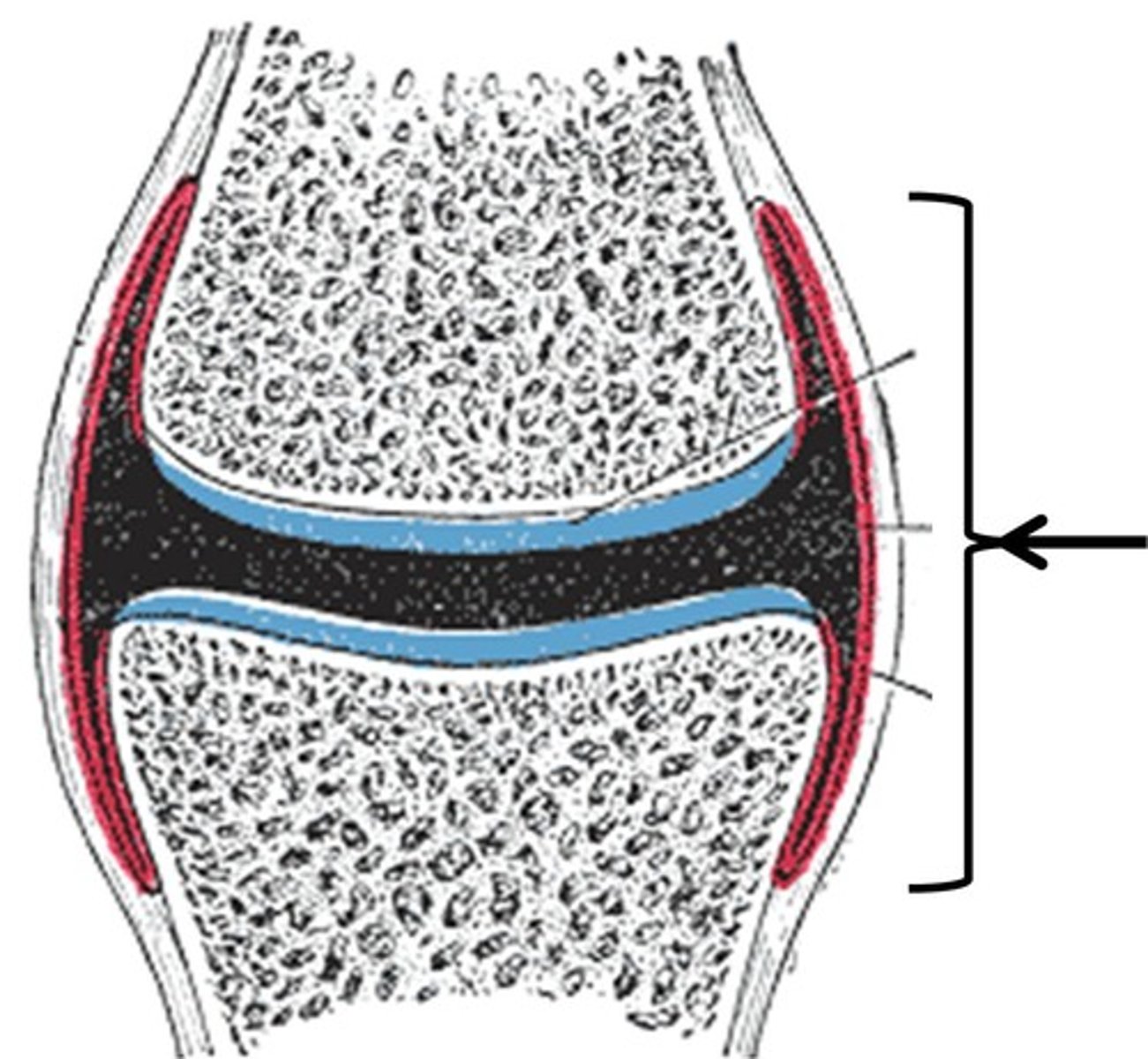
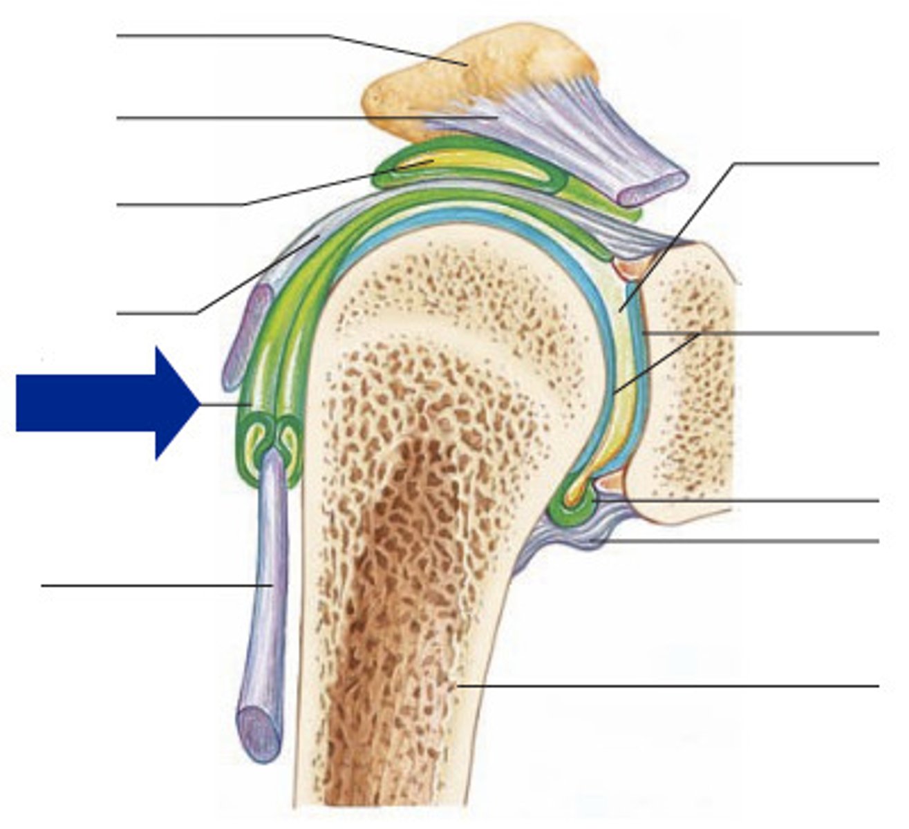
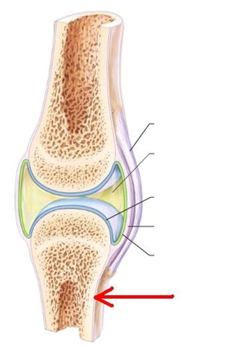
articular capsule
Fibrous envelope that encloses a synovial joint
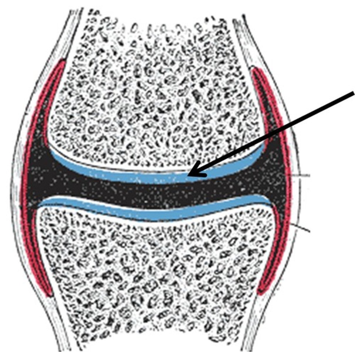
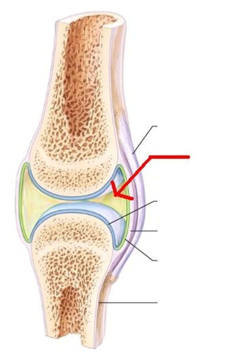
articular cartilage
covers the surfaces of bones where they come together to form joints
synovial membrane
lines the capsule and secretes synovial fluid
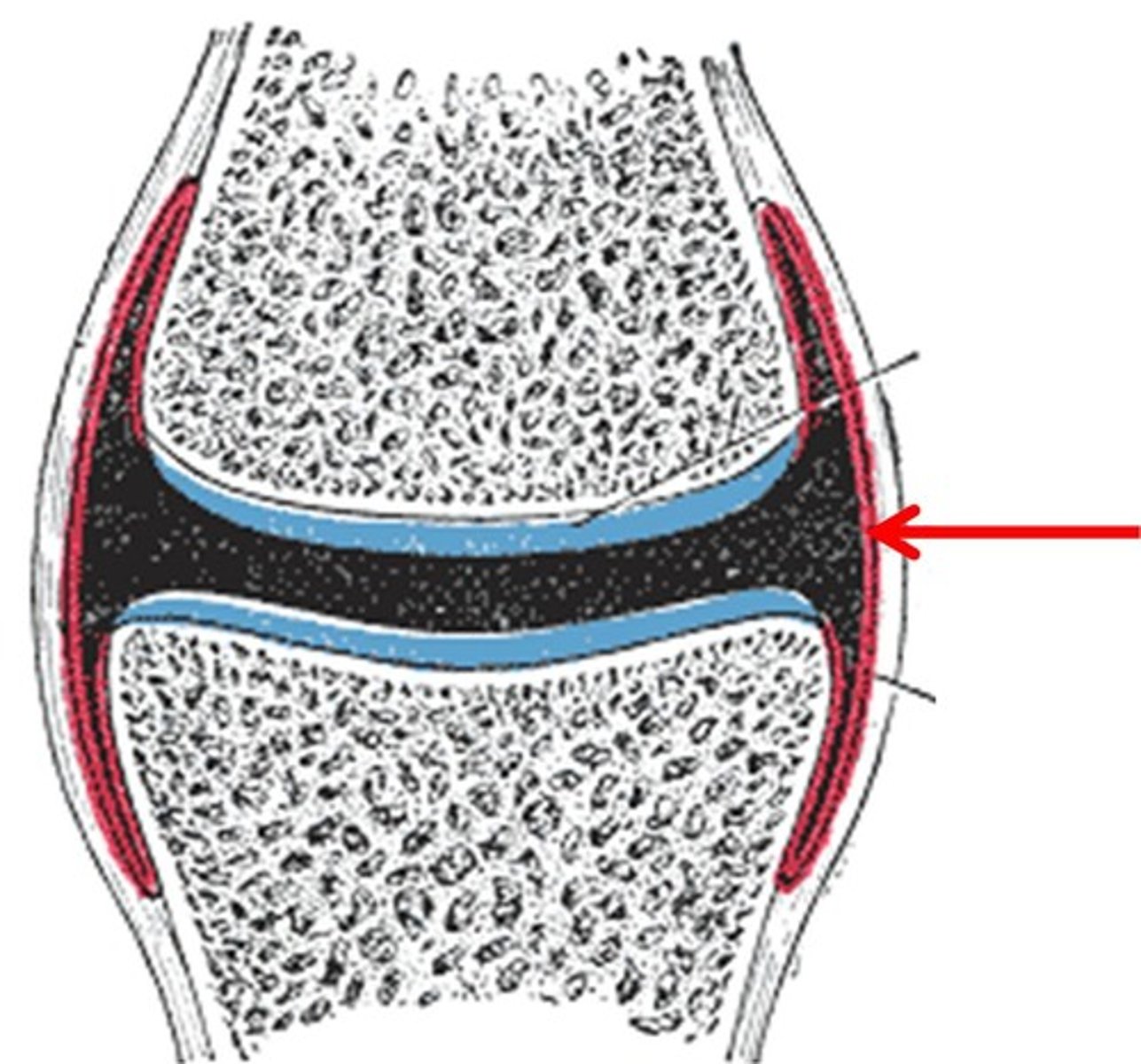
synovial fluid
joint-lubricating fluid secreted by the synovial membrane
Bursa
fluid-filled sac that allows for easy movement of one part of a joint over another
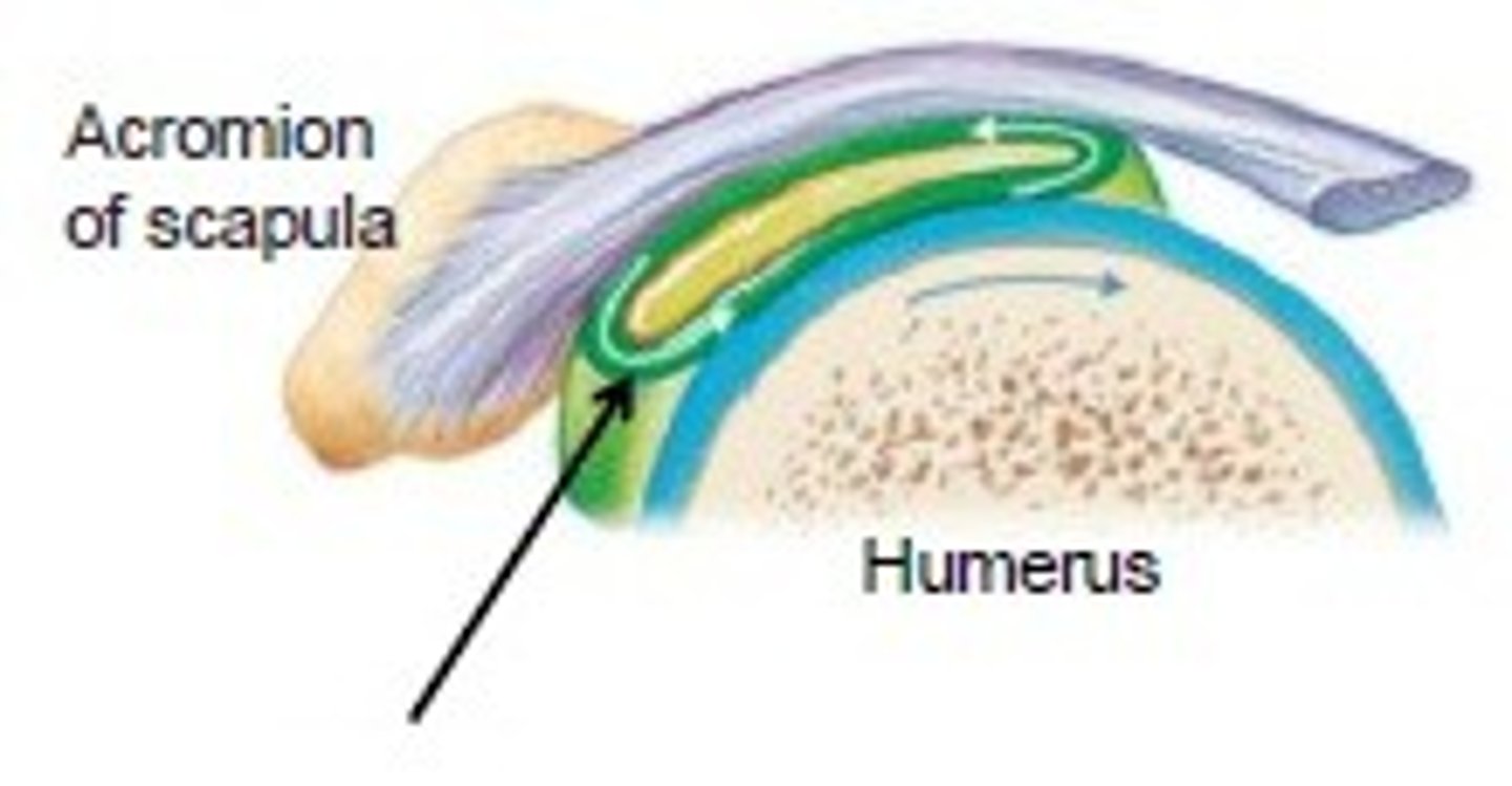
Tendon sheath
elongated bursa that wraps around a tendon
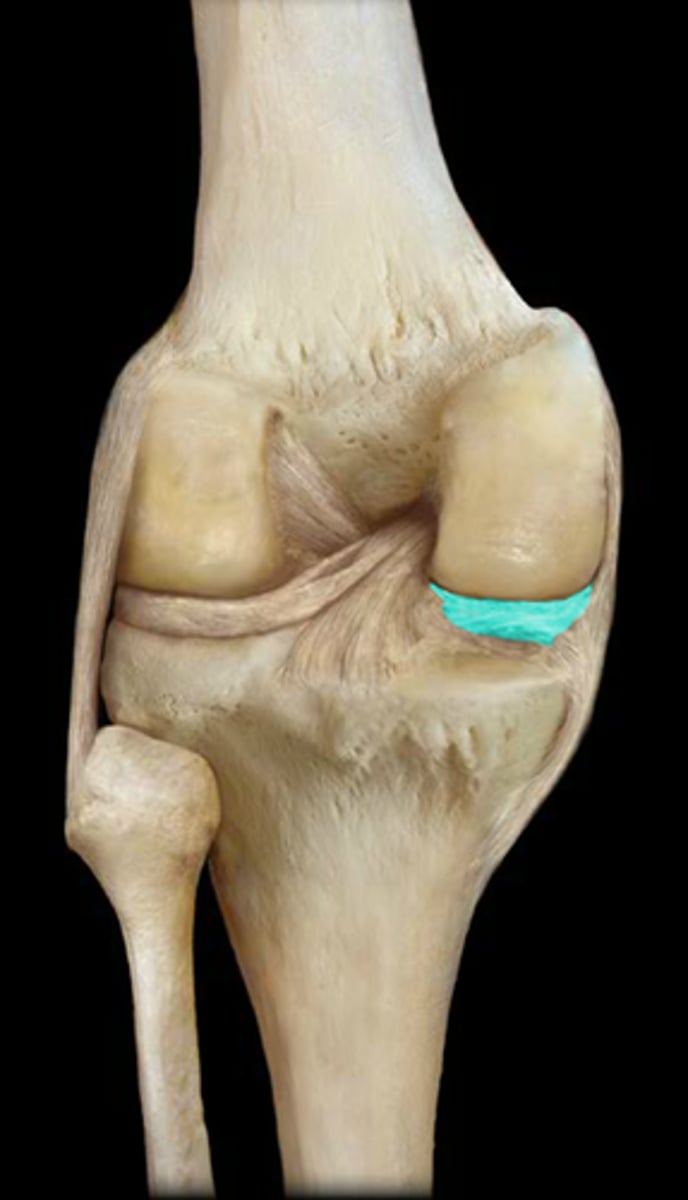
Menisci
flattened, shock-absorbing pads of fibrocartilage between the articulating surfaces of some joints
fibrous layer
Layer of the periosteum, made of dense irregular CT
osteogenis layer
Layer of periosteum, contains osteoblasts and osteoclasts
Osteoblasts
bone forming cells
osteoclasts
bone destroying cells
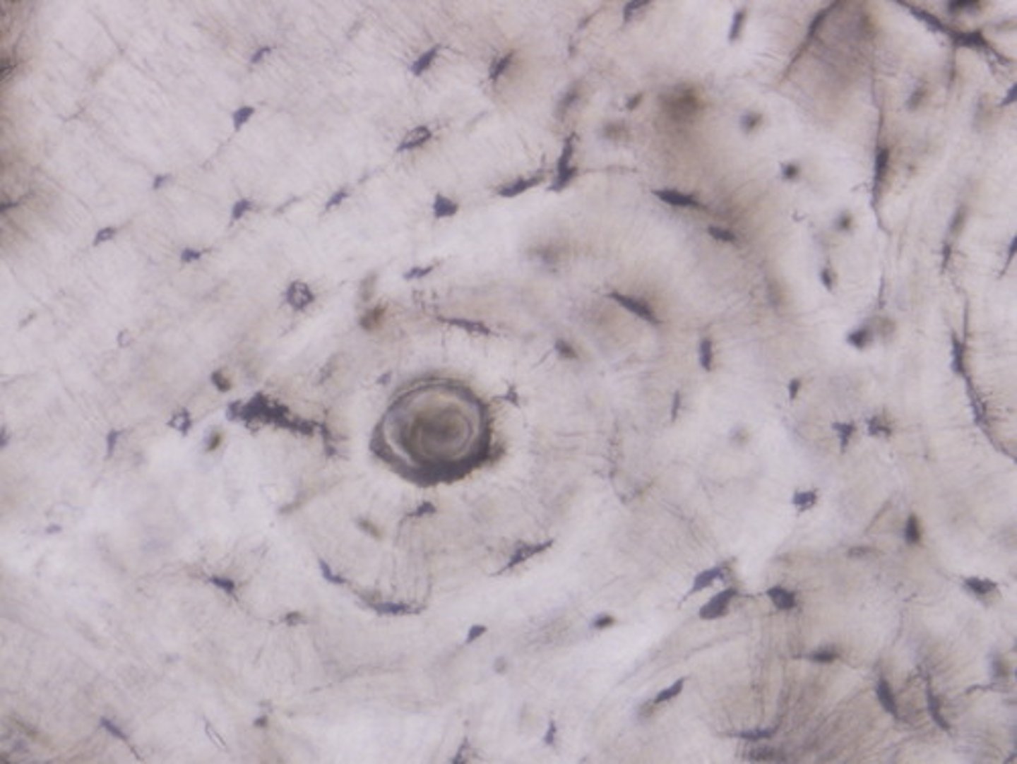
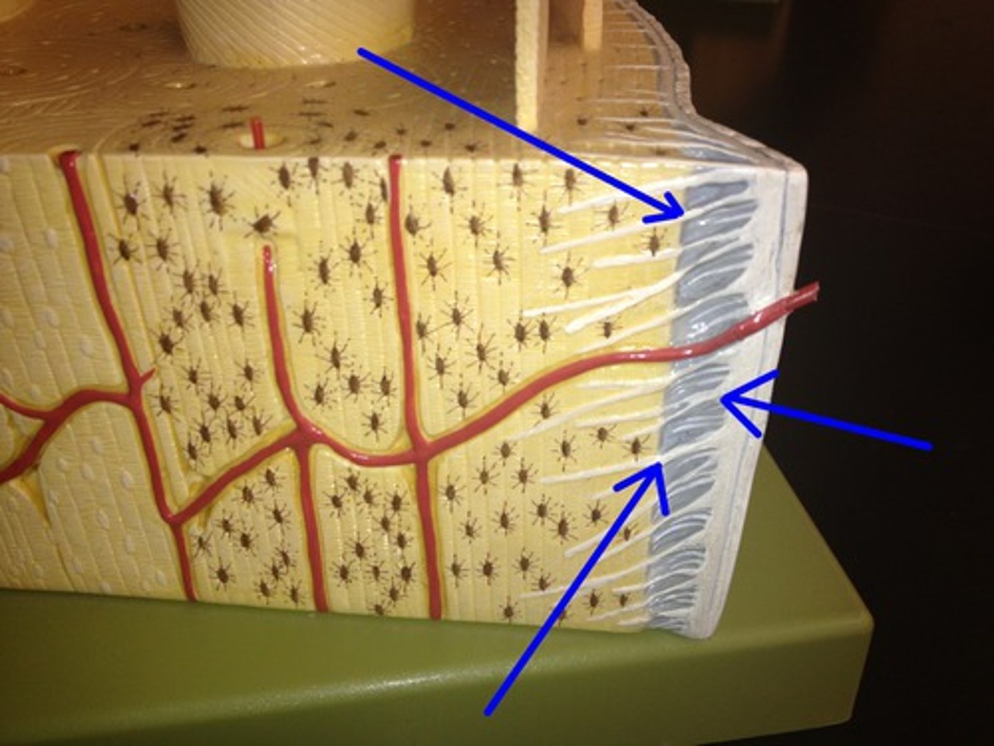
Haversian canal
central canal runs in the core of each osteon, contains blood vessels/nerves
lacunae
shallow cavities in solid bone matrix. Houses osteocytes
lamella
concentric rings of calcified matrix
volkmann's canal
connection between haversian canal and periosteum
Canaliculi
tiny canals connect lacunae to Haversian canal for transfer of substances
Osteons are located in
compact bone
in the fetal skeleton hyaline cartilage is secreted from
chondroblasts
chondroblasts and fibrous cartilage are derived from
mesenchyme
intramembranous ossification
pre natal ossification developed from fibrous tissue
endochondral ossification
pre natal ossification developed from hyaline cartilage
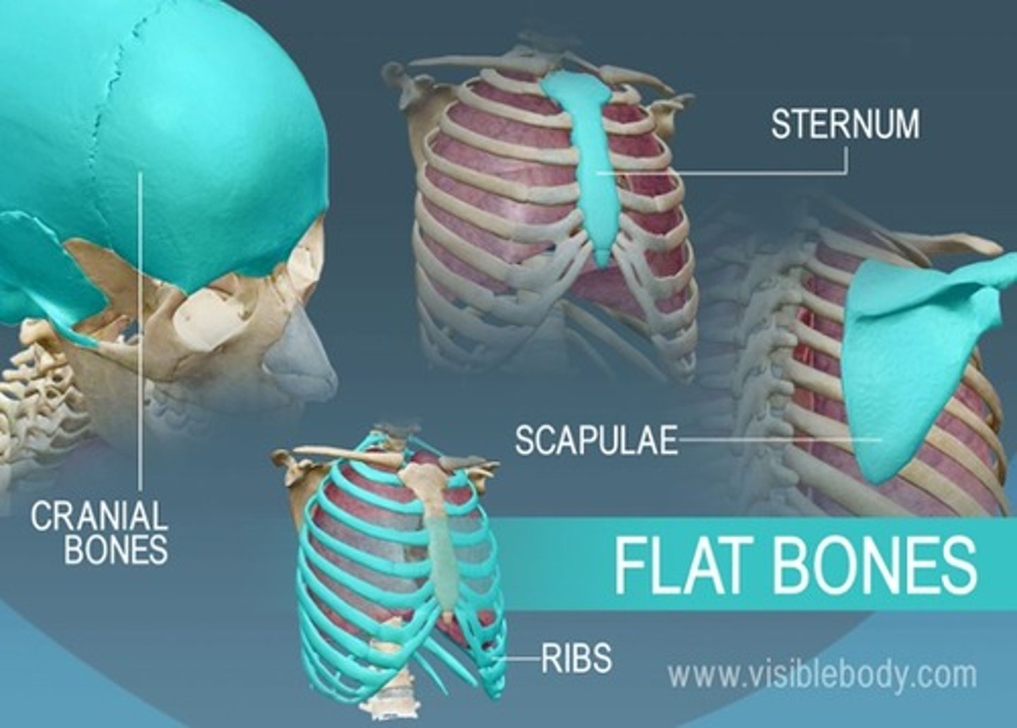
intramembranous ossification forms
cranial and clavicle bones. All membrane bones are flat bones but not all flat bones are membrane bones
endochondral ossification forms
all bones except clavicle and cranial bones
proliferation zone
Chondroblasts divide quickly, increase in secretin of hyaline cartilage
hypertrophic zone
chondrocytes undergo growth by increasing sized
calcification/deterioration zone
hydroxyapatites move into zone calcifying matrix and killing chondrocytes
ossification/osteogenic zone
osteoblasts secrete new bone tissue called osteoid
resorption zone
resorption of the corner of new bone tissue to lengthen medullary cavity
Longitudinal postnatal ossification
indirect, height, at epiphyseal face of plates
appositional postnatal ossification
direct, width, endosteum (osteoclasts) or periosteum (osteoblasts)
normal blood calcium levels
9-11% OR 9-11 mg per 100 cc of blood
parathyroid hormone (PTH)
raises blood calcium level
osteoclast-activating factor
stimulates osteoclasts and produces severe osteoporosis
active form of vitamin D
1,25-dihydroxy vitamin D
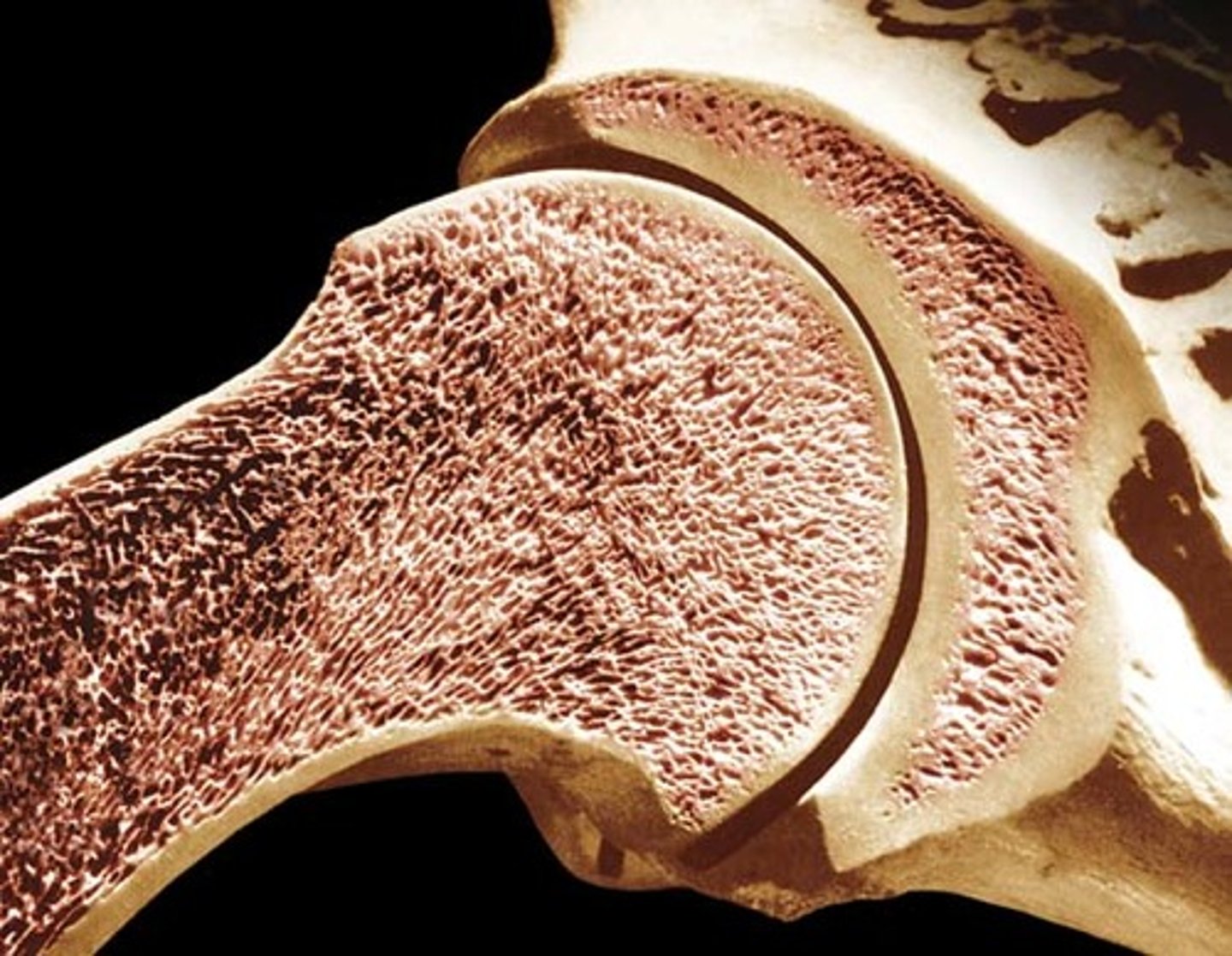
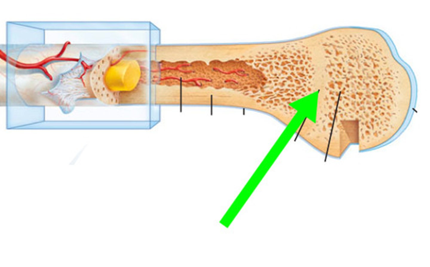
compact bone
Hard, dense bone tissue that is beneath the outer membrane of a bone
spongy bone
Layer of bone tissue having many small spaces and found just inside the layer of compact bone.
flat bones
thin, flattened, and usually curved
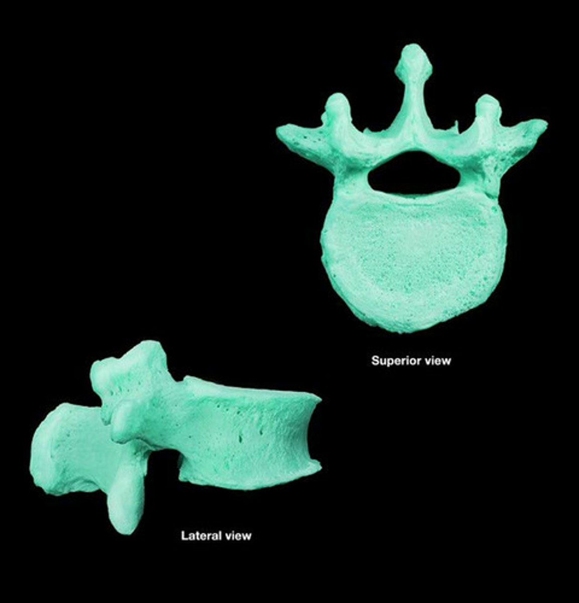
irregular bones
vertebrae and facial bones
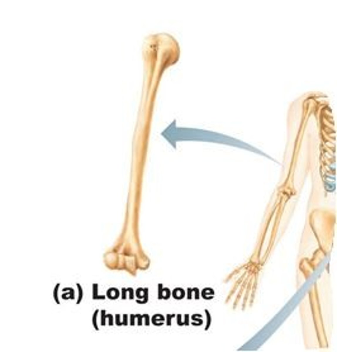
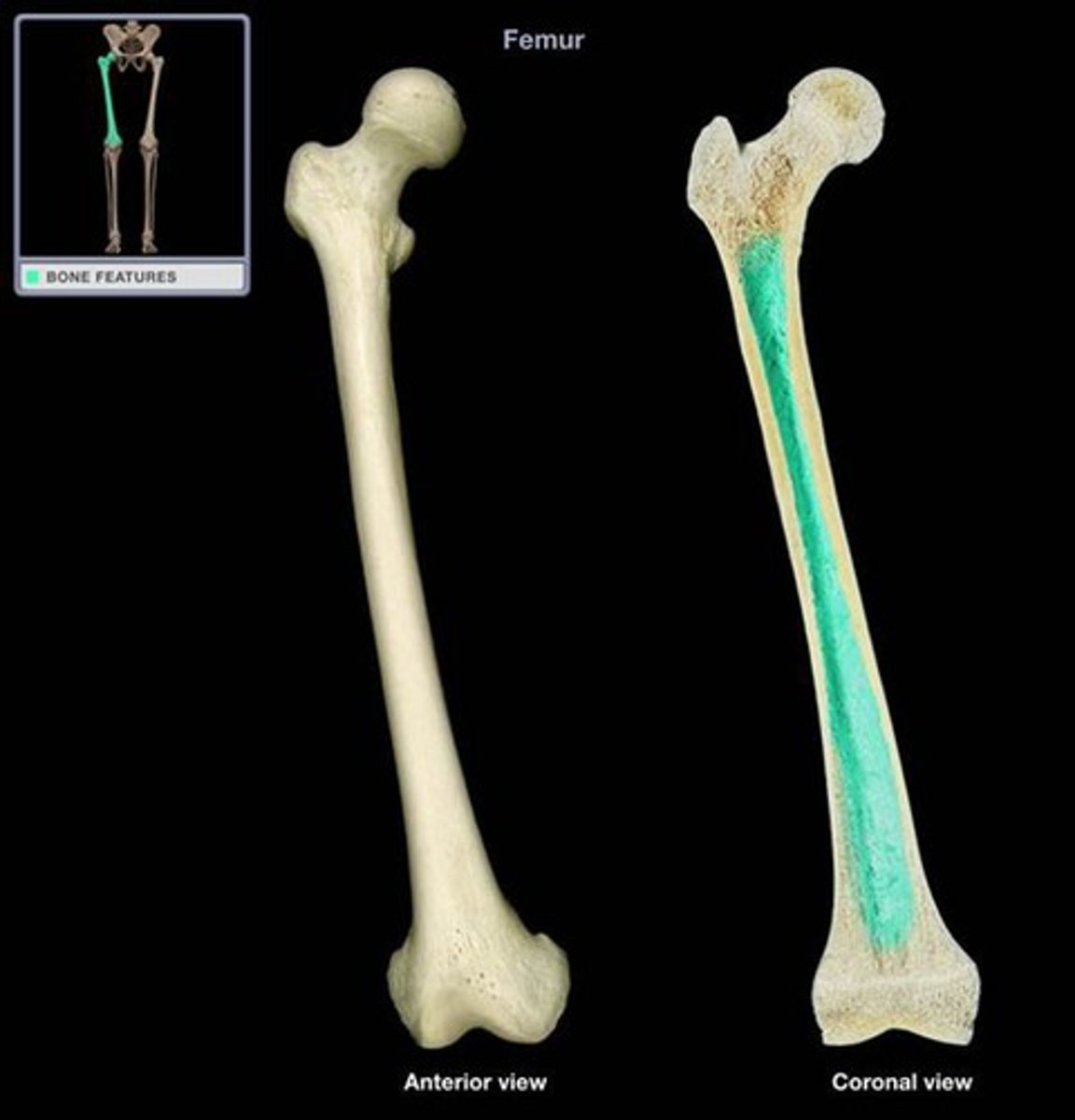
long bones
longer than they are wide
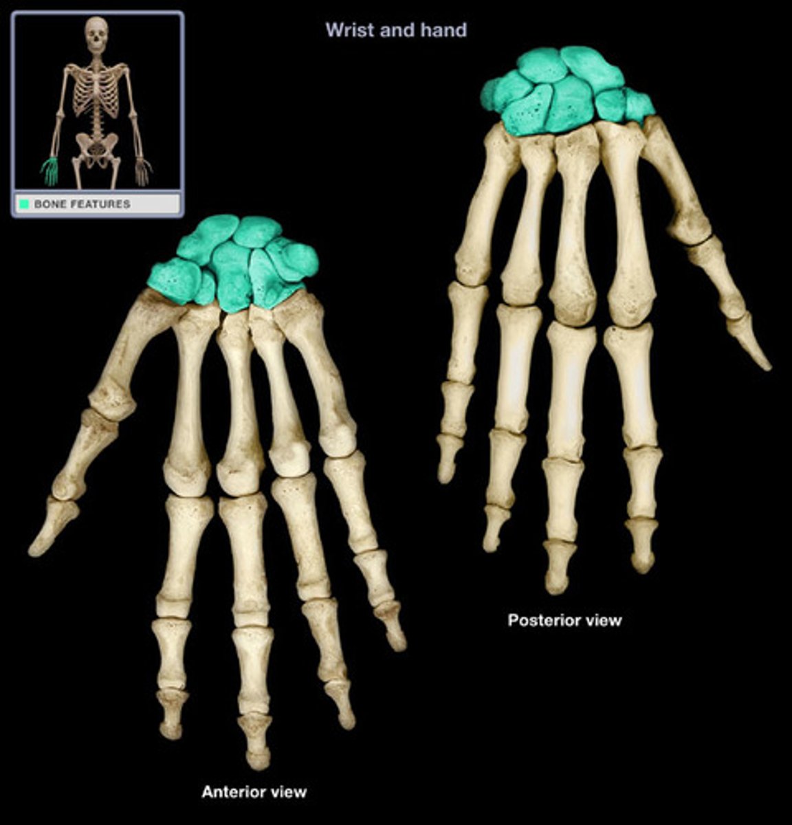
short bones
bones of the wrist and ankles
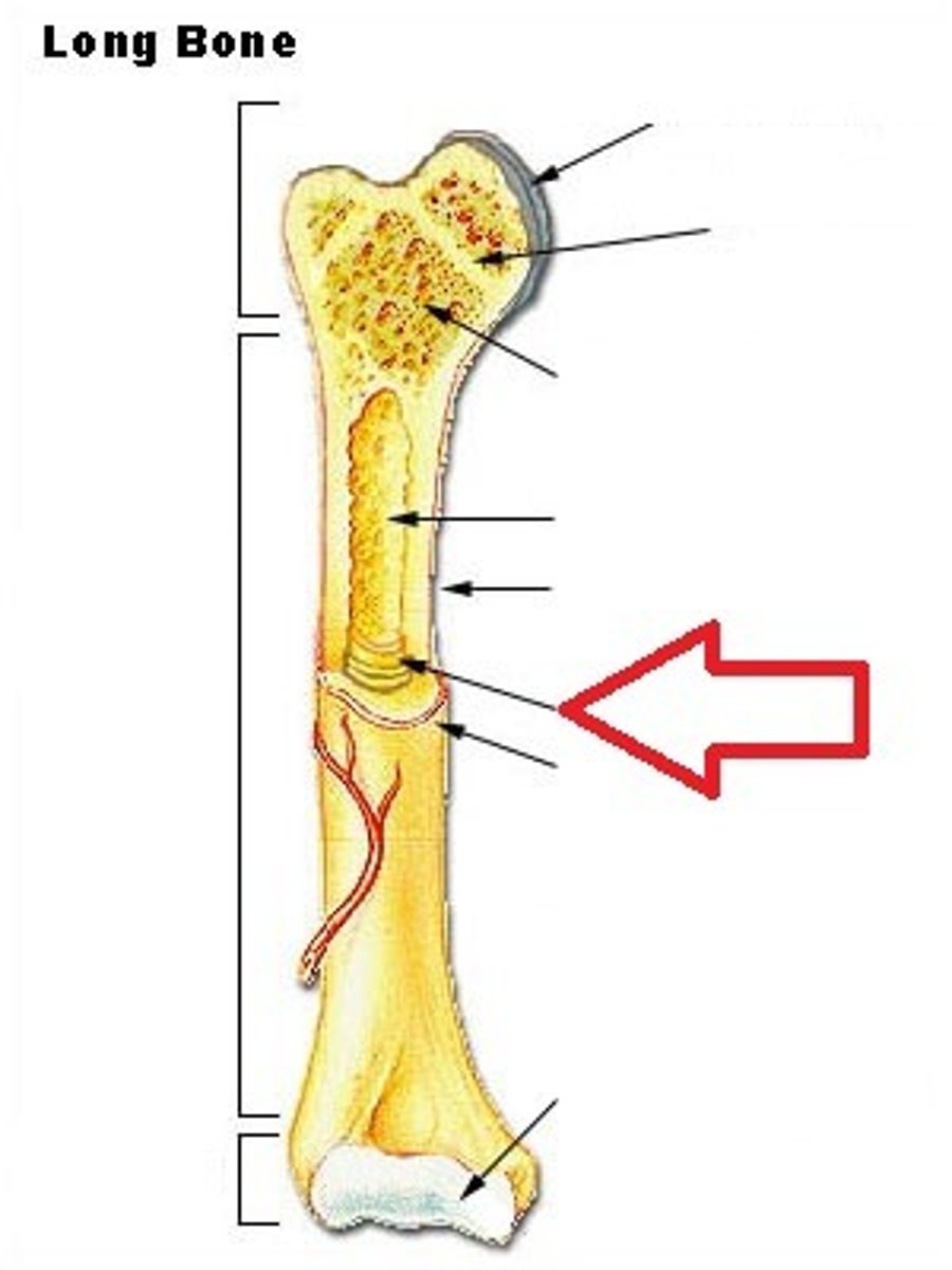
diaphysis
shaft of a long bone
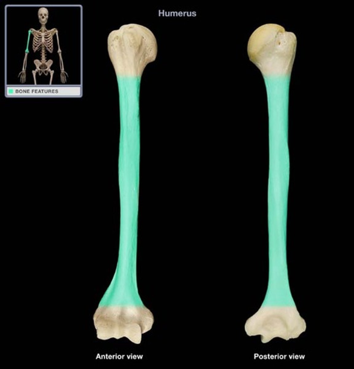
epiphysis
End of a long bone
medullary cavity
central, hollowed-out area in the shaft of a long bone
periosteum
Double-layered connective tissue that covers and nourishes the bone.
endosteum
lines the medullary cavity
sharpey's fibers
secure periosteum to underlying bone
epiphyseal line
remnant of epiphyseal plate
osteoid
unmineralized bone matrix
osteons
Structures in bones that carry the blood supply
epiphyseal plate closure
Bone lengthening ceases; bone of epiphysis and diaphysis fuses
Wolff's Law
A bone grows or remodels in response to forces or demands placed upon it
What does 1,25-dihydroxyvitamin D do?
stimulate increased uptake of calcium from the small intestine to also increase the blood calcium
Calcitonin
Lowers blood calcium levels
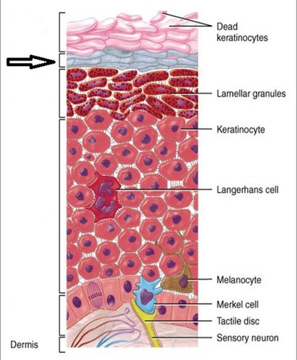
stratum corneum
outermost 20-30 layers of the epidermis, which consists of dead and flattened, keratinized cells.
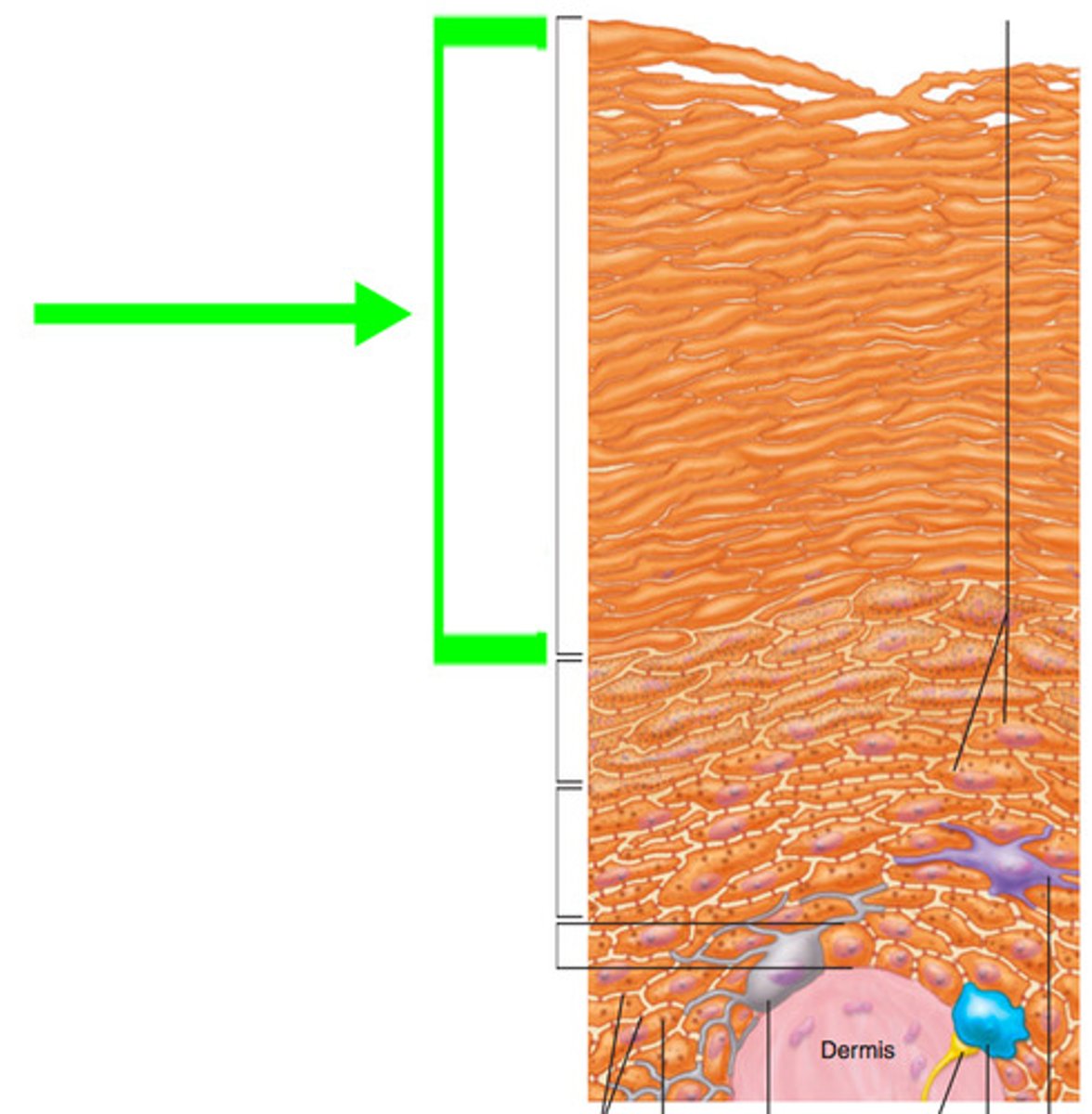
stratum lucidum
4-5 layers of dead cells. ONLY IN THICK SKIN