A&P CH 10-11
1/59
Earn XP
Description and Tags
Muscle Physiology and Neurons
Name | Mastery | Learn | Test | Matching | Spaced | Call with Kai |
|---|
No analytics yet
Send a link to your students to track their progress
60 Terms
Main components of the nervous system
• The brain: the nervous tissue contained within the cranium
• The spinal cord: the extension of nervous tissue within the vertebral column
• Neurons and neuroglia: cellular components of the nervous system
The two structural divisions of the nervous system are:
-central nervous system (brain and spinal cord)
-peripheral nervous system (cranial nerves, spinal nerves and their branches.)
What is the division of the nervous system for body control?
-sensory (Afferent) division: Divided into somatic and visceral sensory division.
-Peripheral nervous system (PNS): Cranial and spinal nerves link CNS and rest of body. Also perform motor and sensory functions.
-Central Nervous system (CNS): Brain and spinal cord integrate information.
-Motor (Efferent) division: Divided into somatic motor division and autonomic nervous system (ANS).
What makes up the Afferent division & their functions
somatic sensory division:
-includes skeletal muscle & skin
-function: Carries general sensory stimuli from muscles , bones, joints, and the skin, as well as special sensory stimuli.
Visceral sensory division:
-includes urinary bladder & stomach
-function: carries stimuli from organs
What makes up the Efferent division & their functions
Somatic motor division:
-Includes skeletal muscle
-function: carries stimuli to skeletal muscles
Autonomic nervous system (ANS):
-includes: cardiac muscle & smooth muscle
-function: carries stimuli to smooth muscle, cardiac muscle, and glands.
What is the general function of the nervous system?
Receive information about the environment around us, generating responses to that information, and integrate sensory input with memories, emotional state, or learning.
What are the three main functions of the nervous system?
Sensory function (detect stimuli)
Response function (produce voluntary/involuntary responses)
Integration (translate sensory info into perceptions)
Describe Sensory function
-detect stimuli that registers a change from homeostasis or a particular event in the environment
Describe Response function
produce a response (voluntary or involuntary) based on the stimuli perceived by sensory structures
Describe Integration function
CNS translate sensations from neural impulses into perceptions (cognition)
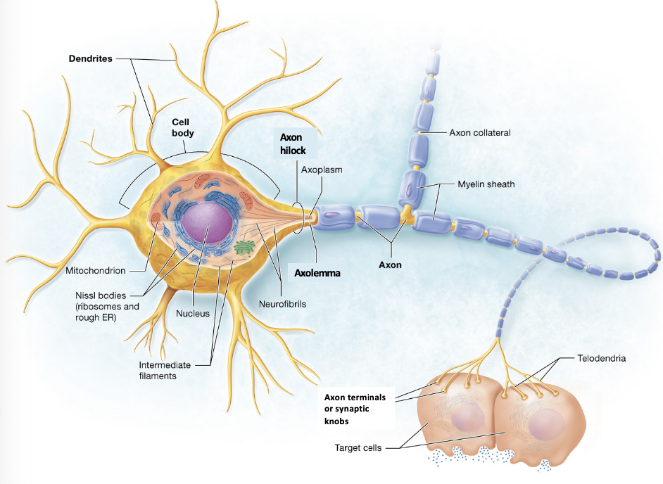
What is a neuron
basic unit of the nervous system
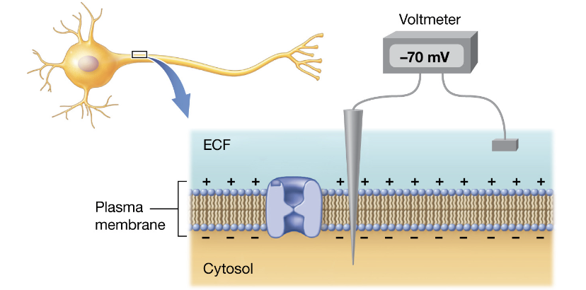
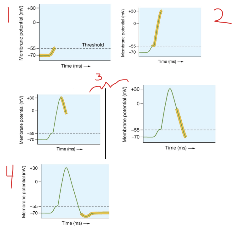
Describe resting potential
-Resting membrane potential (-70mV for neurons).
-the membrane at its negative resting membrane potential, before stimulation.
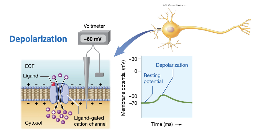
Describe depolarization
-60 mV
gain of positive charges makes the inside of the cell less negative, causing depolarization.
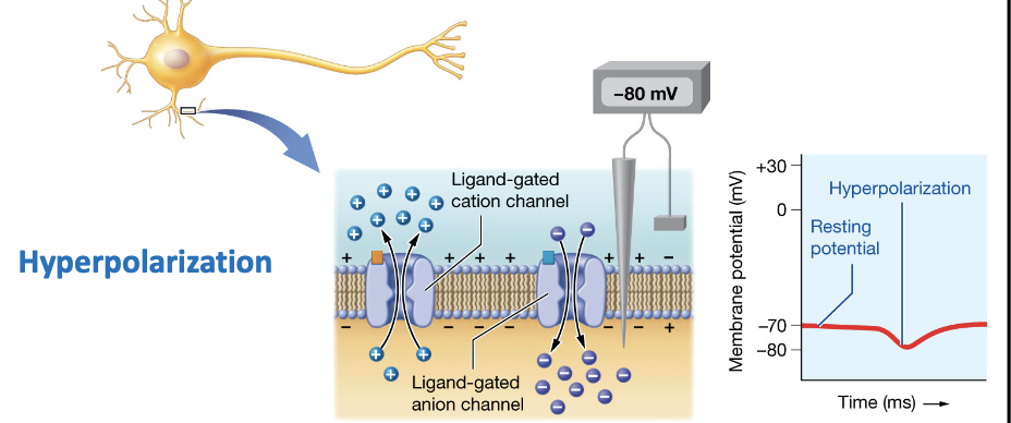
Describe Hyperpolarization
-80 mV
Loss of positive charges (or gain of negative charges) makes the inside of the cell more negative, causing hyper polarization.
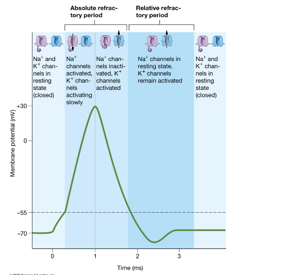
What happens during repolarization?
Na⁺ channels inactivate
Voltage-gated K⁺ channels open
K⁺ leaves the cell → membrane returns toward resting
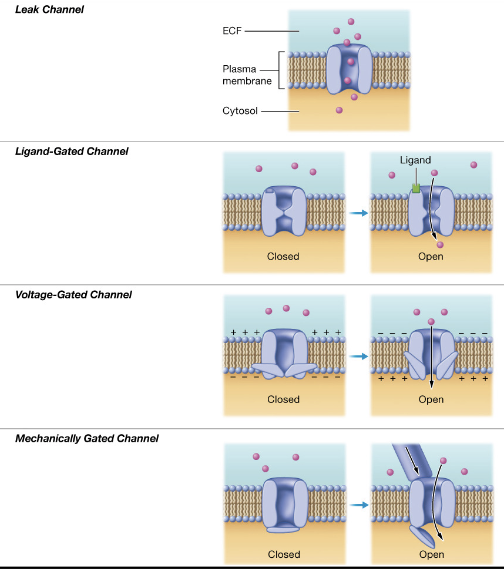
What are all the ion channels important to neurons
Leak channel: stimulus- none, always open
also, Na+/K+ pump works with the leak channels to maintain the resting potential.
Ligand-gated channel: stimulus- binding of a ligand to a receptor associated with the channel.
voltage-gated channel: stimulus- voltage changes across the plasma membrane.
mechanically gated channel: stimulus- mechanical deformations of the channel (by pressure, stretch, etc.)
What kind of potential is specific to dendrites and cell bodies
Dendrites and cell bodies
generate local potentials that spread to the axon hilock
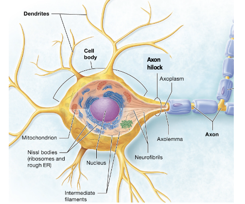
what is action potential
rapid depolarization and repolarization of the membrane potential of a cell.
Only axons generate action
potentials
Action potentials are initiated in the trigger zone of the axon hilock
where are voltage gated channels located in neurons
Voltage gated channels are in the axons and axon terminals of neurons
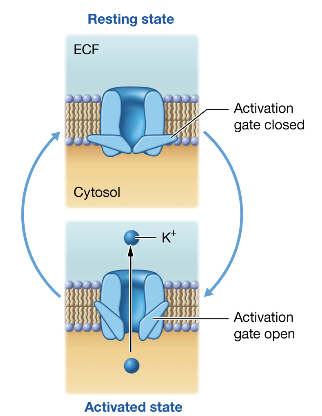
Describe Voltage gated K+ channels
has two states:
resting – activation gate is closed;
channel is closed
and activated – activation gate is
open; channel is open
there is NO inactive sites for K+
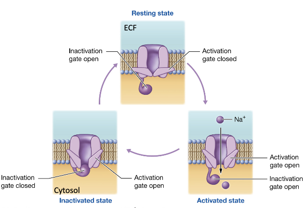
describe voltage gated Na+ channels
has three states:
resting – channel is closed,
although the activation gate is closed while inactivation gate is open.
inactivated – inactivation gate is
closed; activation gate is open
and activated – both the inactivation
and activation gates are open, and the channel is open.
Describe the sequence of events during an action potential.
Threshold reached at -55 mV by local potential depolarizing axolemma.
activation gate for Na⁺ channels open in axon hillock (depolarization)
Inactivation gate for voltage-gated Na+ channels close, K⁺ channels stay open (repolarization). voltage-gated Na+ channels at resting stage.
voltage-gated K+ channels may release additional K+ ions before returning to at resting stage (hyperpolarization). Resting polarization is restored in this segment of the axolemma.
What is the absolute refractory period?
No additional stimuli can produce an additional action potential
-Only one action potential can occur at a time on a given region of a neuron.
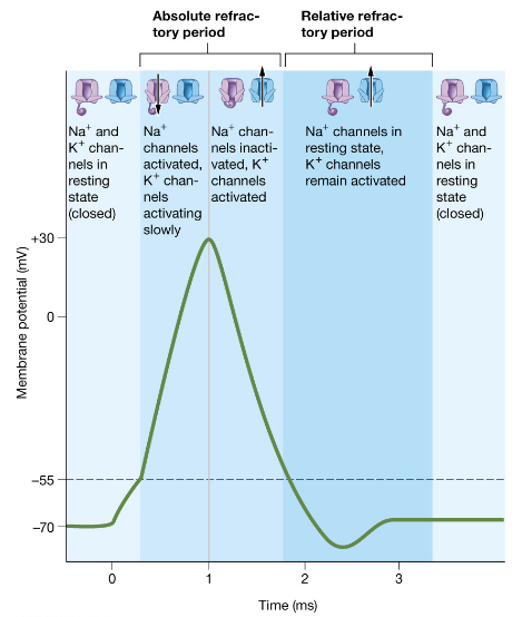
What is the relative refractory period?
Only a strong stimulus can produce an action potential
What is a nerve impulse?
A series of propagating (transmitting) action potentials along a neuron.
How do motor neurons interact with skeletal muscle fibers?
Skeletal muscle fibers are innervated (stimulated) by motor neurons that control membrane potential via synapses (axon terminals).
What neurotransmitter is used by motor neurons to communicate with muscle fibers?
Acetylcholine (ACh)
What activates vesicle exocytosis in motor neurons?
Voltage-gated Ca²⁺ channels must open to allow Ca²⁺ entry, enabling vesicles to bind to the terminal and release ACh.
What is the neuromuscular junction?
The synapse between a motor neuron and a skeletal muscle fiber.
What is the synaptic cleft?
The space between the axon terminal (synapse) of the motor neuron and the skeletal muscle fiber.
What is the motor end plate?
Region of the sarcolemma with numerous ligand-gated receptors specific for acetylcholine (ACh).
What happens in Phase 1: Excitation of muscle contraction?
A neural impulse reaches the synaptic knob, causing ACh to be released into the synaptic cleft.
What else occurs during Phase 1: Excitation?
ACh binds to ligand-gated sodium channels, causing an influx of Na⁺ and generating a motor end-plate potential.
What happens during Phase 2: Excitation-Contraction Coupling (Part 1)?
AChE degrades excess ACh; multiple neural impulses are needed to sustain end-plate potentials in target fibers.
What happens during Phase 2: Excitation-Contraction Coupling (Part 2)?
The action potential spreads down the T-tubules and reaches the triads.
What happens when the T-tubules are depolarized?
Voltage-gated calcium channels in the terminal cisternae of the SR open, releasing Ca²⁺ into the cytosol.
What must happen for a myosin head to bind actin and form a crossbridge?
(preparing for contraction)
Calcium must enter the myocyte cytosol to expose actin binding sites.
What initiates Contraction in a skeletal muscle fiber?
(preparing for contraction)
It begins after Ca²⁺ enters the cytosol and binds to troponin, which causes tropomyosin to shift away from the actin binding sites, allowing crossbridge formation.
Phase 3: Contracting sarcomeres rely on multiple crossbridge cycles
The thick and thin filaments will slide in between each other in the A zone of the contracting sarcomere as the myosin head pulls on actin
Showcases the ability of muscle cells to be distensible.
What role does ATP play in the contraction cycle?
ATP is hydrolyzed by ATPase into ADP + Pi, energizing the myosin head for crossbridge cycling.
What causes the power stroke in crossbridge cycling?
Pi leaves the myosin head, initiating the power stroke, which pulls actin toward the M-line.
What happens after the power stroke in muscle contraction?
ADP leaves, and a new ATP binds to myosin, causing myosin to detach from actin and reset.
What occurs during continued crossbridge cycling?
As long as Ca²⁺ and ATP are present, the cycle repeats, allowing thick and thin filaments to slide past each other in the A zone of the sarcomere.
What is Phase 4 of muscle contraction?
Relaxation, where the muscle returns to its resting state.
What enzyme breaks down excess acetylcholine during relaxation?
Acetylcholinesterase (AChE) breaks down ACh in the synaptic cleft.
Concurrently, no additional neuronal action potentials are occurring.
How is resting membrane potential restored in the sarcolemma during relaxation?
The sarcolemma repolarizes, reducing the fluorescent green signal
How is calcium removed from the myocyte cytosol during relaxation?
Calcium pumps return Ca²⁺ to the sarcoplasmic reticulum (SR).
What happens to actin binding sites during relaxation?
As Ca²⁺ leaves the cytosol, tropomyosin recovers the actin binding sites, ending contraction.
How does smooth muscle contract similarly to skeletal muscle?
It undergoes similar contraction cycles but involuntarily and with different regulatory proteins.
What are some stimuli that activate smooth muscle contraction?
Stretch receptors (mechanical)
Hormonal and neural signals
Pacemaker cells
Where does calcium come from in smooth muscle cells?
Sarcoplasmic reticulum (SR)
Extracellular fluid (ECF)
Why is the role of calcium different in smooth muscle?
Smooth muscle lacks troponin, so Ca²⁺ regulates contraction differently.
What protein does Ca²⁺ bind to in smooth muscle to initiate contraction?
Calmodulin (Cam) in the cytosol.
What does the Ca²⁺/calmodulin complex activate in smooth muscle?
Myosin light-chain kinase (MLCK)
What does MLCK do in smooth muscle contraction?
MLCK activates myosin ATPase, allowing ATP to be hydrolyzed so myosin can bind actin and form a crossbridge.
What happens after crossbridge formation in smooth muscle?
Crossbridge cycling occurs as in skeletal muscle, followed by normal relaxation techniques.
Which part of the nervous system performs motor functions?
a-Entire PNS
b-Afferent division of the PNS
c-Brain portion of CNS
d-Spinal cord portion of CNS
e-Efferent division of the PNS
e
Which part of the nervous system performs sensory functions?
A-Spinal cord portion of CNS
B-Afferent division of the PNS
C-Brain portion of CNS
D-Entire PNS
E-Efferent division of the PNS
B
What must occur to initiate repolarization in a segment of the axolemma if a neuron has experienced depolarization?
A-Voltage-gated K+ channel opens.
B-Voltage-gated K+ channel closes.
C-Both portions of a voltage-gated Na+ channel close.
D-Both portions of a voltage-gated Na+ channel open.
A
What must occur for depolarization to start in a segment of the axolemma if a neuron is at rest?
A-Activation gate of voltage-gated Na+ channel opens.
B-Inactivation gate of voltage-gated K+ channel closes.
C-Activation gate of voltage-gated K+ channel opens.
D-Inactivation gate of voltage-gated Na+ channel closes.
A