Kidneys 2
1/77
There's no tags or description
Looks like no tags are added yet.
Name | Mastery | Learn | Test | Matching | Spaced |
|---|
No study sessions yet.
78 Terms
What does the following describe?
• Do not communicate with collecting system
• Originate from obstructed uriniferous tubules
renal cysts
What is the sonographic appearance of a cyst?
• Anechoic
• Through transmission
• Round with smooth borders
• Posterior enhancement
What type of renal cyst is this? - bulging calyx that appears as cyst
Pylogenic cysts
What type of renal cyst is this? – cortical cysts that bulge into the central sinus of the kidney
Parapelvic cysts
What type of renal cyst is this? – lymphatic cysts in the central sinus, usually multiple
Peripelvic cysts
What type of renal cyst is this? - originates in cortex
Cortical cyst
True/False: Sonographically: different types of cyst cannot be distinguished, only location
true
Where are cortical or parenchymal cysts normally located?
peripheral cysts
Where are peripelvic cysts normally located?
located in center, sinus, of kidney
What are cysts with a single thin septation, minimal wall calcification, internal echoes caused by artifact or lobulated shapes may be associated with benign simple cysts
Atypical Renal Cysts
What are atypical characteristics that may suggests a malignant cystic lesion & need a
biopsy?
– Multiple thick septations
– Irregular walls
– Solid component
– Thick calcifications

What is associated with arterial aneurysms, especially cerebral arterial (Berry) aneurysms
of the circle of Willis?
autosomal dominant (adult) polycystic kidney disease
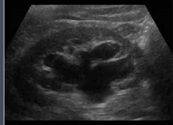
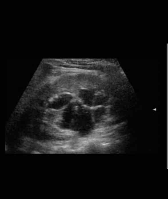
What does the following describe?:
• Bilateral renal enlargement due to development of numerous cysts of varying sizes
• Associated with cysts in the liver, pancreas and spleen
• May be identified early in someone in their 20-30s.Destruction of the residual renal tissue in advanced stages leads to renal failure and hypertension
autosomal dominant (adult) polycystic kidney disease

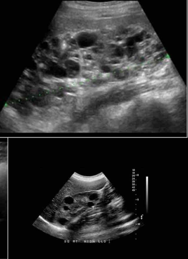
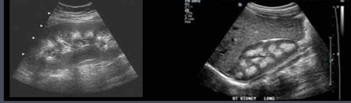
What is the sonographic appearance of ADPKD (autosomal dominant (adult) polycystic kidney disease)?
• Bilateral renal enlargement
• Multiple cyst in both kidneys
What are the ADPKD Associations?
• Liver cysts (50%), Pancreatic cysts (9%), rarely elsewhere
• Aneurysms (berry type in 3-13%; also aortic)
• Mitral valve prolapse
• Colonic diverticulosis
• Hypertension (50-70%)

What is: Multiple microscopic small cysts throughout the kidney, resulting from cystic
dilation of the collecting tubules secondary to hyperplasia of the interstitial portions of the ducts?
Autosomal recessive (infantile) polycystic kidney disease (ARPKD)
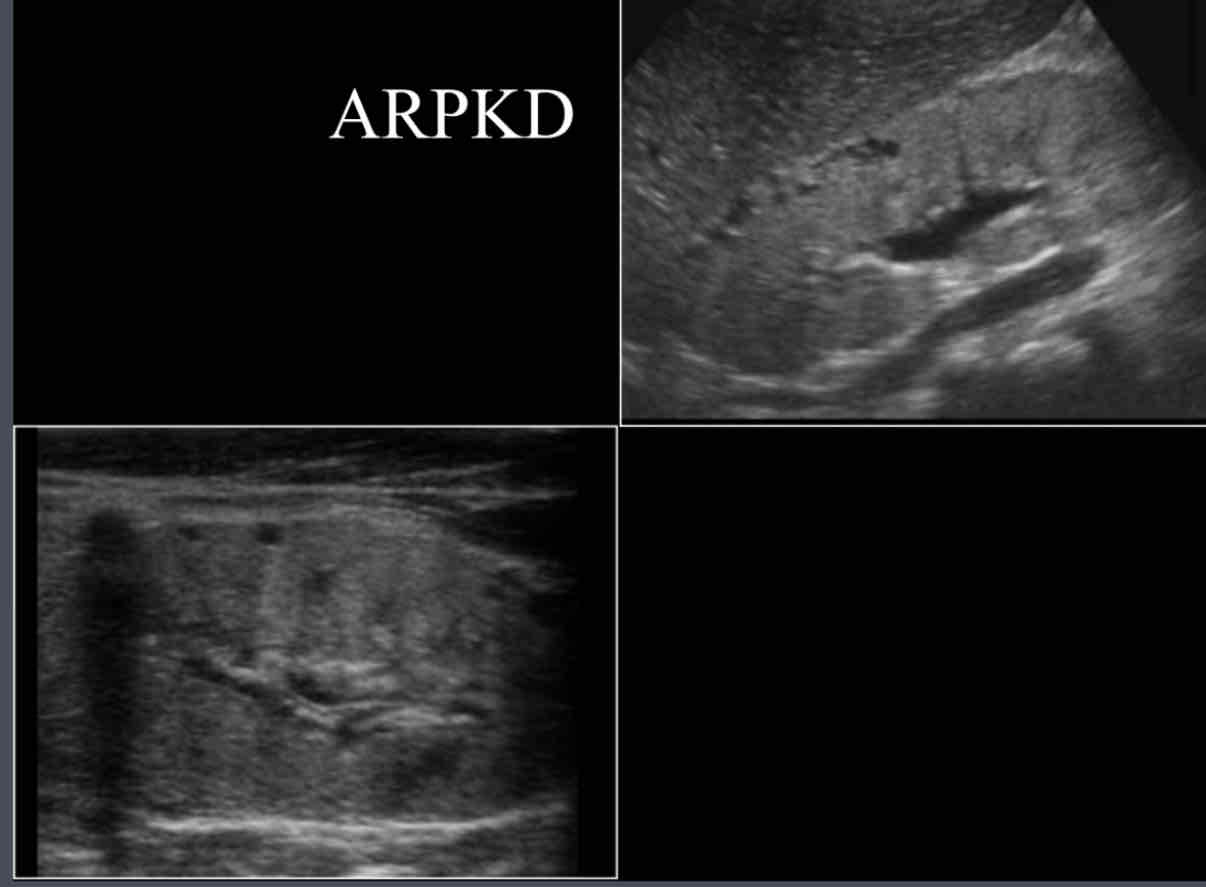
What is the sonographic appearance of ARPKD?
• enlarged bilateral kidneys,
• hyperechoic parenchyma
• loss of cortical medullary distinction
• May be seen in utero along with oligohydramnios.
dominant indicates….
adult
recessive indicates….
infantile
Autosomal recessive (infantile) polycystic kidney disease (ARPKD) is associated with:
Renal dysfunction, pulmonary hypoplasia, periportal fibrosis, and portal hypertension
What refers to the typical physical appearance of a neonate as a direct result of oligohydromnios compression while in utero
Potter Syndrome
Causes of Potter Syndrome include….
A. Bilateral renal agenesis
B. ADPKD
C. ARPKD
D. Multicystic renal dysplasia
E. Obstructive uropathy
D. early rupture of membranes
What is the most common cause of an abdominal mass in newborns?
multicystic dysplastic kidney
What does the following describe?
• Typically unilateral, but may be bilateral.
• Non-functioning kidney consists of non-communicating cysts with the absence of
renal parenchyma
• Usually results from atresia of uretero-pelvic junction during fetal development
Sonographically:
• in area of kidney, there are multiple cyst
• Kidney size usually decreases as cysts replace parenchyma so little or no parenchyma
multicystic dysplastic kidney
What is associated with these renal anomalies:
– Contralateral UPJ obstruction, contralateral renal agenesis or hypoplasia,horseshoe kidney
multicystic dysplastic kidney
What is the differential diagnosis for multicystic dysplastic kidney?
Differential diagnosis is severe hydronephrosis, but with hydronephrosis
parenchyma is seen
What does the following describe?
• Development of multiple cysts in chronically failed kidneys during long-term dialysis
• After 3-4 yrs dialysis
• Hemorrhage often occurs in them, resulting in pain and hematuria
• Associated with increased incidence of RCC
Sonographic Appearance:
• Bilaterally atrophied kidneys
• Little to no visible parenchyma
• Multiple cyst formation
acquired cystic disease
What is the development of multiple cysts in chronically failed kidneys during long-term
dialysis?
acquired cystic disease
What is associated with increased incidence of RCC?
acquired cystic disease

What does the following describe?
• It’s an Autosomal recessive defect.
• Congenital dysplastic cystic dilatation of medullary pyramids due to tubular ectasia
or dysplasia
• Causes: urinary stasis, calcium deposits form in the dilated tubules
medullary sponge kidney
What is caused by urinary stasis, calcium deposits form in the dilated tubules?
medullary sponge kidney

What does the following describe?
Sonographically:
• calcium deposits appear as hyperechoic in medullary pyramids.
• Pyramids dilated and may be echogenic
• Thinned cortex
• With and without shadowing
• Associated with nephrocalcinosis
medullary sponge kidney
What is an inherited disease which usually presents in 2nd-3rd decade of life with serious visual impairment. This disease is characterized by retinal and central nervous system hemangioblastomas
Von Hippel-Lindau Disease
What related tumors can be found with Von Hippel-Lindau Disease?
– Renal Cell Carcinoma
– Pheochromocytomas
– Islet cell tumors
– Renal and pancreatic cysts
True/False: In the presence of Von Hippel-Lindau Disease, imaging should be focused on evaluating the kidneys, adrenal glands and the pancreas.
true
What is a Multi-system genetic disease that causes benign tumors to grow on organs like brain, kidneys, heart, eyes, lungs and skin (Commonly affects CNS.)?
Tuberous Sclerosis
Patients with what condition have increased likelihood of renal cysts & angiomyolipomas?
Tuberous Sclerosis

True/False: Angiomyolipomas are typically unilateral in patients with tuberous sclerosis.
false, Angiomyolipomas are typically bilateral in patients with tuberous sclerosis.
What tumors are benign?
– ANGIOMYOLIPOMA
– ADENOMA
– ONCOCYTOMA
– MESOBLASCTIC NEPHROMA
What renal tumors are malignant?
– RCC
– TCC
– WILMS
– METASTASES

What does the following describe?
• Benign renal tumor composed of fat, blood vessels and smooth muscles tissue
• Also called renal hamartoma.
• More common in females 40-60 years of age
• Asymptomatic if small
angiomyolipoma
Formation of multiple bilateral angiomyolipomas is usually associated with
tuberous sclerosis
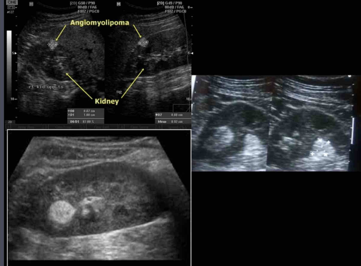
What does this sonographic appearance describe?
1. Solid hyperechoic parenchymal mass.
2. Use color Doppler to assess the intramural flow.
3. Propagation speed artifact may result in posterior displacement of structures due to slow acoustic velocity in the fatty mass
4. 80% involve right kidney
5. Can undergo hemorrhage when large
angiomyolipoma

What is a solid hyperechoic parenchymal mass?
angiomyolipoma

What does the following describe?
• Often an incidental finding. Originating from renal tubular epithelium. Solitary in 75%.
• Usually measuring 1 cm or less and rarely larger than 3 cm in diameter.
Signs and symptoms--
• They are asymptomatic unless they enlarge and then may cause hematuria and severe flank pain due to hemorrhage.
adenoma
What are the most prevalent benign kidney tumors?
Cortical adenomas
What does the following sonographic appearance describe?
• They are small well defined sub capsular
Isoechoic or hypoechoic or echogenic
cortical mass usually 3cm.
• Vascular and attenuates sound
adenoma
What does the following describe?
• They are epithelial tumors of the proximal tubular cells
• Class of very large vascular adenomas that usually occur in middle to old age
• Male to female ratio of 1.7:1.
• Patients are usually asymptomatic but may have pain or hematuria.
Sonographic appearance
• Usually a hypoechoic solid mass; can have varied echogenicity
• May see a central scar
• Cannot be distinguished from typical RCCs
• Difficult to differentiate from renal cell carcinoma; requires biopsy
Oncocytoma or oxyphilic adenomas
What is a rare benign salivary gland tumor?
Oncocytoma or oxyphilic adenomas
What is a pediatric benign parenchymal tumor and is the most common renal tumor diagnosed in the neonatal period?
Mesoblastic nephroma
What does the following describe?
• Referred to as fetal renal hamartoma or
leiomyomatous hamartoma.
• Usually presents as an abdominal neonatal mass.
• Some cases are discovered at prenatal US
examination.
• About 75% of cases are diagnosed during the first 4 months of life.
Signs and symptoms
• The most frequent clinical presentation is a palpable abdominal mass, with hematuria found less frequently.
Mesoblastic nephroma
What does the following sonographic appearance describe?
• At gross analysis, congenital mesoblastic nephroma is an infiltrative mass with ill-defined margins and no capsule;
• US shows a large solid renal mass exhibiting low echogenicity or mixed echotexture.
– The “ring sign” (concentric hyperechoic and hypoechoic rings) is a suggestive pattern, the anechoic ring surrounding the tumor containing abnormal vessels depicted with
Doppler;
• CT scan shows a homogeneous renal mass, or a large tumor with areas of low attenuation representing cystic, hemorrhagic and/or necrotic regions.
• Treatment is nephrectomy.
Mesoblastic nephroma

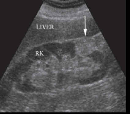
What does the following describe?
• also referred to as hypernephroma or adenocarcinoma, represents 1% to 3%
of all visceral cancers.
• are the most common malignant tumor of the kidney, accounting for approximately 80% to 90% of all renal malignancies in adults.
• The lesion occurs most often after age 50 and has a 2:1 male to female ratio.
Renal cell carcinoma (RCC)

In what does the patient may present with flank pain, hematuria (most common) and palpable mass?
Renal cell carcinoma (RCC)
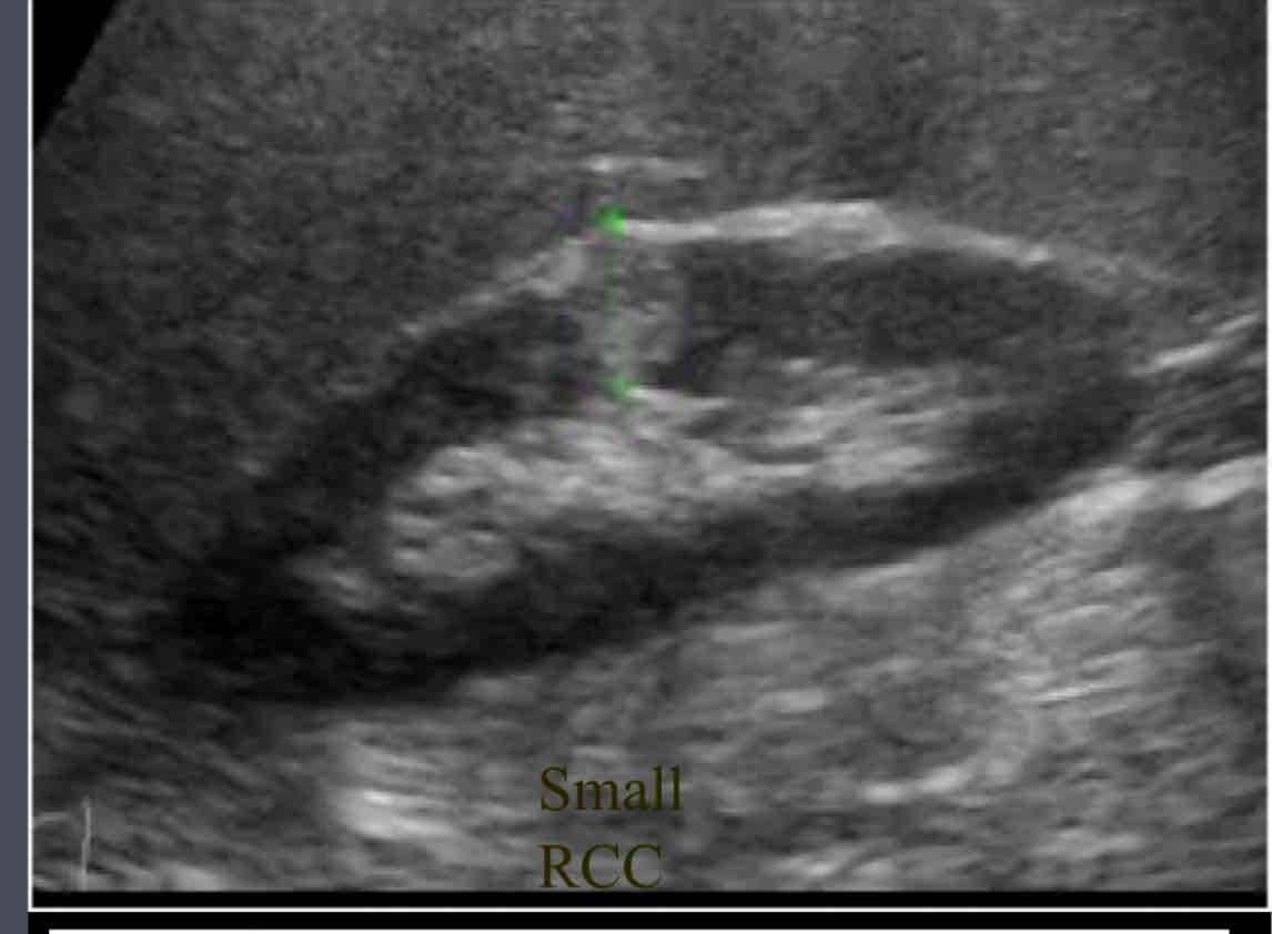
Sonographic appearance
• The mass is seen with varied sonographic appearances. Compared to normal adjacent renal parenchyma, 50% appear hyperechoic, 40% appear more echogenic, and 12% appear markedly hyperechoic, similar to the echogenicity of the renal sinus or an AML.
• Larger tumors typically isoechoic/ hypoechoic.
• Masses often demonstrate low resistance hyperemia with high systolic and diastolic arterial flow.
Renal cell carcinoma (RCC)
What is the most common site for mets?
lungs
What is recommended for RCC?
Nephrectomy

What is a tumor extension into renal veins and IVC common, and contralateral kidney?
Renal cell carcinoma (RCC)
What is increased incidence associated with RCC?
1.Acquired cystic disease(chronic dialysis)
2.Von hippel Lindau syndrome
3.Tuberous sclerosis
4.ADPKD
What stage is the following? - RCCs are 7 cm or smaller and confined to the kidney.
stage 1
stage 2
stage 3
stage 4
stage 1
What stage is the following? - RCCs are larger than 7 cm but still confined to the kidney.
stage 1
stage 2
stage 3
stage 4
stage 2
What stage is the following? - tumors extend into the renal vein or vena cava, involve the ipsilateral adrenal gland or perinephric fat or both, or have spread to one local lymph node.
stage 1
stage 2
stage 3
stage 4
stage 3
What stage is the following? - tumors extend beyond Gerota's fascia to more than one local node or have distant metastases.
stage 1
stage 2
stage 3
stage 4
stage 4
What is it called when transitional cells line the renal pelvis, ureter and bladder?
urothelial carcinoma (transitional cell carcinoma)
Urothelial tumors are malignant tumors of the lining of the…..
renal pelvis, calyces, ureter, and bladder
Urothelial carcinoma (transitional cell carcinoma) accounts for 85% of all tumors of the….
collecting system lining
What is the most common symptom of Urothelial carcinoma (transitional cell carcinoma)?
Painless hematuria
What does this sonographic appearance describe?
• Hypoechoic mass ,may invade parenchyma.
• Distorts internal architecture
• Ureteral involvement can lead to hydronephrosis
Urothelial carcinoma (transitional cell carcinoma)
True/False: Because of its profuse blood flow, the kidney is frequently the site of metastasis of carcinomas and sarcomas that arise in other organs.
true
What are the main sources renal metastases?
Lung and breast tumors
What is the only malignancy that selectively metastasizes to the opposite kidney?
RCC of the kidney
Malignant cells from what can metastasize here (renal mets)?
leukemia and lymphoma
What does the following describe?
• AKA Nephroblastoma
• Most common malignant mass in the abdomen in children 8 years of age and younger
• Mean diagnosis age is 3 ½ years
• 90% survival rate with chemotherapy
• Tumor extension can be seen into renal vein and IVC
wilm’s tumor
What is the most common malignant childhood renal tumor?
wilm’s tumor
Mets can be to where?
lungs, liver, bone, lymph nodes, and retroperitoneum
What do the following signs and symptoms describe?
• Patient presents with large, asymptomatic flank mass.
• 33% show sporadic aniridia (severe hypoplasia of the iris)
• Also hypertension, fever and hematuria
Wilm’s tumors
Wilm’s tumors must be differentiated from what?
adrenal neuroblastomas

What does the following sonographic appearance describe?
• Solid mass
• Varying echogenicity
• Areas of necrosis or hemorrhage
• Distorts renal parenchyma and disrupts contour but neuroblastoma(Adrenal tumor) does NOT distort/disrupt renal contour
• Can invade IVC and renal vein
Wilm’s tumors