B3.1: Gas Exchange | IB Biology HL
1/37
Earn XP
Description and Tags
Name | Mastery | Learn | Test | Matching | Spaced | Call with Kai |
|---|
No study sessions yet.
38 Terms
Define gas exchange.
B3.1.1: Gas exchange as a vital function in all organisms
Gas exchange is the exchange of carbon dioxide and oxygen gases at cells and tissues through the process of diffusion.
State the role of diffusion in gas exchange.
B3.1.1: Gas exchange as a vital function in all organisms
In gas exchange, carbon dioxide and oxygen diffuse through membranes along a concentration gradient.
Explain the need for structures of larger organisms to maintain a large enough surface area for gas exchange.
B3.1.1: Gas exchange as a vital function in all organisms
As the size of an organism increases, its surface area to volume ratio decreases. In other words, its volume will increase at a faster rate than its surface area. Its volume will be much greater than its surface area. Therefore, the volume will have a lot of chemical reactions, but it won’t have a lot of surface area to regulate what goes in and out. For gas exchange, larger organisms will have many cells (large volume), but not enough space for gases (oxygen and carbon dioxide) to diffuse, and thus, not enough oxygen. Therefore, larger organisms need greater surface area for diffusion. They need structures to maintain a large enough surface area for gas exchange.
Outline the function of the following properties of gas-exchange surfaces: permeability, thin tissue layer, moisture and large surface area.
B3.1.2: Properties of gas-exchange surfaces.
Specialized exchange surfaces are parts of an organism over which they exchange substances with their environment.
A large surface area increases the quantity of gas particles exchanged. It allows many more molecules to diffuse across the membrane at the same time, which increases the rate of diffusion.
Very thin tissue layers reduce the distance gases must travel so that diffusion can take place more quickly. Oftentimes, these tissue layers are one cell thick.
They also have permeable membranes so gases can diffuse across them.
Exchange surfaces are covered in a layer of moisture to allow gases to dissolve and diffuse rapidly.
Concentration gradients allow gases to diffuse from an area of high to low concentration.
State the reason why concentration gradients must be maintained at exchange surfaces.
B3.1.3: Maintenance of concentration gradients at exchange surfaces in animals
Concentration gradients must be maintained at exchange surfaces so that gases can diffuse across membranes rapidly.
Explain dense networks of blood vessels, continuous blood flow, and ventilation with air for lungs and with water for gills as mechanisms for maintaining concentration gradients at exchange surfaces in animals.
B3.1.3: Maintenance of concentration gradients at exchange surfaces in animals
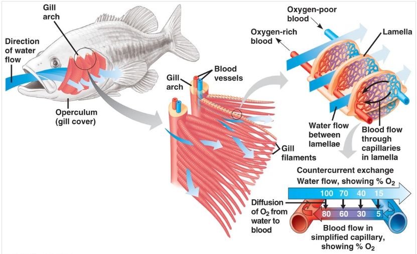
A dense network of blood vessels (capillaries) surround tissues involved in gas exchange because there is a continuous blood flow through capillaries. This allows for a constant maintenance of concentration gradient at the exchange surfaces. There is always deoxygenated blood.
Animals have lungs that are ventilated with air so that a high concentration of oxygen is brought to the alveoli. This creates a concentration gradient where there is more oxygen in the alveoli than in the blood, so oxygen will diffuse into the capillaries. Furthermore, there is more carbon dioxide in the blood than in the alveoli, so carbon dioxide will diffuse out of the capillaries, into the lungs.
Animals with gills move water through the gills. This brings in a high concentration of oxygen, creating a concentration gradient. Carbon dioxide will also move away from the gills (due to the concentration gradient).
State the locations of gas exchange in humans.
B3.1.4: Adaptations of mammalian lungs for gas exchange
Gas exchange in the lungs in humans.
Outline the structures of mammalian lungs that are adapted to maximizing gas exchange.
B3.1.4: Adaptations of mammalian lungs for gas exchange
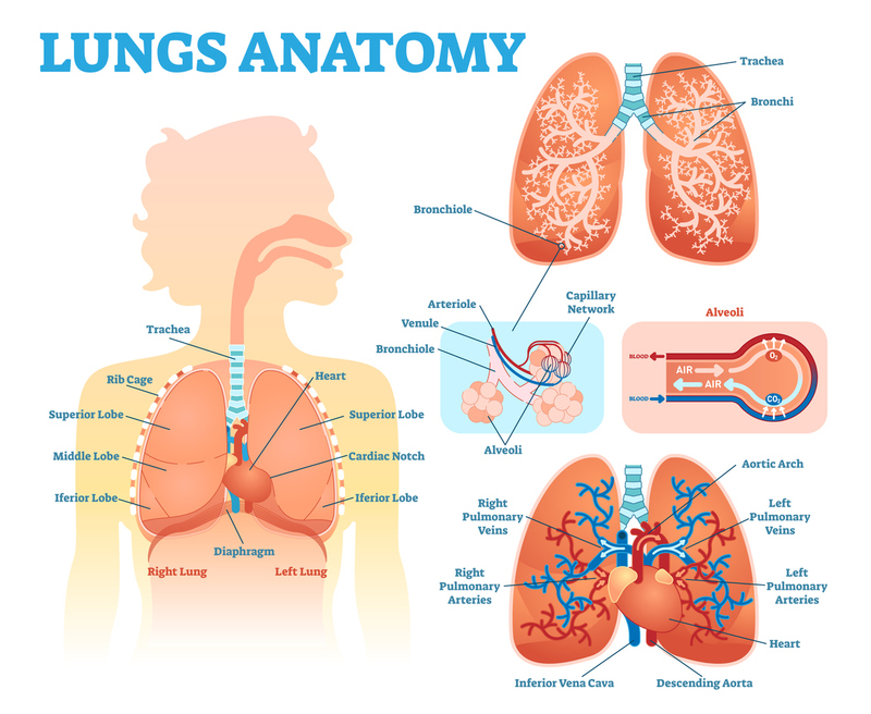
The branching bronchioles connect the bronchi to many alveoli.
The alveoli provides a very large surface area for gas exchange.
Surfactant (a fatty substance secreted by alveoli) prevents the walls of the alveoli from sticking to each other and provides a moist surface for gas exchange.
Alveoli are surrounded by an extensive capillary bed, which maintains high Oxygen and Carbon dioxide concentrations between the blood and alveoli (which allows for faster diffusion).
The capillaries provide a continuous supply of blood with low Oxygen concentration and high Carbon dioxide concentrations to the alveoli.
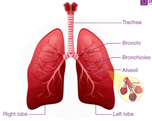
Draw and label a diagram showing the structure of the lungs.
B3.1.4: Adaptations of mammalian lungs for gas exchange
Include trachea, bronchi, bronchioles, left and right lobes, alveoli.
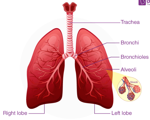
Draw a diagram showing the structure of an alveolus and an adjacent capillary.
B3.1.4: Adaptations of mammalian lungs for gas exchange

Identify the structure of the airway that connects the lungs to the outside of the body.
B3.1.5: Ventilation of the lungs
The trachea connects to the lungs. This splits into the bronchi and more bronchioles.
Define ventilation, inspiration and expiration.
B3.1.5: Ventilation of the lungs
Ventilation is the supply of air to the lungs.
Inspiration is a stage of ventilation where air (oxygen) is brought into the lungs by breathing in.
Expiration is a stage of ventilation where carbon dioxide is exhaled from the lungs by breathing out.
State the relationship between gas pressure and volume.
B3.1.5: Ventilation of the lungs
Gas moves from an area of high pressure to an area of low pressure (down a pressure gradient).
There is an inverse relationship between pressure and volume. This means that if there is more volume, the pressure is lower. If there is less volume, the pressure is higher.
Outline the direction of movement of the diaphragm and thorax during inspiration and expiration.
B3.1.5: Ventilation of the lungs
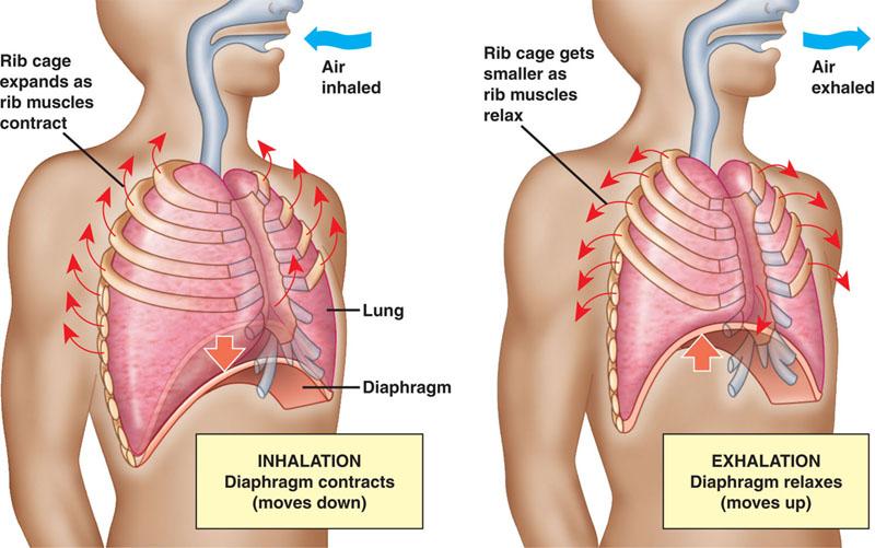
During inspiration, the diaphragm contracts and moves downward. The external intercostal muscles contract, so the rib cage moves up and out. As a result, the volume in the thorax increases.
During expiration, the abdominal muscles contract so the diaphragm is pushed outwards. The external intercostal muscles relax while the internal intercostal muscles contract, so the rib cage moves down and inwards. Therefore, the volume in the thorax decreases.
Outline the pressure and volume changes that occur during inspiration and expiration.
B3.1.5: Ventilation of the lungs
During inspiration, the volume in the thorax increases. Therefore, there is a decrease in pressure in the lungs. Air moves passively from the outside air (higher pressure) into the lungs (lower pressure).
During expiration, the volume in the thorax decreases. Therefore, there is increased pressure in the lungs. The gas will move from out of the lungs (higher pressure) to outside air (lower pressure).
Define tidal volume, vital capacity, and inspiratory and expiratory reserve.
B3.1.6: Measurement of lung volumes
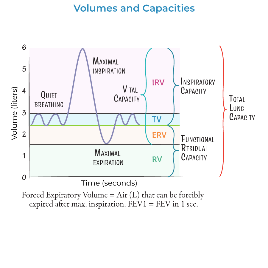
Tidal Volume (TV) is the volume of air that moves in and out of the lungs with normal, quiet breathing.
Inspiratory Reserve Volume (IRV) is the additional volume of air that can be inhaled with maximum effort after a normal breath.
Expiratory Reserve Volume (ERV) is the additional volume of air that can be exhaled with maximum effort after a normal breath.
Residual Volume (RV) is the amount of air remaining in the lungs after maximum exhalation.
Vital Capacity (VC) is the greatest volume of the deepest breath that lungs can handle (VC = TV + IRV + ERV).
List methods for measuring tidal volume, vital capacity, and inspiratory and expiratory reserve.
B3.1.6: Measurement of lung volumes
Spirometry
Body plethysmography
Nitrogen washout
Helium Dilution
State the direction of movement of gasses exchanged in leaves.
B3.1.7: Adaptations for gas exchange in leaves
Carbon dioxide is taken in through the stomata and oxygen is released into the atmosphere.
Outline adaptations for gas exchange in leaves, including epidermis, waxy cuticle, stoma, guard cells, air spaces, spongy mesophyll, and veins.
B3.1.7: Adaptations for gas exchange in leaves
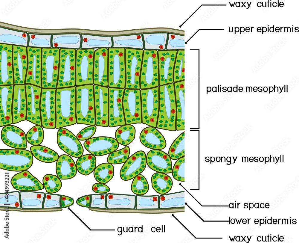
The waxy cuticle is a thin layer of lipids that covers the epidermis cells. It reduces the evaporation of water from the leaf.
The upper epidermis provides protection for the mesophyll cells in the leaf. Epidermal cells are transparent, which allows light from the sun to pass through and reach the mesophyll cells, where photosynthesis is carried out.
Palisade Mesophyll cells are more compact than spongy mesophyll and have lots of chloroplast for photosynthesis.
Spongy Mesophyll cells have an irregular shape, which increases the surface area for gas exchange.
Spongy Mesophyll cells are surrounded by air spaces, which facilitate the diffusion of gases between the surrounding atmosphere and mesophyll cells.
The stomata are pores that allows gases to enter and exit the leaf. They are typically on the lower epidermis of the leaf because it is more shaded, so there is less water loss.
Guard cells control water loss in the cell by opening and closing the stomata. At night, when photosynthesis cannot occur, the guard cells close. During the day, stomata try to be open as short as possible to prevent water loss. If there is a lot of water, guard cells are turgid (well-hydrated), so lots of carbon dioxide can pass through. If the plant is short of water, the guard cells lose water (due to osmosis and become flaccid), so the stomata close, don’t take in carbon dioxide, and conserve water vapor.
The veins provide support for the leaf. They contain the xylem, which transports water and minerals from the roots, and the phloem, which transports nutrients and sugar from photosynthesis up and down the plant.
State that a plan diagram shows the distribution of tissues, but not individual cells.
B3.1.8: Distribution of tissues in a leaf
A plan diagram shows the distribution of tissues, but not individual cells.
Draw and label a plan diagram to show the distribution of tissues in a transverse section of a leaf. Include upper and lower epidermis, palisade and spongy mesophyll, xylem and phloem.
B3.1.8: Distribution of tissues in a leaf
Define transpiration.
B3.1.9: Transpiration as a consequence of gas exchange in a leaf
Transpiration is the movement of water through a plant and its evaporation from aerial parts of the plant (like the leaves).
Outline the relationship between water evaporation and transpiration.
B3.1.9: Transpiration as a consequence of gas exchange in a leaf
The main way for water to return to the atmosphere is through transpiration. Transpiration, essentially, is when water evaporates through plants.
Discuss the effect of abiotic factors on the rate of transpiration; including temperature and humidity.
B3.1.9: Transpiration as a consequence of gas exchange in a leaf
Light Intensity: As light Intensity increases, more stomata open. Therefore, more water can diffuse out of the leaf, and there is an increased rate of transpiration.
Temperature: As temperature increases, water particles gain kinetic energy and move faster. These faster moving particles can diffuse through the stomata at a faster rate, which increases the rate of transpiration. Higher temperatures also increase the rate of evaporation, also increasing the rate of transpiration.
Humidity: As the humidity increases, the concentration of water (vapor) outside the leaf increases. Therefore, the concentration gradient between the inside and the outside of the leaf decreases. As a result, water particles diffuse slower, and there is a slower rate of transpiration.
Air flow (wind): As air flows past the leaf, water vapor is moved away from the leaf. The concentration of water outside the stomata, therefore, decreases. Thus, the concentration gradient is higher, so there is an increased rate of transpiration.
Discuss the advantages of opening and closing stomata at different times of day.
B3.1.9: Transpiration as a consequence of gas exchange in a leaf
Opening and closing stomata at different times of day prevents the plant from losing too much water.
During the day, the stomata can open and close according to light intensity and temperature so that it can take in more carbon dioxide for photosynthesis or prevent useless water loss.
At night, when photosynthesis doesn’t occur, the stomata can close to prevent water loss.
Calculate stomatal density from a leaf cast or micrograph.
B3.1.10: Stomatal density
Stomatal Density: The number of stomata per unit area of a leaf
Calculate the radius of the field of view (in mm)
Calculate the area of the field of view
Area = (pi)r2
Divide the mean number of stomata by the area of hte field of view to calculate the density
Density = (number of stomata) / area
Round to the nearest whole number
Describe the structure and function of hemoglobin.
B3.1.11: Adaptations of fetal and adult hemoglobin for the transport of oxygen.
Hemoglobin is a protein molecule found within erythrocytes (red blood cells). They are responsible for carrying most of the oxygen within the bloodstream.
Each RBC has cytoplasm with hemoglobin.
Each hemoglobin has 4 polypeptides and has a quaternary structure. Each polypeptide has a haem group near its center and each haem group has an iron atom within it.
Define affinity.
B3.1.11: Adaptations of fetal and adult hemoglobin for the transport of oxygen.
Affinity is the relationship between hemoglobin oxygen saturation and bonding.
Outline how cooperative binding alters hemoglobin's affinity for oxygen.
B3.1.11: Adaptations of fetal and adult hemoglobin for the transport of oxygen.
When an oxygen molecule bonds to hemoglobin, there is an increased affinity (attraction) for oxygen. The iron atom within the haem group bonds with oxygen in the hemoglobin.
Hemoglobin molecules that carry 3 O2 molecules have the greatest affinity for oxygen.
Hemoglobin molecules that carry no O2 molecules have the least affinity for oxygen.
Hemoglobin molecules that carry 4 O2 molecules have no affinity for oxygen (because it can carry a maximum of 4 oxygen molecules).
Compare the oxygen affinity of adult and fetal hemoglobin.
B3.1.11 (AHL): Adaptations of fetal and adult hemoglobin for the transport of oxygen.
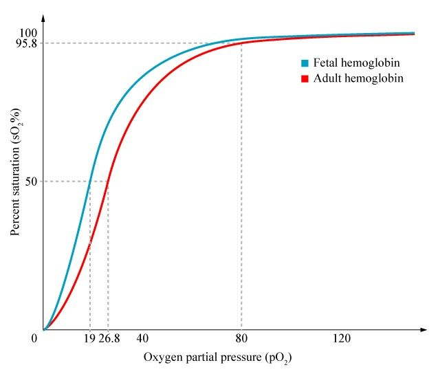
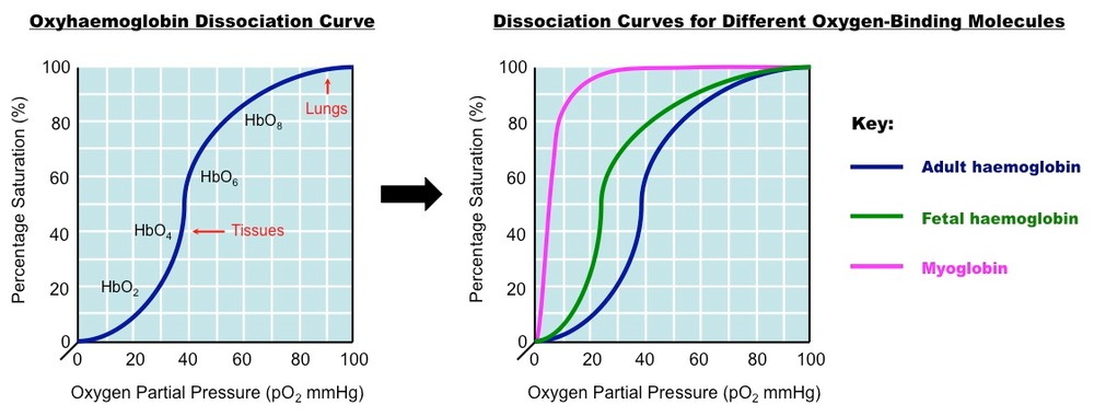
Fetal hemoglobin have a higher affinity for oxygen than adult hemoglobin.
In the placenta of a pregnant woman, capillaries are very close to the capillaries of the fetus, so molecular gas exchanges between the mother and fetus are fast.
Describe allosteric binding of CO2 to hemoglobin and the consequences for oxygen transport.
B3.1.12 (AHL): Bohr shift
Allosterity is the binding of carbon dioxide to the polypeptide chains of hemoglobin and the resulting change in hemoglobin’s affinity for O2.
CO2 binds to the polypeptide regions of the molecule while O2 binds to iron of haem group. This decreases the hemoglobin’s affinity for oxygen.
Define the Bohr shift.
B3.1.12 (AHL): Bohr shift
Bohr shift is the binding of CO2 to hemoglobin and the increase in release of oxygen molecules.
Explain the mechanism and benefit of the Bohr shift.
B3.1.12 (AHL): Bohr shift
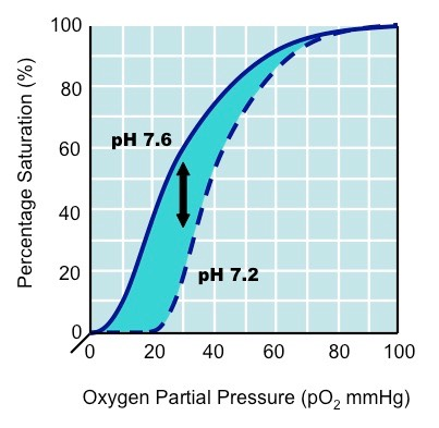
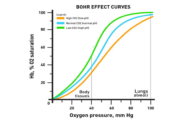
When respiration increases, the amount of carbon dioxide in the blood increases. The pH decreases from a normal 7.6 to 7.2. The increase in acidity makes hemoglobin have affinity for oxygen (highly respiring tissues would want more oxygen). Hemoglobin is more likely to increase its oxygen stores in the tissues.
The Bohr shift decreases hemoglobin’s affinity for oxygen. Therefore, the unloading of oxygen into tissues increases.
State the effect of the Bohr shift on the oxygen dissociation curve.
B3.1.12 (AHL): Bohr shift
The Bohr shift causes a rightward shift in the oxygen dissociation curve.
Define partial pressure.
B3.1.13 (AHL): Oxygen dissociation curves as a means of representing the affinity of haemoglobin for oxygen at different oxygen concentrations
Partial pressure is the pressure exerted by a single gas within a mixture gases.
State the relative partial pressures of oxygen in the atmosphere at sea level, in the alveoli, in alveoli blood capillaries, and in respiring tissue.
B3.1.13 (AHL): Oxygen dissociation curves as a means of representing the affinity of haemoglobin for oxygen at different oxygen concentrations
Oxygen in the atmosphere at sea level is 20.9% of the atmosphere, so it is 158.8 mm Hg (total atmospheric pressure is 760.0 mm Hg).
Oxygen in the alveoli is 13.7% of total composition, so it is 104 mm Hg.
In the pulmonary veins, oxygen is about 100 mm Hg.
The partial pressure of oxygen in tissues is low, about 40 mm Hg, because it is continuously used for cellular respiration.
Draw the oxygen dissociation curve to show affinity of hemoglobin for oxygen at different partial pressures of oxygen.
B3.1.13 (AHL): Oxygen dissociation curves as a means of representing the affinity of haemoglobin for oxygen at different oxygen concentrations
Explain difference in the oxygen dissociation curves of adult and fetal hemoglobin.
B3.1.13 (AHL): Oxygen dissociation curves as a means of representing the affinity of haemoglobin for oxygen at different oxygen concentrations
Because fetal hemoglobin has a higher affinity for oxygen than normal hemoglobin, the fetal hemoglobin curve is shifted to the right. It has greater affinity for oxygen at almost every partial pressure of oxygen
