module 14: sensory pathways 1: vision (unfinished)
1/55
There's no tags or description
Looks like no tags are added yet.
Name | Mastery | Learn | Test | Matching | Spaced | Call with Kai |
|---|
No analytics yet
Send a link to your students to track their progress
56 Terms
amplitude of light
intensity of light
eg large amplitude = bright light
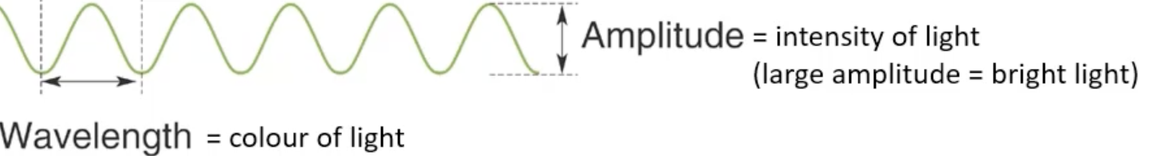
wavelength of light
colour of light
the shorter the distance is between peaks = the more energetic the light is
becomes more blue as well (opposite = red)

visible light wavelength
380 (violet) -750 (red) nm
diopter (D)
unit
power of lens/ ability to bend light
lens
crystalline protein structure = composed crystallin proteins which give the lens its transparency + ability to focus light
absorbs, focuses and directs incoming light to the retina
fine-tunes focus by changing shape
12 D
cornea
transparent, curved front layer of the eye
focuses light and protects the eye
42 D - bends (refracts) light the most
iris
muscle
controls size of the pupil - regulates light entry
retina
layer at the back of the eye
also a part of the brain/CNS
contains photoreceptors (rods and cones)
optic nerve
transmits visual info from the retina to the brain
lateral geniculate in the thalamus
visual vortex
accommodation (lens adjustment)
distant objects = lens flattens to reduce refraction
near objects = lens thickens to increase refraction
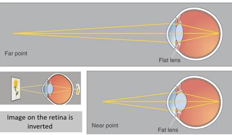
image inversion
light passing through the lens produces inverted + reversed image on the retina
the brain (visual cortex) reorientates the image correctly
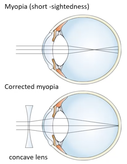
myopia
short-sightedness
lens is too strong or the eye is too long => light focuses before it reaches retina (ie focuses too soon)
correction = concave lens to spread out light before reaching the eye + into the retina
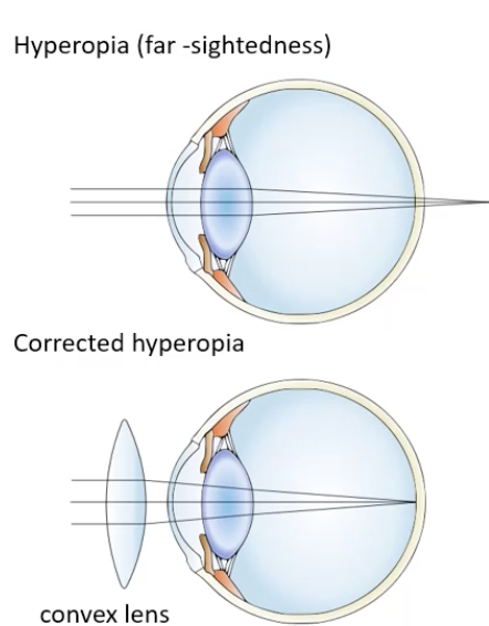
hyperopia
far/long-sightedness
lens too weak or eye too short => light focuses behind the retina
correction = convex lens to converge light before it reaches the eye + into the retina
photoreceptors in the retina
found at the back of the retina - makes light pass thru all cells types
rods:
function in low light (night vision)
highly sensitive
don’t detect colour
cylindrical shape
cones:
function in bright light (day vision)
detect colour (red, blue, green cones)
why cant see colour well at night
rods used for night vision
rods cant detect colour
therefore little colour visible
Retina as Part of the Brain
• Retina is considered an extension of the central nervous system (CNS).
• Directly connected to the brain via the optic nerve.
Optic Pathway
Optic Nerve → Lateral Geniculate Nucleus (in thalamus) → Visual Cortex
Retinal Cell Layers (Information Pathway)
Photoreceptors (Rods & Cones) → Bipolar Cells → Retinal Ganglion Cells → Optic Nerve → Brain
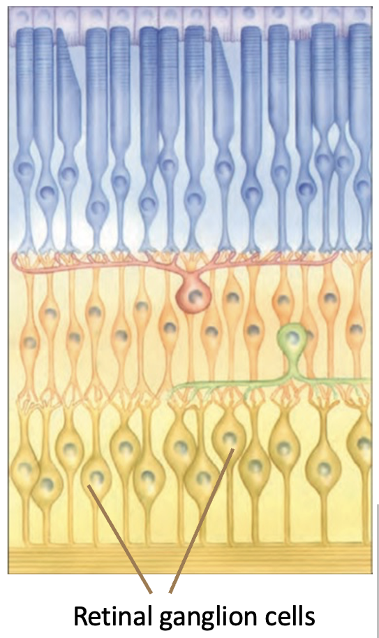
Retinal Ganglion Cells
Convert light signals into action potentials and send them to the optic nerve to be processed in brain
neurotransmitter used by photorecetpors
glutamate (glutamatergic excitatory neurones)
Horizontal cells
Found between photoreceptors and bipolar cells
involved in adapting to light intensity and spatial + colour processing.
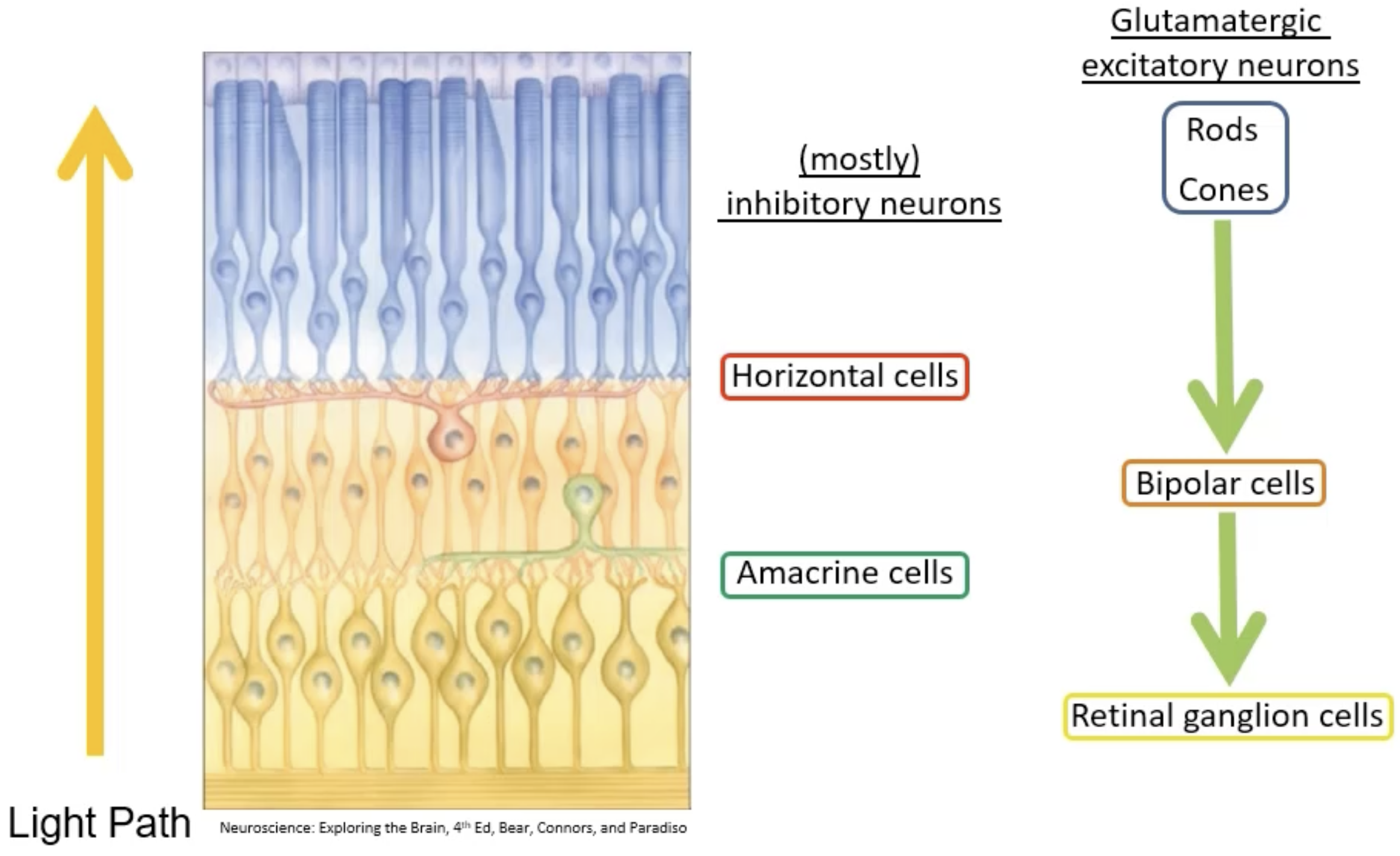
Amacrine Cells
Found between bipolar cells and retinal ganglion cells
help extract visual features such as motion direction and background light adaptation
modulate circadian rhythm

Retinal Ganglion Cells
Neurons that receive input from photoreceptors and transmit action potentials to the brain via the optic nerve.
intrinsically photosensitive retinal ganglion cells (ipRGC)
type of retinal ganglion cell
Contain melanopsin so can detect and respond to light- help regulate circadian rhythms and pupil size
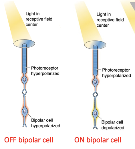
Bipolar Cells
Intermediate neurons in the retina that transmit signals from photoreceptors to ganglion cells.
types:
on bipolar cells = Depolarize when light is detected by photoreceptors
off bipolar cells = Hyperpolarize when light is detected by photoreceptors
type of bipolar cell it is is dependent on the type of glutamate receptor it has - determines the way in which it responds to glutamate
pigmented layer at back of retina
supports photoreceptors by regenerating retinaldehyde - maintains rod and cone function
reduces oxidative stress of rods and cones
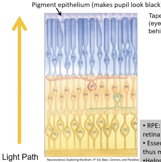
retinaldehyde
important cofactor in light-sensing proteins / opsins found in rods / cones
reflective layer behind RPE in some animals
enhances night vision by reflecting light back through the retina.
Light-sensitive proteins in photoreceptors that, when bound to retinal, detect light
maximises rods/cones sensitivity to light
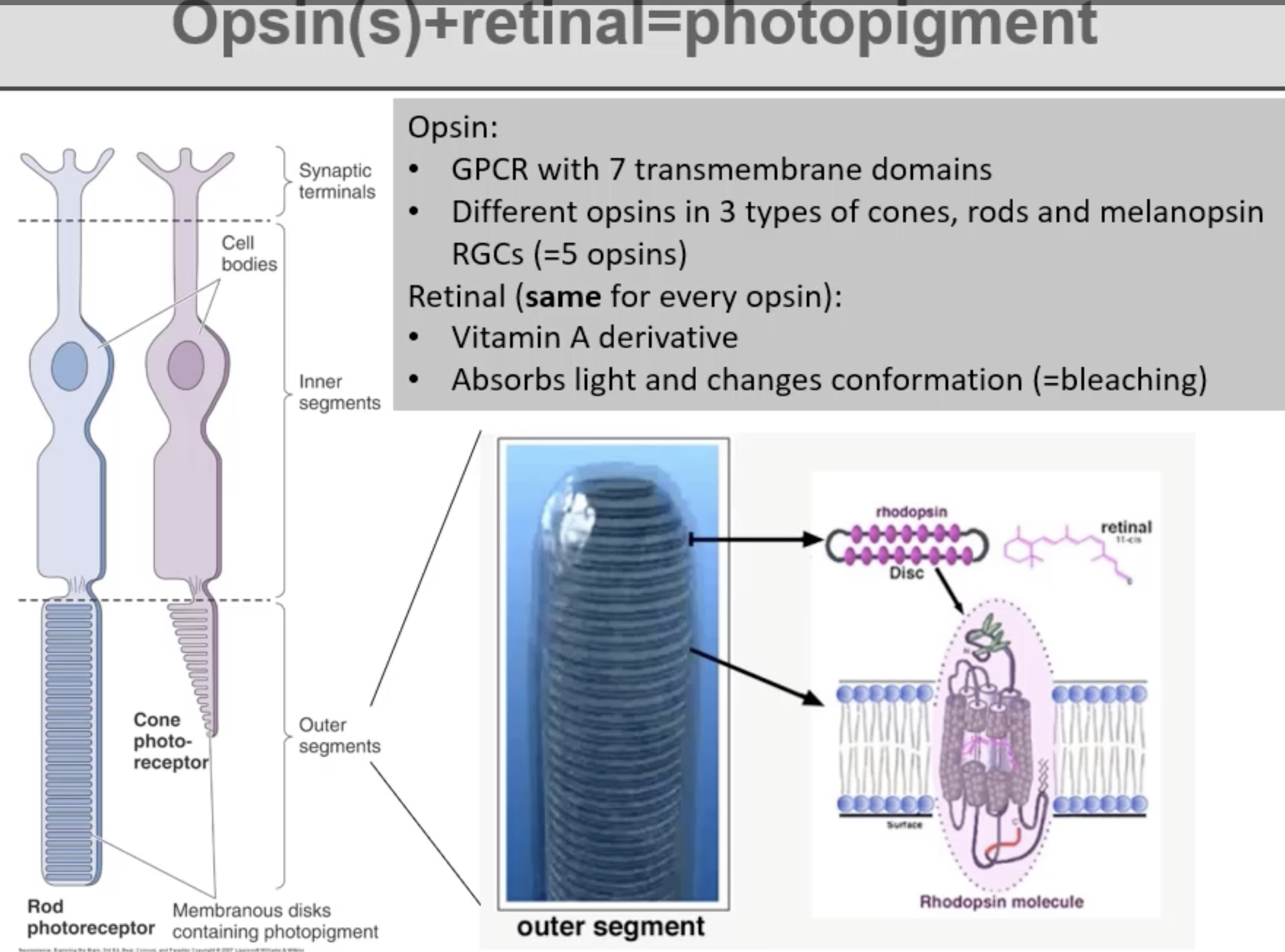
retinal
vitamin A derivative
absorbs light and changes conformation => phototransduction occurs - allows us to see light
Rhodopsin
opsin + retinal = rhodopsin
The opsin in rods responsible for night vision
opsin in cones responsible for colour vision
3 types:
detect red, blue, green
An opsin found in some retinal ganglion cells
mainly involved in:
• Regulating the biological clock (day-night cycle)
• Pupil constriction in bright light.
Retinal
Vitamin A Derivative
absorbs light
types:
11-cis retinal
all-trans retinal (active form)
11-cis becomes all-trans via a conformational change when light is absorbed
A molecule that keeps sodium channels open in photoreceptors (in the dark)
Depolarisation in Darkness
Photoreceptors remain depolarised in the dark due to open sodium channels
presence of cGMP in photoreceptors keeps sodium ion channels open => depolarisation
depolarisation leads to release of glutamate - photoreceptors are active
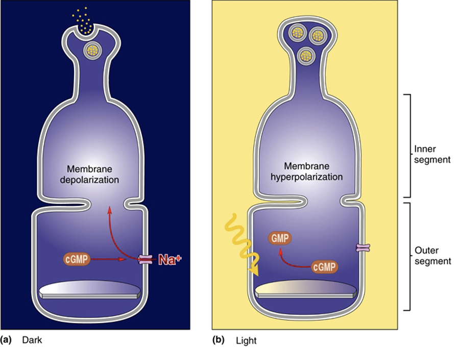
Hyperpolarisation in Light
Light causes sodium channels in photoreceptors to close
light causes cGMP to become GMP => sodium channels remain closed and so photoreceptors are hyperpolarised
hyperpolarised state means glutamate is not released

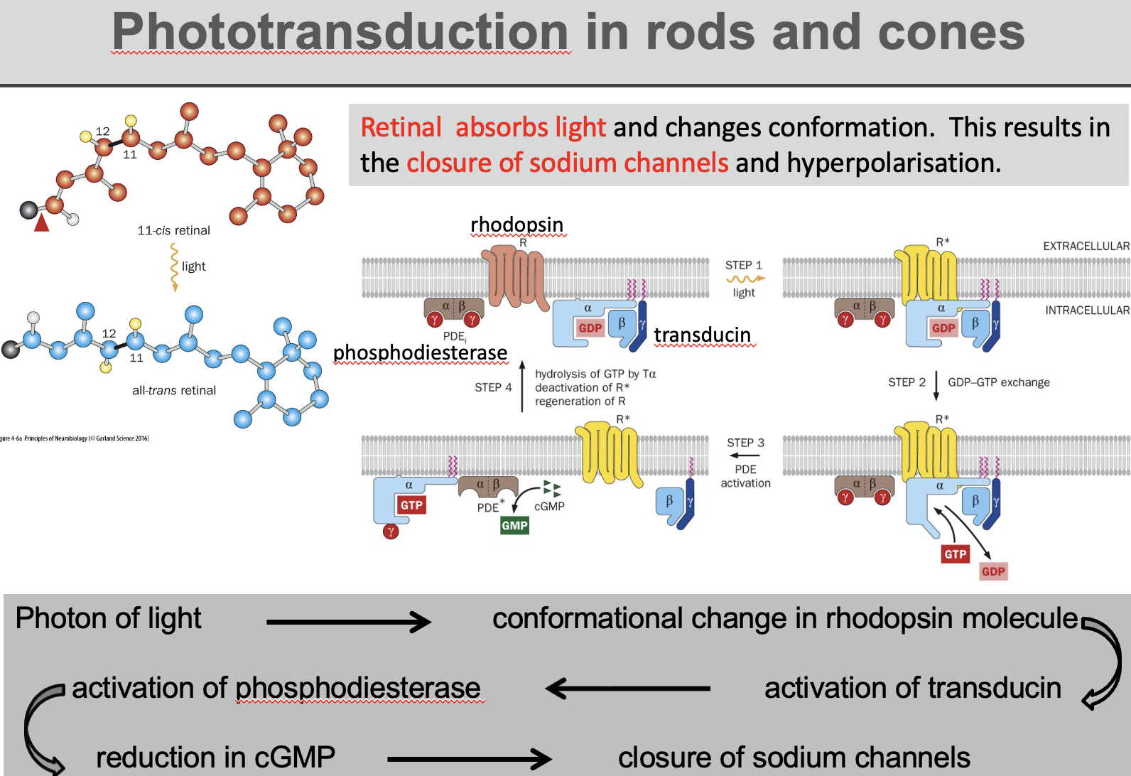
phototransduction
process by which light is converted into electrical signals in the retina
When light hits rhodopsin, retinal changes from 11-cis retinal → all-trans retinal
this activates opsin, triggering a G-protein signaling cascade: transducin → phosphodiesterase → cGMP breakdown
leads to an electrical signal sent to the brain.
After activation, retinal recycled back to 11-cis form to reset rhodopsin
Transducin
A G-protein that activates phosphodiesterase when light is detected
swaps GDP for GTP when activated, allowing activation of phosphodiesterase
Phosphodiesterase (PDE)
Enzyme that breaks down cyclic GMP
inhibited when 0 light absorbed
active in the presence of light
trichromatic vision
normal color vision
ability to perceive colors by sensing three primary wavelengths of light: red, green, and blue in retina
done by 3 cones in mammals
Monochromatic Vision
Vision in animals with only one type of cone photoreceptor.
common in aquatic animals
Dichromatic Vision
Vision in animals with two types of cone photoreceptors
eg dogs, cats, horses
Color Blindness
A condition caused by mutations in opsin genes
red and green opsin genes found on X chromosome, blue found on chromosome 7
means men more likely to be (red-green) colourblind cos only have 1 X chromosome - if mutation found on X chromosome then condition manifests
women can be tetrachromatic
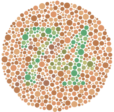
Ishihara Test
A color vision test using colored dot patterns to identify color blindness.
Lateral Geniculate Nucleus (LGN)
key relay station in the visual pathway
located in the thalamus
receives visual information from the retina and relays it to the primary visual cortex (V1).

Visual Cortex (V1)
main processing center for vision, located at the back of the brain (occipital lobe)
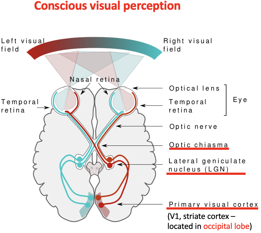
Optic Chiasm
The point where nasal retina signals cross to the opposite side of the brain

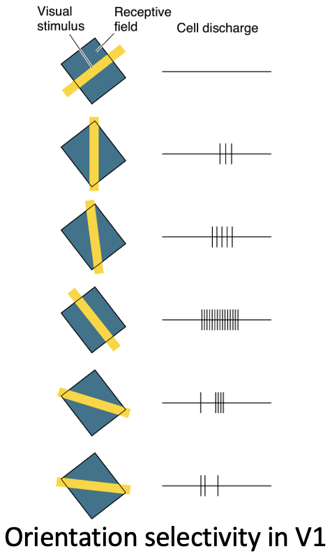
Orientation Selectivity in V1
Neurons in the visual cortex respond to specific orientations of visual stimuli.
The brain has specialized neurons in the visual cortex (V1) that respond to different line orientations.
Some neurons detect vertical lines, others detect horizontal or diagonal lines.
When a pattern rotates, different neurons activate, helping the brain break down shapes and recognize objects, letters, and patterns.