NPB: The Cardiovascular System Lecture Set #3
1/91
Earn XP
Description and Tags
Cardiac Cycle
Name | Mastery | Learn | Test | Matching | Spaced | Call with Kai |
|---|
No analytics yet
Send a link to your students to track their progress
92 Terms
Blood Flow Cycle =.9 seconds ≈ 900 miliseconds
Diastole: 0.6sec
atria is relaxed and in a repolarized state and pushing the blood passively into the →
repolarized relaxed ventricles →
the atria come out of relaxed state →
begin to depolarize and push the last of the blood into the ventricles →
Systole: 0.3sec
Then atria resume back to repolarized state as they relax →
the ventricles then go into a depolarized state and contract
Cardiac Cycle Goal
Fill ventricles to their end-diastole volume
Cardiac Output:
Heart Rate x Stroke Volume
increase in heart rate or stroke volume: CO increase
decrease in heart rate or stroke volume: CO decreases
Which factor(s) would decrease cardiac output?
A) increased end diastolic volume
B) increased SNS input
C) decreased SNS input
D) increased venous return
E) decreased vagal stimulation of the heart
C) decreased SNS input
bc this would decrease heart rate so therefore decrease CO
If the cardiac output is 4,800 ml/min and the heart rate is 60 beats per minute, the stroke volume averages ____ ml.
A) 70
B) 60
C) 80
D) 140
E) 120
C) 80
CO = Heart Rate x Stroke Volume
CO / HR = SV
4,800 / 60 = 80
Diastole
ventricles are relaxed
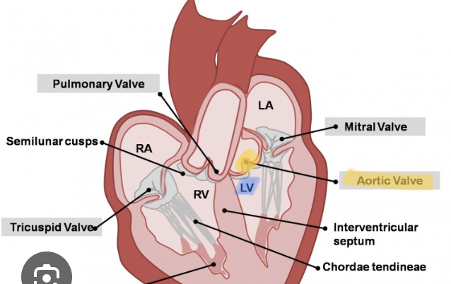
AV valves
Left:
Bicuspid
Right:
Tricuspid
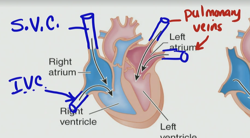
S.V.C - Superior Vena Cava
the big gigantic vein where the return of our blood will drain into → deoxygenated
IVC - Inferior Vena Cava
Which type of blood is carried from the lungs to the left atrium of the heart?
A. Blood low in oxygen and high in carbon dioxide
B. Blood high in nitrogen waste
C. Blood rich in oxygen
D. Blood high in glucose from the liver
C. Blood rich in oxygen
The blood in the superior and inferior vena cava is low in:
A. Carbon dioxide
B. Oxygen
C. Sodium
D. Glucose
B. Oxygen
Why is the blood in the superior and inferior vena cava low in oxygen content?
A. Because it hasn't passed through the lungs yet
B. Because it's returning from body tissues like the brain and muscles that have already used the oxygen
C. Because the vena cava connects directly to the lungs
D. Because it carries nutrients from the digestive tract
B. Because it's returning from body tissues like the brain and muscles that have already used the oxygen
Where does the blood entering the left atrium come from?
A. Superior vena cava
B. Pulmonary veins
C. Pulmonary artery
D. Aorta
B. Pulmonary veins
What is the primary function of veins in the circulatory system?
A. To carry blood away from the heart
B. To pump oxygen into the bloodstream
C. To bring blood back to the heart
D. To deliver nutrients to the lungs
C. To bring blood back to the heart
What determines whether a blood vessel is classified as a vein?
A. Whether the blood it carries is deoxygenated
B. Whether the blood is flowing slowly
C. Whether it carries blood toward the heart
D. Whether it contains carbon dioxide
C. Whether it carries blood toward the heart
Which of the following statements about veins is correct?
A. Veins always carry oxygenated blood
B. Veins carry blood to the heart
C. Oxygen content doesn’t matter (deo, or ox)
D. Two or more of the above are correct
D. Two or more of the above are correct
B. Veins carry blood to the heart
C. Oxygen content doesn’t matter (deo, or ox)
What primarily drives blood flow through the veins back to the heart?
A. High pressure generated by the left ventricle
B. Muscle contraction and pressure gradient toward the heart
C. Constriction of capillaries
D. Blood being pulled by gravity
B. Muscle contraction and pressure gradient toward the heart
Which chamber determines whether ventricular filling is passive or active?
A. The atria, based on their contraction timing
B. The pulmonary veins, based on blood return
C. The ventricles, based on whether they are relaxed or contracted
D. The aorta, based on systemic pressure
C. The ventricles, based on whether they are relaxed or contracted
Passive filling of the ventricles occurs when the ventricles are:
A. Contracted
B. Depolarized
C. Relaxed
D. Repolarized and contracted
C. Relaxed
When does passive ventricular filling primarily occur?
A. When the ventricles are contracted and depolarized
B. When the ventricles are repolarized and relaxed
C. When the atria are repolarized and relaxed
D. When the semilunar valves are open
B. When the ventricles are repolarized and relaxed
What causes atrial pressure to be greater than ventricular pressure during diastole, allowing the AV valves to open?
A. Continuous contraction of the atria
B. The 20% of blood actively pushed by atrial contraction after 80% passive filling
C. High pressure in the pulmonary artery
D. Ventricular contraction increasing suction force
B. The 20% of blood actively pushed by atrial contraction after 80% passive filling
Where is the pacemaking action potential generated in the heart?
A. AV Node
B. Purkinje fibers
C. SA Node
D. Bundle of His
C. SA Node
What is the role of the SA Node in the cardiac cycle?
A. Initiates ventricular contraction
B. Acts as the secondary pacemaker
C. Initiates the action potential that spreads across the atria
D. Closes the AV valves during systole
C. Initiates the action potential that spreads across the atria
Which of the following correctly describes the sequence of electrical conduction from the SA node to atrial contraction?
A. Atrial myocytes → AV node → SA node
B. AV node → SA node → Ventricles
C. SA node → Internodal & Interatrial pathways → Atrial myocytes
D. SA node → Ventricles → Interatrial pathways
C. SA node → Internodal & Interatrial pathways → Atrial myocytes
Pathway L & R atrium Electric Flow w/ diastole and systole
Right Atrium:
Diastole:
SA node (in the right atrium)
➝ Generates the pacemaker AP➝ Signal spreads through right atrial myocytes
➝ Travels via the internodal pathway
➝ Reaches the AV node (still in right atrium, near septum)
➝ AV nodal delay
➝ Allows both atria to finish contracting before ventricles are activated
Systole
➝ Signal passes to the Bundle of His
➝ Then to the right and left bundle branches
➝ Then to the Purkinje fibers
➝ Both ventricles contract simultaneously
Left Atrium:
Diastole
SA node (located in the right atrium)
➝ Generates the pacemaker action potential➝ Signal spreads to the left atrium via the interatrial pathway (aka Bachmann’s bundle)
➝ Depolarization spreads through left atrial myocytes
➝ This causes the left atrium to contract, pushing blood into the left ventricle
❌ No AV node in the left atrium
➝ The AV node (in the right atrium) still handles the signal delay before passing it to the ventricles
Systole
➝ Signal continues from AV node
→ AP spread to ventricular conductive pathways:
→ Bundle of His
→ Left & Right bundle branches
→ Purkinje fibers
➝ Both ventricles contract simultaneously
Isovolumetric
Iso= same
same volume is unchanging
During isovolumetric contraction, which of the following is true?
A) The semilunar valves are open and blood is ejected
B) The ventricles are relaxed and filling with blood
C) The ventricles contract but all heart valves remain closed
D) The atrioventricular valves open to allow ventricular ejection
C) The ventricles contract but all heart valves remain closed
During isovolumetric contraction...
A) The ventricles relax but do not change volume
B) The heart valves open
C) The ventricles contract, but do not eject blood
D) The ventricles fill with blood
C) The ventricles contract, but do not eject blood
volumetric refers to volume in ventricles
This can only happen ( not ejecting blood, but contracting) if
AV valves are shut ≠ atria
semilunar valves are still close ≠ arteries
the pressure is building with contraction of ventricles, but here the semilunar valves (atrium pressure) isnt low enough, we want
ventricular pressure > arterial trunks
Cardiac Cycle
STEP (a) Diastole – Passive Ventricular Filling
State: Atria & ventricles relaxed
Valves: AV valves open, semilunar valves closed
Flow: Blood flows passively into ventricles from veins
Right side: from SVC & IVC: deoxygenated
Left side: from pulmonary veins: oxygenated
Why? Atrial pressure > ventricular pressure
drive blood from veins to atrium so > ven pr
this will push blood into the ventricles & open up AV Valves
STEP (b) Diastole – Atrial Contraction
AP generated at SA node (1º Pacemaker) fires →
spreads to atria via interatrial & internodal pathways
Atria contract, pushing last ~20% of blood into ventricles
Ventricular filling completes → this is EDV (~135 mL)
AV nodal delay (~200 ms) allows atria to finish contracting
STEP (c) Systole – Isovolumetric Contraction
AP spreads from AV node → Bundle of His → L/R bundle branches → Purkinje fibers
Ventricles begin contracting → squeeze blood
Ventricular pressure > atrial pressure BECAUSE
→ AV valves close
semilunar valves are closed
Pulmonary valve
Aorta Valve
Builds pressure inside ventricles
No blood leaves yet = isovolumetric contraction (same volume unchanging)
STEP (d) Systole – Ventricular Ejection
Because of this Ventricular pressure > arterial pressure (aortic & pulmonary trunk)
AV valves still closed
Semilunar valves swing open
Blood comes out of ventricles
ejected into arteries
Right side → pulmonary artery/trunk
Left side → aorta
NO blood going backwards to atria
Blood only eject out as long as pressure is higher within ventricles compared to arteries
STEP (e) Diastole – Isovolumetric Relaxation
Pressure in ventricle falls below aorta or pulmonary trunk
Ventricular pressure < arterial pressure → semilunar valves close
Isovolumetric relaxation - bc all valves r closed
arteries valves closed = no blood leaving ventricle
AV valves still closed = no filling yet
Once ventricular pressure < atrial pressure, AV valves open again → back to step a
What is the purpose of the AV nodal delay in the cardiac conduction system?
A. To allow the SA node to repolarize
B. To ensure the atria and ventricles contract at the same time
C. To give the ventricles time to fully relax
D. To allow both atria to contract fully before the ventricles are stimulated
D. To allow both atria to contract fully before the ventricles are stimulated
What initiates the contraction of the ventricles at the start of systole?
A. Closing of semilunar valves
B. Action potentials traveling through the Purkinje fibers
C. Relaxation of atrial muscle
D. Passive ventricular filling
B. Action potentials traveling through the Purkinje fibers
AP → Bundle of His → L & R bundle branches → Purkinje Fibers → contraction of ventricles
During isovolumetric contraction, which of the following is TRUE?
A. Both AV and semilunar valves are open
B. Blood is being ejected into the arteries
C. Ventricular pressure is rising but no blood is moving
D. The atria are actively contracting
What causes the AV valves to close during the start of systole?
A. The ventricles begin to relax
B. Blood pressure in the atria exceeds that in the ventricles
C. Ventricular pressure becomes greater than atrial pressure
D. The semilunar valves open
C. Ventricular pressure becomes greater than atrial pressure
What is the purpose of isovolumetric contraction?
A. To open the AV valves
B. To allow blood to fill the atria
C. To increase ventricular pressure without changing volume
D. To complete atrial depolarization
Which structure does the electrical impulse travel through immediately before reaching the Purkinje fibers?
A. SA node
B. AV node
C. Bundle branches
D. Atrial myocytes
C. Bundle branches
Bundle of His → bundle branches → Purkinje Fibers
During Systole ___ (relaxing/contracting) is occurring
Contracting → Depolarization
During Diastole ___ (relaxing/contracting) is occurring
Relaxing → Repolarization
What is occurring here?
Relaxation?
Contraction?
Also what is the special name for this?
Contraction is occurring so → Isovolumetric Contraction

What is occurring here?
Relaxation?
Contraction?
Also what is the special name for this?
Relaxation is occurring so → Isovolumetric Relaxation
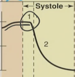
Why did the volume go down?
A. Ventricular ejection of blood into the arteries
B. Passive filling of the ventricles from the atria
C. Atrial contraction adding the final 20% of blood to the ventricle (atrial kick)
D. Closure of the semilunar valves preventing blood entry
A. Ventricular ejection of blood into the arteries
What causes the first heart sound ("Lub")?
A. Opening of the semilunar valves
B. Closure of the AV valves
C. Contraction of the atria
D. Ejection of blood from the ventricles
B. Closure of the AV valves
When does the "Lub" sound occur?
A. At the start of diastole
B. During atrial contraction
C. At the beginning of systole
D. When ventricular volume reaches its lowest point
C. At the beginning of systole
What causes the second heart sound ("Dub")?
A. Closure of the AV valves
B. Opening of the AV valves
C. Closure of the semilunar valves
D. Atrial relaxation
C. Closure of the semilunar valves
T/F: You can hear sound when the AV valves open up
False, you cannot hear sound when AV valves open up, when they are closed you can hear a “Lub” noise though
Which of the following is true about heart sounds?
A. The "Dub" is caused by the AV valves opening
B. The "Lub" is associated with semilunar valve closure
C. The "Dub" is heard when the semilunar valves close
D. The "Lub" occurs at the end of systole
C. The "Dub" is heard when the semilunar valves close
What event occurs during ventricular systole?
A. The ventricles relax and fill with blood
B. Blood is ejected from the atria into the ventricles
C. The ventricles contract and eject blood into the arteries
D. The atria contract and push blood into the ventricles
C. The ventricles contract and eject blood into the arteries
What event occurs during ventricular diastole?
A. The ventricles contract and increase pressure
B. Blood flows passively from the ventricles into the arteries
C. The ventricles relax and fill with blood from the atria
D. The semilunar valves open and blood exits the heart
C. The ventricles relax and fill with blood from the atria
What does End-Diastolic Volume (EDV) represent?
A. The blood left in the ventricles after they squeeze
B. The blood pushed out of the heart when the ventricles contract
C. The total blood collected in the ventricles right before they contract
D. The blood inside the atria during atrial contraction
C. The total blood collected in the ventricles right before they contract
The 80% that passively flowed in from atria → ventricle
and 20% that the atria contracted “kick” cus it got pushed in when the atria contracted, atria → ventricle
What is the typical End Diastolic Volume (EDV) in the ventricles?
A. 40–50 mL
B. 65–70 mL
C. 135 mL
D. 200 mL
C. 135 mL
What does End-Systolic Volume (ESV) refer to?
A. The volume of blood ejected from the ventricles during contraction
B. The total blood collected in the atria before contraction
C. The volume of blood remaining in the ventricles after systole
D. The blood pressure in the arteries during ventricular contraction
C. The volume of blood remaining in the ventricles after systole
What is the typical End Systolic Volume (ESV)?
A. 30–40 mL
B. 65–70 mL
C. 125–135 mL
D. 100 mL
B. 65–70 mL
During which phase of the cardiac cycle is the stroke volume ejected from the ventricles?
A. Early diastole
B. Late diastole
C. Systole
D. Isovolumetric relaxation
C. Systole
the ventricles will be contracting/ ejecting the blood into the arteries
What does Stroke Volume (SV) refer to?
A. The volume of blood left in the ventricles after contraction
B. The total blood pumped by both atria
C. The volume of blood ejected from the ventricles
D. The total blood returned to the heart via veins
C. The volume of blood ejected from the ventricles
What is the typical Stroke Volume (SV) ejected by the ventricles during systole?
A. 30–40 mL
B. 65–70 mL
C. 120–130 mL
D. 135–140 mL
B. 65–70 mL
T/F: You usually eject out the entire end diastolic volume (EDV)
False, you do not eject the entire EDV, because the heart needs a buffer to keep the cardiac cycle going
Why is it important for the ventricles to fill completely during diastole?
A. To prevent blood from flowing into the atria
B. To reduce the pressure in the arteries
C. To ensure there is enough blood to eject during systole
D. To stop the heart from overfilling
C. To ensure there is enough blood to eject during systole
Venous Return
This is the blood returning to the heart, specifically into the atria, from the veins (like the superior/inferior vena cava and pulmonary veins).
What happens if venous return is lower than normal?
A. EDV increases
B. Stroke volume decreases
C. Heart rate slows
D. ESV increases significantly
B. Stroke volume decreases
Stroke volume decreases when venous return is low,
because the ventricles fill with less blood, due to the atria, not ejecting enough blood to the ventricles, so then the ventricles eject less into the arteries during contraction
Why does some blood remain in the ventricles after systole (ventricular contraction)?
A. To prevent overfilling of the arteries
B. To allow time for the atria to repolarize
C. To maintain efficient pressure and allow continuous filling during the next cycle
D. Because the AV valves are closed during systole
C. To maintain efficient pressure and allow continuous filling during the next cycle
Which of the following situations is most likely to result in a decreased stroke volume?
A. A person lying down after drinking plenty of fluids
B. An athlete during peak exercise with high venous return
C. A dehydrated hiker who has been vomiting and hasn’t had water in hours
D. A person who just received IV fluids in a hospital setting
C. A dehydrated hiker who has been vomiting and hasn’t had water in hours
This is because dehydration lowers your blood volume,
Which will reduce the venous return to your heart
With less blood returning, the atria send less blood to the ventricles
So leading to a lower EDV
& since SV depends on how much blood is ejected into ventricles & how much is left in ventricles a lower EDV means decrease stroke volume
EDV - ESV = SV
Which of the following best explains how dehydration can lead to decreased stroke volume?
A. Dehydration increases heart rate, which reduces blood pressure.
B. Dehydration decreases End-Systolic Volume (ESV), reducing blood flow to the lungs.
C. Dehydration reduces blood volume, lowering venous return and End-Diastolic Volume (EDV), which decreases Stroke Volume (SV).
D. Dehydration causes more blood to remain in the atria, increasing EDV and stroke volume.
C. Dehydration reduces blood volume, lowering venous return and End-Diastolic Volume (EDV), which decreases Stroke Volume (SV).
The Frank-Starling Law
Specifically about ventricles
The more the heart fills with blood during diastole (i.e., the greater the end-diastolic volume, or EDV),
like a balloon filling with air
the stronger/forcefully it contracts during systole, and the more blood it pumps out (stroke volume).
so the more air in a balloon the more air being pushed out forcefully when you release it
Intrinsicly regulated - property of the heart
According to the Frank-Starling Law, what happens when End-Diastolic Volume (EDV) increases?
A. Stroke volume decreases due to overstretching
B. Contractile force and stroke volume increase
C. The atria contract harder
D. The semilunar valves close sooner
B. Contractile force and stroke volume increase
Frank Starling Law refers to ventricles
the contractile force in EDV is the force of contraction/pressure of blood built up in ventricles before systole (release of blood to arteries)
The SV refers to the volume of blood that came out of ventricles so that contractile force leads to more blood ejected out so = SV inc
What is the primary determinant of End-Diastolic Volume (EDV)?
A. Stroke volume
B. Atrial contraction
C. Venous return
D. Ventricular pressure
C. Venous return
What is the primary factor that determines End-Diastolic Volume (EDV)?
A. The strength of ventricular contraction
B. Atrial depolarization
C. Venous return to the heart
D. Pressure in the aorta
C. Venous return to the heart
The Frank-Starling Law describes what kind of property of the heart?
A. Extrinsic (neural control)
B. Metabolic property
C. Intrinsic (within the heart muscle itself)
D. Hormonal property
C. Intrinsic (within the heart muscle itself)
According to the Frank-Starling law of the heart,
A) shortening cardiac muscle fibers prior to contraction causes more forceful contractions
B) the left ventricle must pump more blood than the right ventricle since the left ventricle must pump blood to more regions of the body
C) increasing venous return increases EDV, which leads to an increased stroke volume
D) the greater the stroke volume, the smaller the subsequent ESV, because as more blood is squeezed out, the heart cannot fill as completely.
E) as cardiac output decreases, blood pools in the vasculature and increases arterial blood pressure
C) increasing venous return increases EDV, which leads to an increased stroke volume
If EDV increases from 135 mL to 200 mL, what is the expected effect on stroke volume?
A. Increase due to greater stretch and force
B. Decrease due to overstretch
C. No change
D. Decrease due to reduced venous return
A. Increase due to greater stretch and force
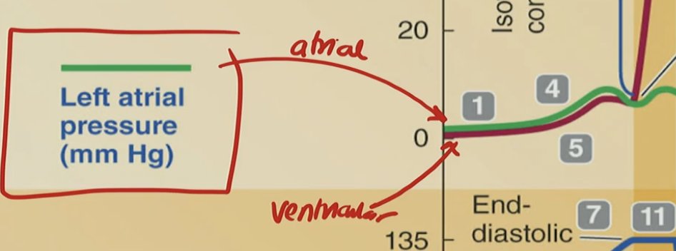
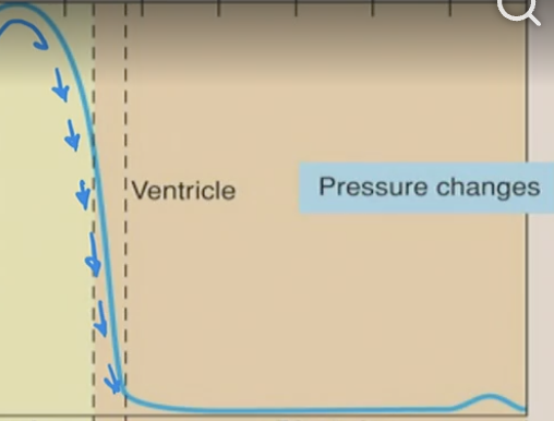
Why is the ventricular pressure lower than atrial?
Because the volume is increasing but not necessarily the pressure because blood is passively going into ventricles

Why is atria pressure higher than ventricle especially at the 4?
because that is where the atria does its last 20% of contraction, but instead of the other 80% the 20% will nto be passive pressure/contraction
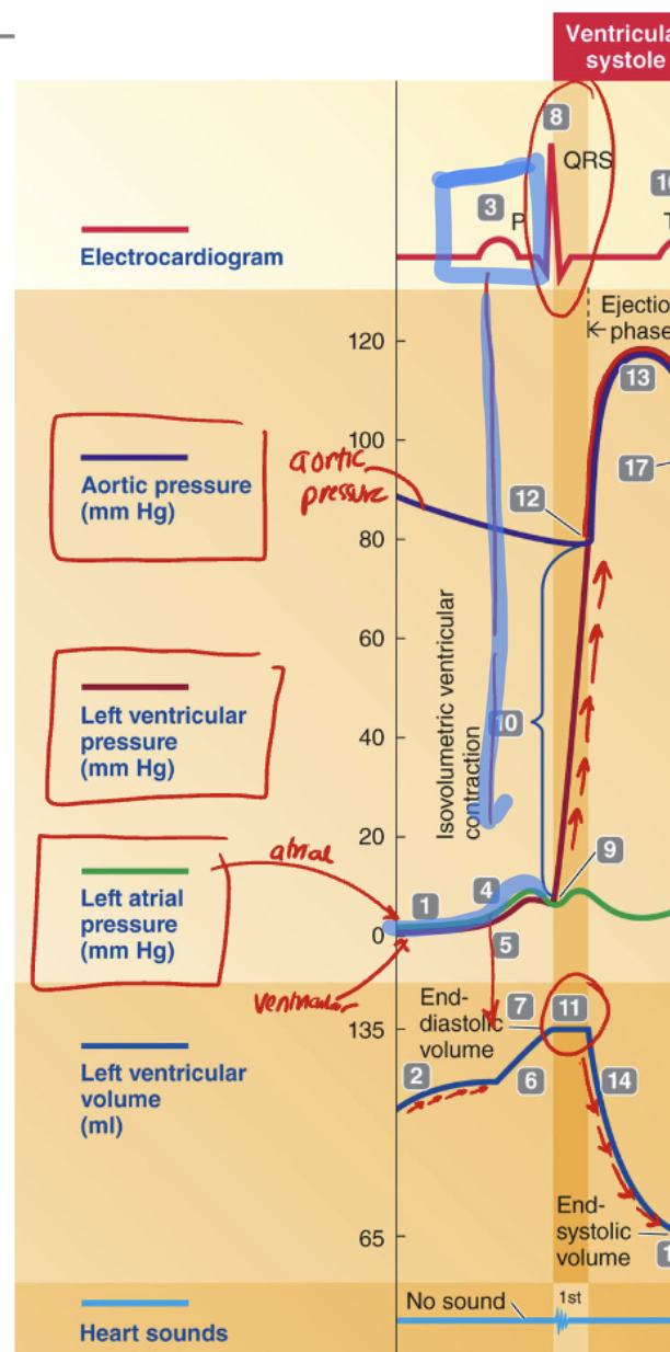
Which EKG waveform is associated with atrial contraction?
A. QRS complex
B. T wave
C. ST segment
D. P wave
D. P wave
Contraction → Depolarization → Atrial Depolarization → end of diastole → P wave
Contraction needs depolarization → happens in end-diastole → so it must be the P wave
In terms of the left ventricular pressure being higher than the left atrial pressure what happens to the AV Valves here?
A. They open, allowing blood to flow from the ventricle back into the atrium
B. They remain open to maintain ventricular filling
C. They close to prevent backflow into the atrium
D. They open to allow ejection into the aorta
C. They close to prevent backflow into the atrium

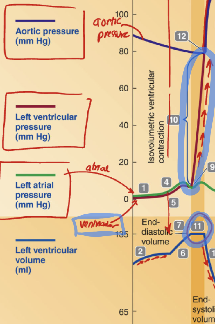
What is the name of the phase when ventricular pressure rises above atrial pressure, causing the AV valves to close, but no blood is ejected yet and ventricular volume remains constant (as seen at point 11)?
A. Ventricular ejection
B. Atrial systole
C. Isovolumetric contraction
D. Passive ventricular filling
C. Isovolumetric contraction
Isovolumetric is constant pressure in ventricle
the ventricle is contracting (pressure being built up)
Which valve separates the left ventricle from the aorta?
A. semilunar valve
B. Tricuspid valve
C. Mitral (bicuspid) valve
A. semilunar valve
Which semilunar valve separates the left ventricle from the aorta?
A. Pulmonary semilunar valve
B. Tricuspid valve
C. Aortic semilunar valve
D. Mitral (bicuspid) valve
C. Aortic semilunar valve
What occurs when aortic pressure becomes greater than ventricular pressure?
A) The AV valve opens
B) The semilunar (aortic) valve opens
C) The semilunar (aortic) valve closes
D) Blood is ejected into the atria
C) The semilunar (aortic) valve closes
aortic pressure > ventricular pressure

Why is the aortic semilunar valve closed when aortic pressure is greater than ventricular pressure?
A) Because the valve is damaged and cannot open
B) To prevent blood from flowing backward into the ventricle
C) Because atrial pressure is higher than aortic pressure
D) To allow blood to move into the ventricles from the atria
B) To prevent blood from flowing backward into the ventricle

What condition keeps the aortic semilunar valve closed during early ventricular diastole?
A) Higher pressure in the aorta than in the ventricle
B) Higher pressure in the atria than in the aorta
C) Depolarization of the atria
D) Closure of the AV valves
A) Higher pressure in the aorta than in the ventricle

What pressure relationship is occurring at Point 12?
A. Aortic pressure is higher than ventricular pressure
B. Atrial pressure is greater than aortic pressure
C. Ventricular pressure is higher than aortic pressure
D. Ventricular pressure is equal to atrial pressure
C. Ventricular pressure is higher than aortic pressure

What valve opens at Point 12 as a result of pressure changes?
A. Atrioventricular valve
B. Semilunar (aortic) valve
C. Tricuspid valve
D. Mitral valve
B. Semilunar (aortic) valve
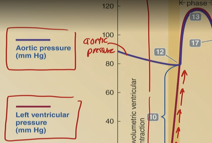
What is the result of the semilunar valve opening at Point 12?
A. Ventricles fill with blood
B. Blood flows from atria to ventricles
C. Aortic pressure drops rapidly
D. Blood is ejected from the ventricle to the aorta
D. Blood is ejected from the ventricle to the aorta
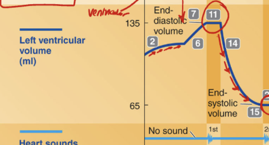
What cardiac state is represented at Point 11, where ventricular volume is actively decreasing?
A. End-Diastole
B. Isovolumetric Contraction
C. Ventricular Systole (Ejection Phase)
D. End-Systole
C. Ventricular Systole (Ejection Phase)
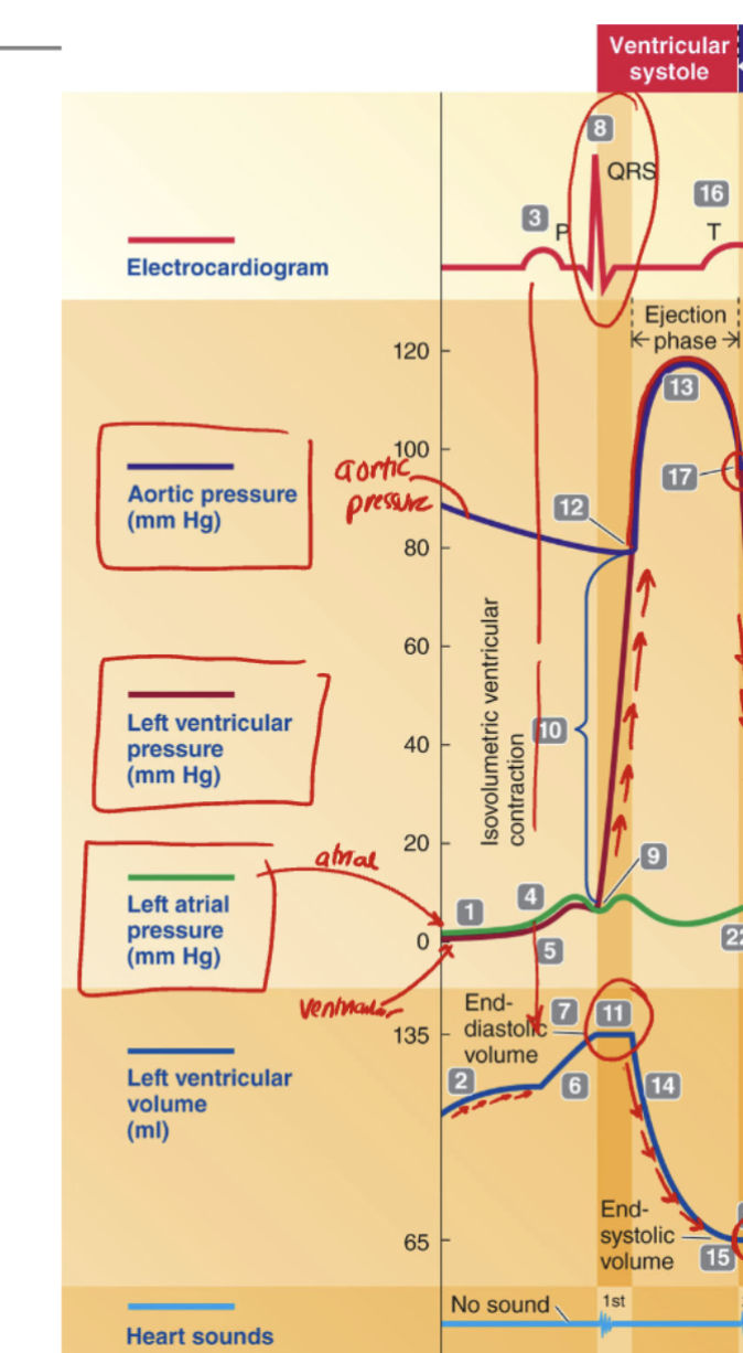
How can we tell that ventricular systole is occurring in the cardiac cycle graph?
A. The T wave on the EKG marks atrial contraction
B. The QRS complex indicates ventricular depolarization and contraction
C. The P wave initiates ventricular systole
D. The second heart sound signals ventricular filling
B. The QRS complex indicates ventricular depolarization and contraction
Stroke Volume is measured during which phase of the cardiac cycle?
A. Atrial systole
B. Isovolumetric relaxation
C. Ventricular systole (ejection phase)
D. Ventricular diastole (passive filling)
C. Ventricular systole (ejection phase)
The total that was ejected

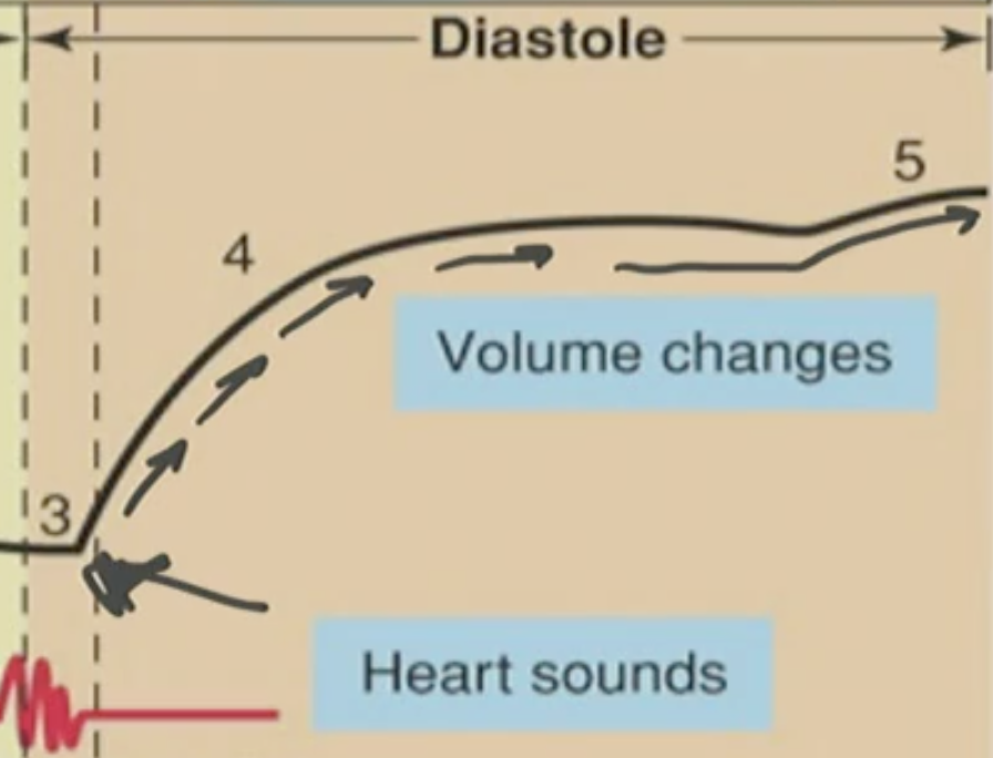
What is occurring between Point 11 and Point 15 on the ventricular volume curve?
A. Ventricles are filling with blood
B. Ventricular pressure is decreasing
C. Blood is being ejected from the ventricles
D. Atrial contraction is pushing blood into the ventricle
C. Blood is being ejected from the ventricles
Blood is being ejected from ventricle to aorta because the ventrical pressure was greater than aortic pressure so the semilunar valve opened up and ventricle blood volume decreased as the blood left ventricle to go into aorta

Why does the left ventricular volume decrease after Point 11?
A. The atrioventricular valve opens
B. The semilunar valve closes
C. Blood flows into the ventricle from the atria
D. The semilunar valve opens, allowing blood to leave
D. The semilunar valve opens, allowing blood to leave
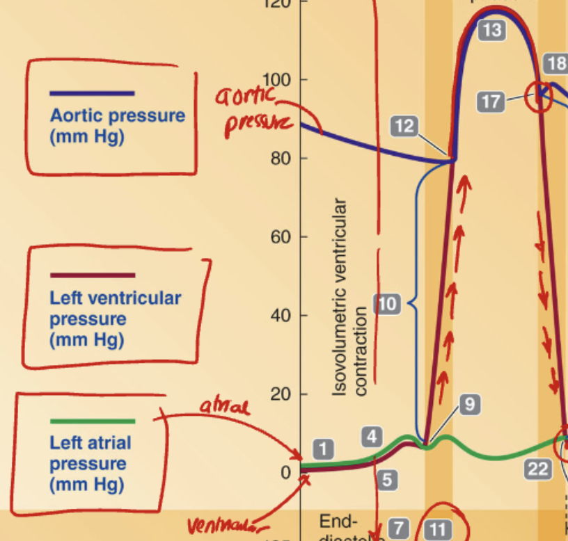
What causes the drop in ventricular pressure after Point 18, where aortic pressure becomes greater than ventricular pressure?
A. The atria begin to contract
B. The semilunar valve closes due to pressure reversal
C. The ventricles begin to fill with blood
D. The AV valves open and allow backflow
B. The semilunar valve closes due to pressure reversal
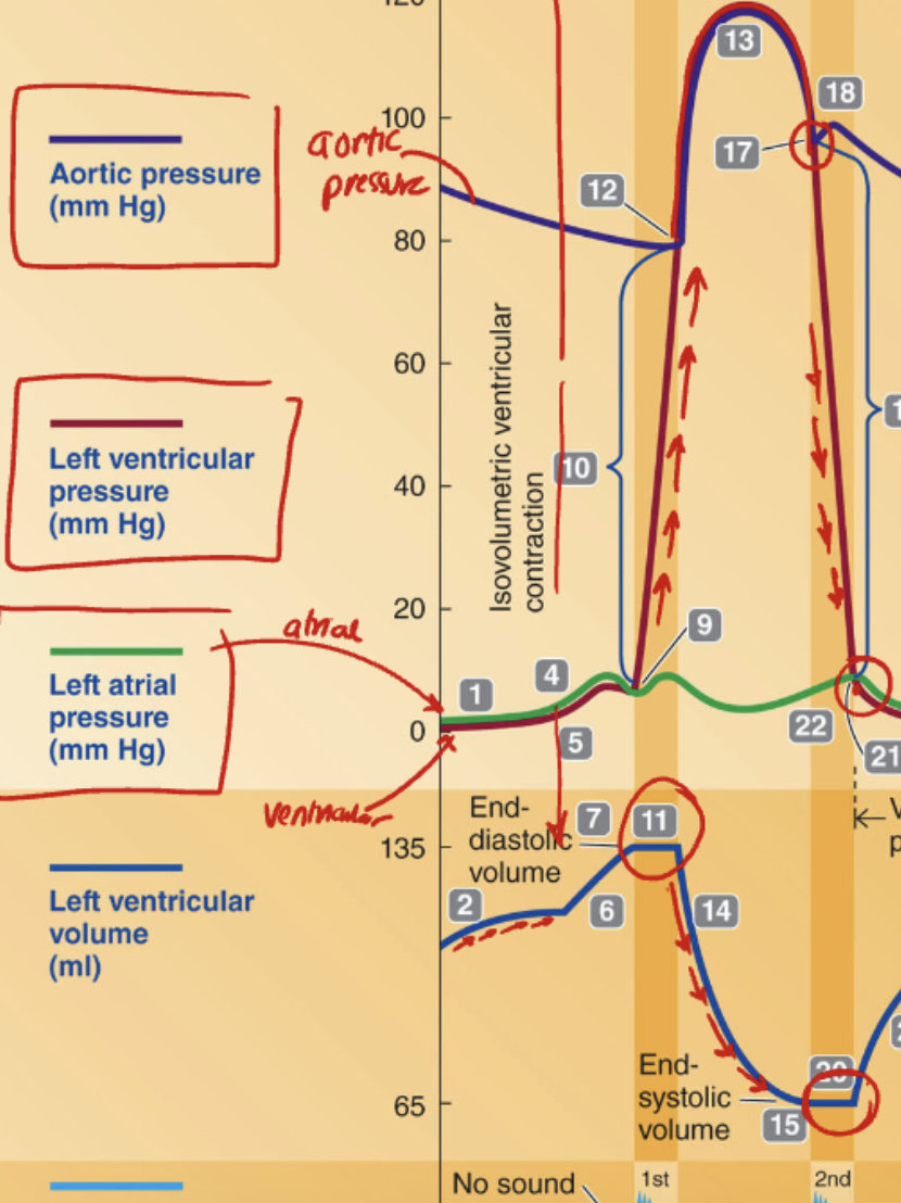
What phase of the cardiac cycle occurs between Point 18 and Point 20 when ventricular pressure is falling but volume remains unchanged?
A. Isovolumetric contraction
B. Rapid ventricular filling
C. Isovolumetric relaxation
D. Ventricular ejection
C. Isovolumetric relaxation
✅ There's still blood in the ventricle.
❌ But no new blood is coming in yet (AV valves closed).
❌ And no more blood is leaving (semilunar valves closed).
👉 So volume = unchanged, but the muscle is relaxing, so pressure drops.
So during isovolumetric relaxation, what happens to ventricular pressure and volume?
A. Pressure increases, volume decreases
B. Pressure and volume both increase
C. Volume decreases, pressure remains constant
D. Pressure decreases, volume remains constant
D. Pressure decreases, volume remains constant
Funny Analogy:
Which of the following best describes the physiological state of isovolumetric relaxation?
A. Like a fart: pressure is released, but nothing leaves, so the volume stays the same
B. Like filling a balloon: both pressure and volume are increasing
C. Like squeezing a sponge: pressure and volume are both decreasing
D. Like gulping air: pressure rises because volume increases
A. Like a fart: pressure is released, but nothing leaves, so the volume stays the same
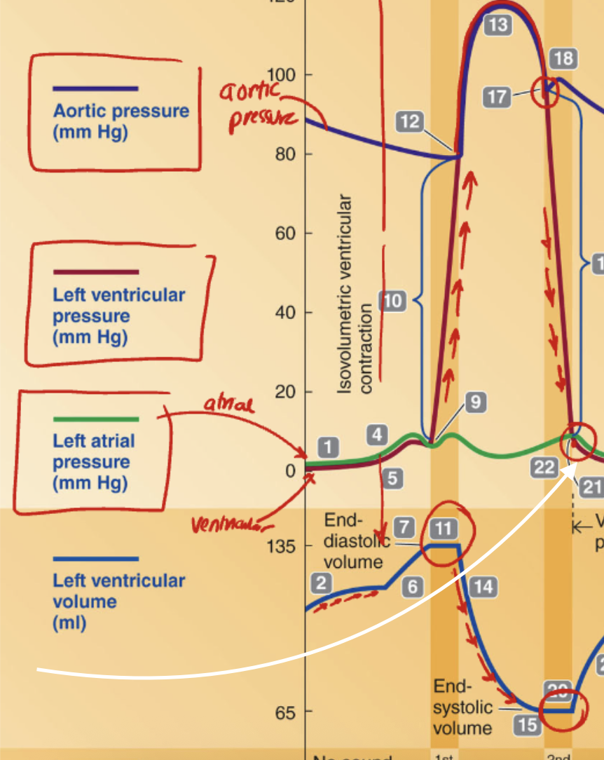
What is happening at the point where the white arrow is pointing?
A. The semilunar valve opens due to higher ventricular pressure
B. The AV valve opens due to higher atrial pressure than ventricular pressure
C. The AV valve closes due to rising ventricular pressure
D. Ventricular ejection begins as ventricular pressure exceeds aortic pressure
B. The AV valve opens due to higher atrial pressure than ventricular pressure
Using the graph, calculate the stroke volume (SV) if the End-Diastolic Volume (EDV) is 135 mL and the End-Systolic Volume (ESV) is 65 mL.
A. 65 mL
B. 70 mL
C. 135 mL
D. 200 mL
B. 70 mL
EDV - ESV = SV
135 - 65 = 70