Appendicular skeleton
1/72
There's no tags or description
Looks like no tags are added yet.
Name | Mastery | Learn | Test | Matching | Spaced |
|---|
No study sessions yet.
73 Terms
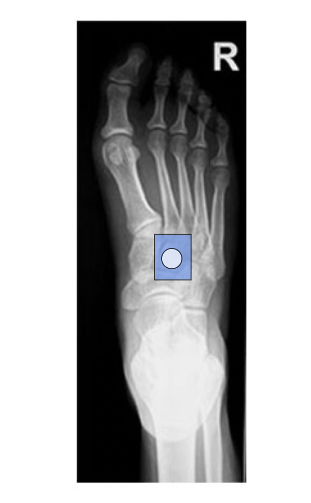
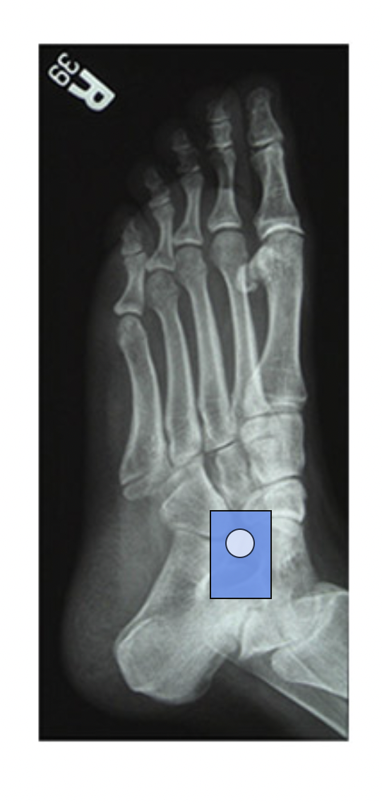
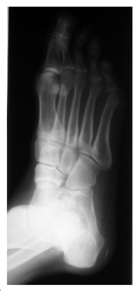
In the dorsoplantar foot projection at right, place the cursor on the second (intermediate) cuneiform and left click the mouse.
Which overhead projections should the radiographer obtain for the vertical ray method of knee arthrography?
a. Anteroposterior (AP) and lateral
b. Posteroanterior (PA) and lateral
c. Anteroposterior (AP) and 20-degree right and left oblique
d. Posteroanterior (PA) and 45-degree right and left oblique
c. Anteroposterior (AP) and 20-degree right and left oblique
A radiographer prepares to perform an anteroposterior (AP) projection of the distal femur on a patient with a suspected femoral fracture. Which represents the best approach for positioning both the patient and the x-ray tube?
a. Do not rotate the leg; place the anode end of the tube toward the foot
b. Do not rotate the leg; place the cathode end of the tube toward the foot
c. Rotate the leg 15 degrees medially; place the anode end of the tube toward the foot
d. Rotate the leg 15 degrees medially; place the cathode end of the tube toward the foot
a. Do not rotate the leg; place the anode end of the tube toward the foot
A radiographer prepares to position a patient with a suspected hip injury. The patient is positioned on a backboard with the right leg and foot lying on its lateral aspect. Which should the radiographer do to successfully complete the examination?
a. Ask the physician if the leg can be rotated
b. Perform the exam without repositioning the leg
c. Medially rotate both feet and lower legs 15 to 20 degrees
d. Request that pain medication be given prior to rotating the leg
b. Perform the exam without repositioning the leg
How is the central ray directed when performing an anteroposterior (AP) knee projection for a patient with thin thighs and buttocks?
a. 3 to 5 degrees caudad
b. 5 to 7 degrees caudad
c. 3 to 5 degrees cephalad
d. 5 to 7 degrees cephalad
a. 3 to 5 degrees caudad
A request for a shoulder exam comes from the emergency room. The patient has a large deformity anterior to the shoulder and the physician suspects an anterior dislocation. Which are the most appropriate views to take on this patient?
a. Anteroposterior (AP) projection in neutral rotation and posteroanterior (PA) oblique scapular Y
b. Anteroposterior (AP) projections in internal, external, and neutral rotation
c. Posteroanterior (PA) neutral and external rotation views
d. Posteroanterior (PA) internal and external rotation views
a. Anteroposterior (AP) projection in neutral rotation and posteroanterior (PA) oblique scapular Y
Which statement accurately describes the appearance of a correctly positioned anteroposterior (AP) hip image?
a. The femoral head is at the center of the collimated field
b. The femoral neck is demonstrated without foreshortening
c. The proximal two-thirds of the femur is visualized
d. The lesser trochanter is demonstrated in profile
b. The femoral neck is demonstrated without foreshortening
Which elbow projection will show the radial head freest of superimposition?
a. Lateral
b. Anteroposterior (AP)
c. Anteroposterior (AP), internal oblique
d. Anteroposterior (AP), external oblique
d. Anteroposterior (AP), external oblique
Which carpal bone is most commonly fractured?
a. Lunate
b. Cuboid
c. Scaphoid
d. Triquetrum
c. Scaphoid
The radiographer uses the Holmblad method to perform a tunnel view of the knee. What is the primary advantage of utilizing this method instead of the Camp Coventry method?
a. The Holmblad method is quick to position
b. The Holmblad method shows no distortion longitudinally or laterally
c. The Holmblad method is easier for the radiographer to properly position
d. The Holmblad method is easier for the patient than the Camp Coventry method
b. The Holmblad method shows no distortion longitudinally or laterally
A carpenter smashes his index finger with a hammer. Which type of fracture might be shown on the subsequent radiographs of the finger?
a. Torus
b. Spiral
c. Depressed
d. Comminuted
d. Comminuted
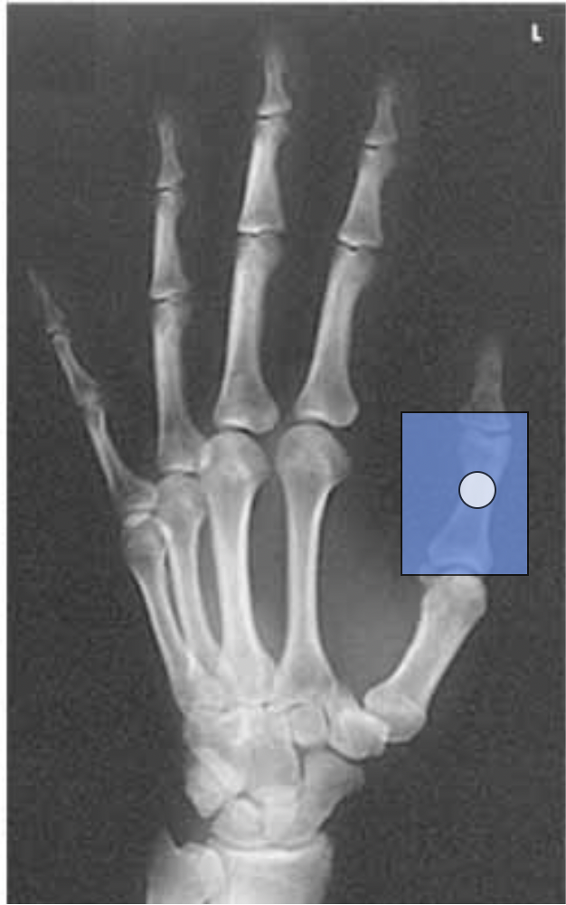
Which projection of the thumb requires digits two through five to be extended, with the palmar surface of the hand flat against the image receptor (IR) as if for a posteroanterior (PA) hand projection?
a. Posteroanterior (PA) Oblique
b. Posteroanterior (PA)
c. Anteroposterior (AP)
d. Lateral
a. Posteroanterior (PA) Oblique
An anteroposterior (AP) oblique mortise joint projection demonstrates the distal fibula without talar superimposition and an open talofibular joint. What should the radiographer do next?
a. No correction needed
b. Increase internal rotation
c. Increase external rotation
d. Increase cephalic angulation
a. No correction needed
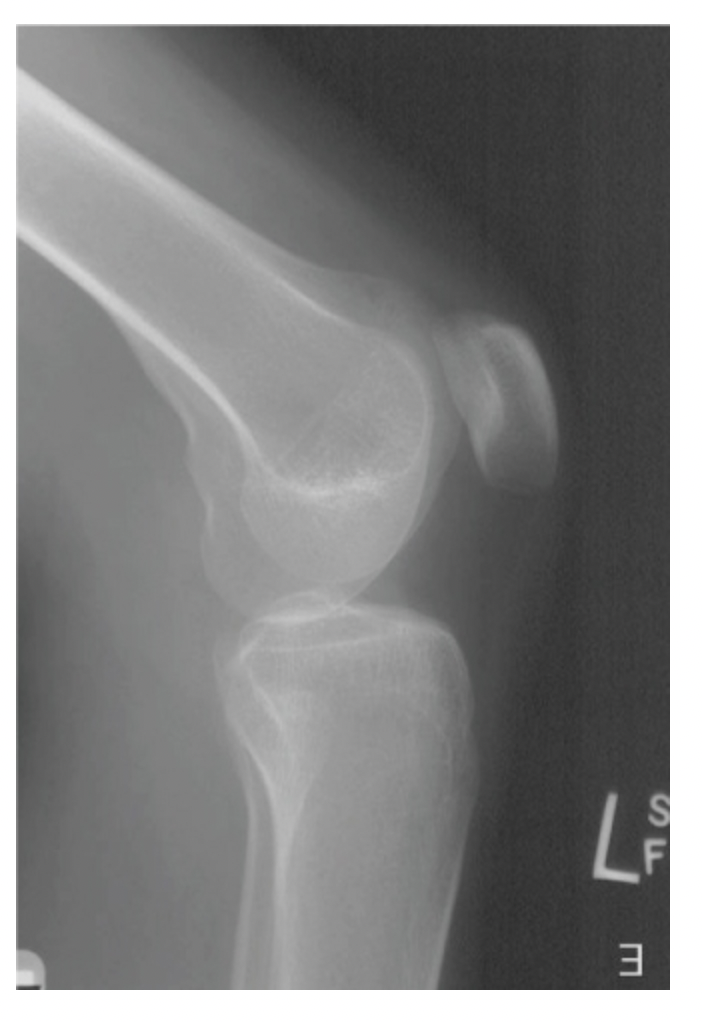
An emergency room physician suspects a 14-year-old male patient may have Osgood-Schlatter’s disease. Which radiographic exam might the ER physician order to evaluate this?
a. Foot
b. Ankle
c. Femur
d. Tibia-fibula
d. Tibia-fibula
The tarsal sinus is visualized when the foot is seen in what projection?
a. Lateral
b. Dorsoplantar
c. 30 degree lateral oblique
d. 30 degree medial oblique
d. 30 degree medial oblique
The only saddle joint in the body is found between which two bones?
a. Scaphoid and trapezium
b. Trapezium and first metatarsal
c. Navicular and first metatarsal
d. Trapezium and first metacarpal
d. Trapezium and first metacarpal
Where is the central ray (CR) directed for an anteroposterior (AP) projection of the lower leg?
a. Mid-lower leg
b. 0.5 inch inferior to patellar apex
c. At the level of the medial malleolus
d. Two inches distal to the medial condyle of the tibia
a. Mid-lower leg
Which joint is the central ray (CR) entrance point for a posteroanterior (PA) finger projection?
a. Proximal interphalangeal (PIP)
b. Metacarpophalangeal (MCP)
c. Carpometacarpal (CMC)
d. Interphalangeal (IP)
a. Proximal interphalangeal (PIP)
Which central ray (CR) orientation is recommended for an anteroposterior (AP) knee projection if the measurement from the tabletop to the patient's anterior superior iliac spine (ASIS) is 22 cm?
a. 3-5 degrees caudad
b. 3-5 degrees cephalad
c. Perpendicular to the image receptor (IR)
d. Perpendicular to the long axis of the patella
c. Perpendicular to the image receptor (IR)
How does the central ray (CR) for an anteroposterior (AP) scapula differ from an AP shoulder x-ray?
a. One inch lower
b. CR is the same
c. One inch higher
d. Same level, medial
a. One inch lower
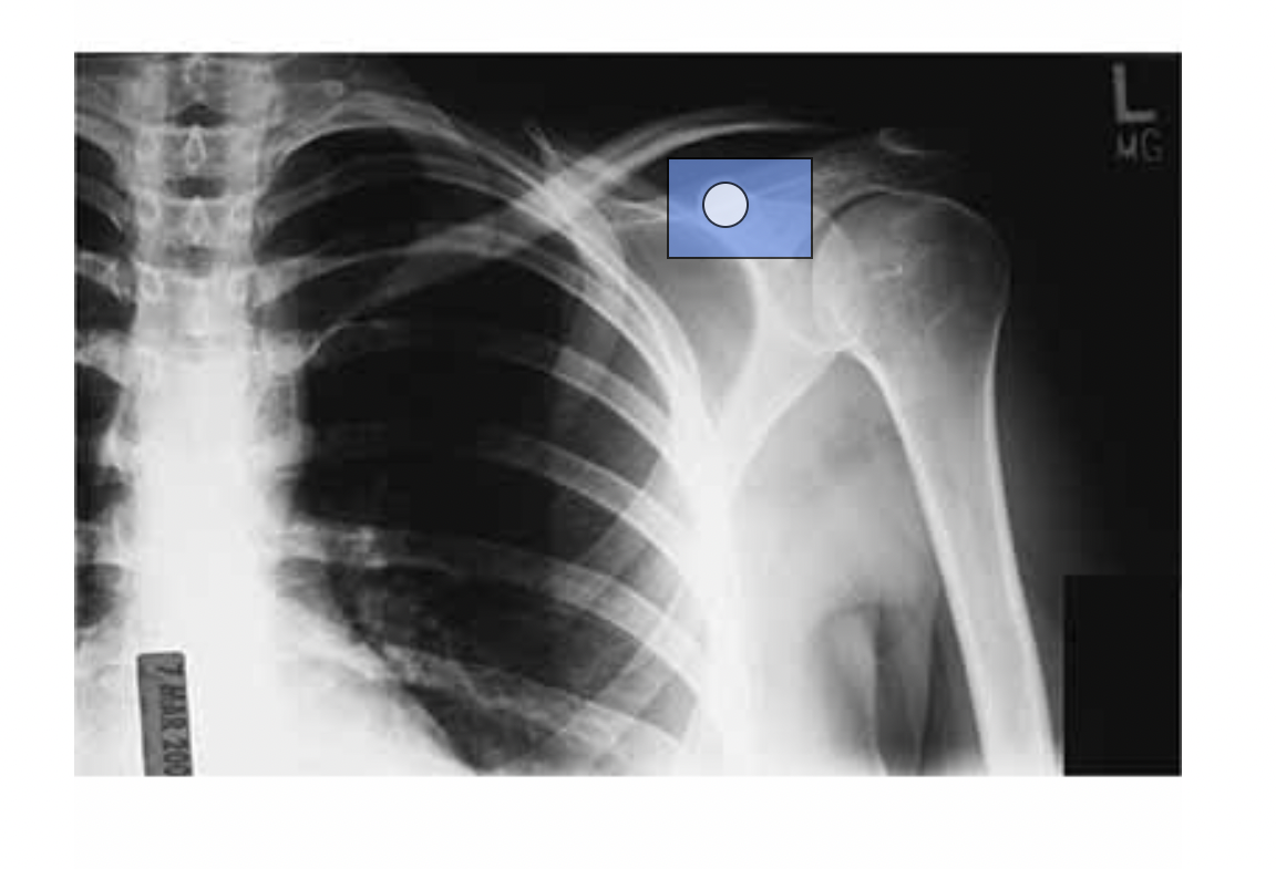
Place the cursor on the left coracoid process, and left click. (Click the chosen location. To change, click on the new location.)
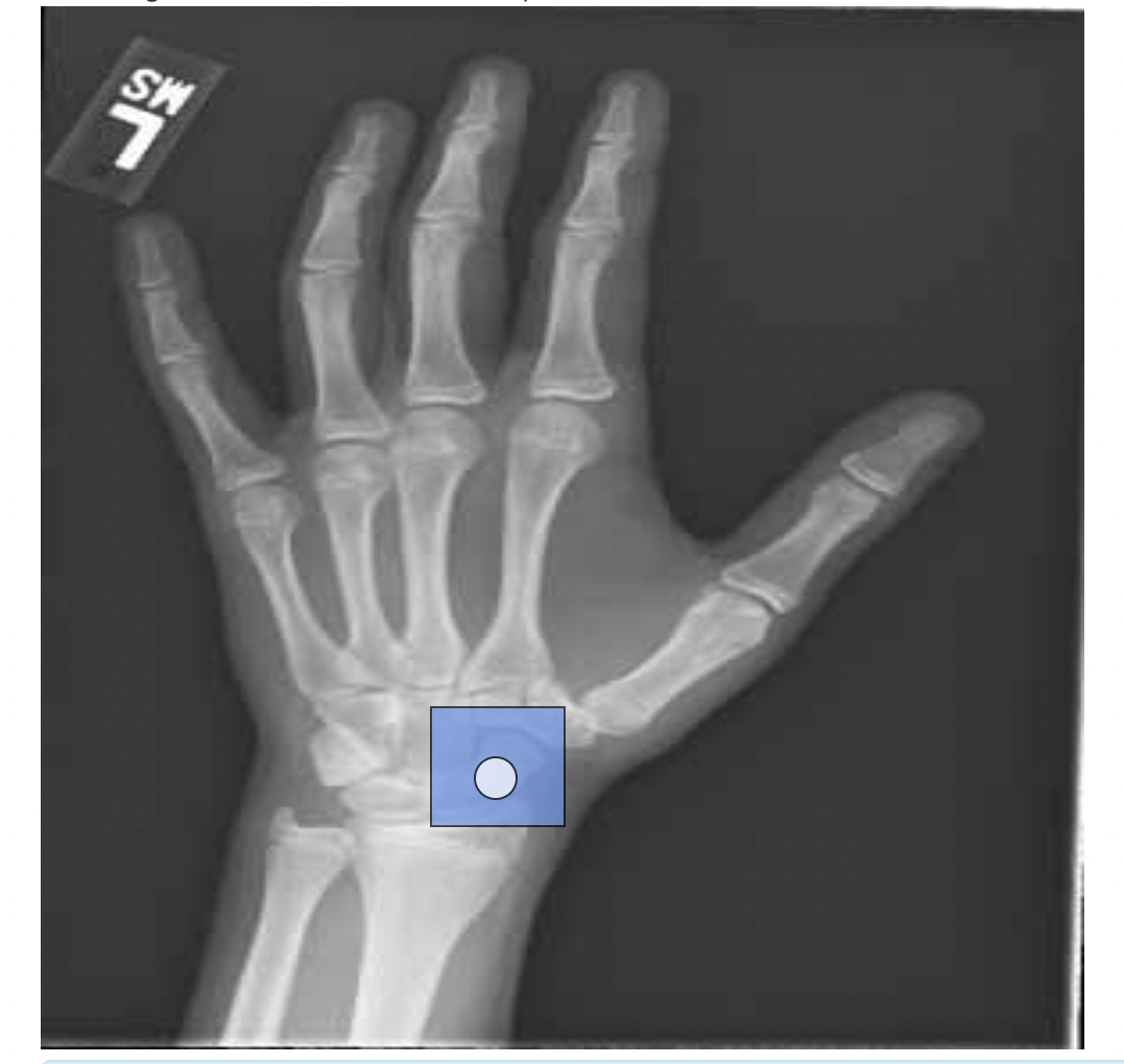
Place the cursor on the most commonly fractured carpal bone, and left click. (Click the chosen location. To change, click on the new location.)
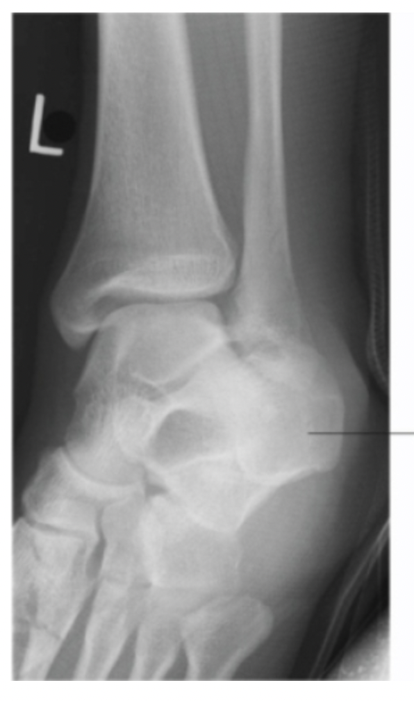
Place the cursor on the sinus tarsi. (Click the chosen location. To change, click on the new location.)
A radiographer critiques an anteroposterior (AP) ankle projection. The medial mortise joint is open while the lateral mortise joint is closed. How can the radiographer adjust the ankle to correct this image?
a. Dorsiflex the foot
b. No correction needed
c. Increase internal rotation
d. Increase external rotation
b. No correction needed
Which projection is a radiographer performing if the patient’s hand is turned in extreme internal rotation, with the central ray directed perpendicular to the first metacarpophalangeal joint?
a. Lateral thumb
b. Oblique thumb
c. Posterior-anterior (PA) hand
d. Anteroposterior (AP) thumb
d. Anteroposterior (AP) thumb
While performing an anteroposterior (AP) projection of a scapula, what should be done to the patient's arm to move the patient’s scapula in a lateral direction?
a. External rotation
b. Internal rotation
c. Abduction
d. Adduction
c. Abduction
On a lateral radiograph of a patient’s leg, where will the patient’s tibia be in relation to the fibula?
a. Completely superimposed
b. Posterior to fibula
c. Superior to fibula
d. Partially superimposed
d. Partially superimposed
On a radiograph of a shoulder, the greater tubercle is visualized in profile. Which projection was taken?
a. Anteroposterior (AP), internal rotation
b. Anteroposterior (AP), external rotation
c. Scapular-Y
d. Transthoracic lateral (Lawrence method)
b. Anteroposterior (AP), external rotation
For a lateral femur, if a patient is rotated too far anteriorly, what will happen to the condyles on the distal femur?
a. They will superimpose
b. Medial condyle will be inferior to the lateral condyle
c. Medial condyle will be anterior to the lateral condyle
d. Medial condyle will be posterior to the lateral condyle
c. Medial condyle will be anterior to the lateral condyle
Which projection of the foot demonstrates the cuboid bone in profile with the least bony superimposition?
a. Lateral
b. Anteroposterior (AP)
c. Anteroposterior (AP), lateral oblique
d. Anteroposterior (AP), medial oblique
d. Anteroposterior (AP), medial oblique
Which central ray (CR) angulation is recommended for an anteroposterior (AP) axial projection of the toes?
a. 5 degrees toward the heel
b. 15 degrees toward the heel
c. 30 degrees toward the heel
d. 40 degrees toward the heel
b. 15 degrees toward the heel
An image demonstrates the patella superimposing the medial condyle of the knee and little superimposition between the head of the fibula and the proximal tibia. Which projection of the knee has been correctlly performed?
a. Anteroposterior (AP) with medial rotation
b. Anteroposterior (AP)
c. Anteroposterior (AP) with lateral rotation
d. Mediolateral lateral
a. Anteroposterior (AP) with medial rotation
Which position most accurately describes the Camp Coventry method for demonstrating the intercondylar fossa of the knee?
a. Supine with the image receptor (IR) under the knee
b. Kneeling with the lower leg parallel to the image receptor (IR)
c. Standing with the lower leg parallel to the image receptor (IR)
d. Prone with the femur parallel to the image receptor (IR)
d. Prone with the femur parallel to the image receptor (IR)
When imaging the toes, how much is the central ray (CR) angled for the anteroposterior (AP) axial image?
a. No angle
b. 5 degrees
c. 15 degrees
d. 25-30 degrees
c. 15 degrees
Which represents the proper amount of internal leg rotation for the ankle mortise joint to appear open?
a. 0 degrees
b. 15 to 20 degrees
c. 30 to 35 degrees
d. 45 degrees
b. 15 to 20 degrees
Which is the proper amount and direction of heel rotation recommended for a posteroanterior (PA) projection of the patella?
a. 5 to 10 degrees laterally
b. 5 to 10 degrees medially
c. 10 to 15 degrees laterally
d. 10 to 15 degrees medially
a. 5 to 10 degrees laterally
Which is the best projection to evaluate the coronoid process in profile?
a. Scapular Y view
b. Medial oblique elbow
c. Anteroposterior (AP) elbow
d. Inferosuperior axial shoulder
b. Medial oblique elbow
Correct positioning for an anteroposterior (AP) pelvis requires the lower limbs to be internally rotated until which structures are parallel with the image receptor (IR)?
a. Pubic rami
b. Sacral alae
c. Femoral necks
d. Innominate bones
c. Femoral necks
What should be done to correct positioning on an anteroposterior (AP) elbow with lateral rotation, when the radial head is slightly superimposed over the proximal ulna on the first effort?
a. The elbow joint should be rotated laterally
b. The elbow joint should be rotated medially
c. The forearm should be pronated to correct the error
d. The humerus and forearm should be placed in the same horizontal plane
a. The elbow joint should be rotated laterally
An 82-year-old female patient is transported to the x-ray department for an examination of the right hip. The ordering physician informs the radiographer that the patient fell at home, and there is high suspicion for a femoral neck fracture. Which positioning guideline is best for the radiographer to follow?
a. Abduct the affected leg
b. Do not move the affected leg
c. Internally rotate the affected leg
d. Externally rotate the affected leg
b. Do not move the affected leg
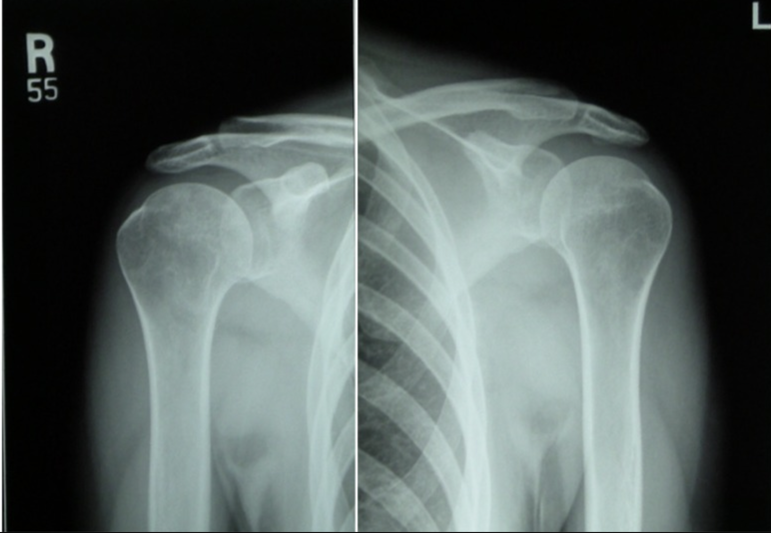
Study the four digital images of both shoulders. What best describes the quality of these images?
a. All four images are diagnostic
b. The degree of internal rotation was inadequate on both sides
c. The degree of external rotation was inadequate on both sides
d. The collimator field is too large in the left internal rotation view
b. The degree of internal rotation was inadequate on both sides
Which part of the scapula is used as a positioning landmark due to its location at the level of the seventh thoracic vertebra?
a. Coracoid process
b. Inferior angle
c. Lateral angle
d. Acromion
b. Inferior angle
Which exam may be used to assess cruciate ligament pathology?
a. Knee arthrogram
b. Shoulder arthrogram
c. Three view elbow series
d. Wrist including navicular views
a. Knee arthrogram
On a fan lateral radiograph of a left hand, which group of bones is best visualized?
a. Tarsals
b. Carpals
c. Phalanges
d. Metacarpals
c. Phalanges
What should the radiographer do to correct this image?
a. No correction needed
b. Increase internal rotation
c. Increase external rotation
d. Roll patient slightly to left side
a. No correction needed
A lateral scapula image demonstrates the lateral border of the scapula next to the ribs and the vertebral border of the scapula is demonstrated posterolaterally. How should the radiographer correct this image?
a. Elevate arm
b. No correction needed
c. Increase patient obliquity
d. Decrease patient obliquity
d. Decrease patient obliquity
A lateral knee image reveals that the femoral condyles are not superimposed with the medial condyle situated posteriorly. How should the radiographer correct this image?
a. No correction needed
b. Increase cephalic angulation
c. Position patella closer to the image receptor (IR)
d. Position patella further away from the image receptor (IR)
c. Position patella closer to the image receptor (IR)
This 45 degree oblique ankle image demonstrates the calcaneus obscuring the distal aspect of the lateral mortise and distal fibula. How should the radiographer correct this image?
a. Increase internal rotation
b. No correction needed
c. Depress distal tibia
d. Dorsiflex foot
d. Dorsiflex foot
How should the radiographer correct this image of a medial oblique foot?
a. Dorsiflex foot
b. No correction needed
c. Increase medial rotation
d. Increase angle toward heel
c. Increase medial rotation
Consider this oblique hand image. Which statement correctly describes this image?
a. The hand is correctly rotated
b. The hand is under-rotated
c. The hand is over-rotated
d. The hand was rotated the wrong way
b. The hand is under-rotated
Which statement describes a properly positioned lateral foot image?
a. Superimposition of all metatarsals
b. Equidistance between all metatarsals
c. Superimposition of the first and fifth metatarsal heads
d. The cuboid and fifth metatarsal demonstrated with minimal overlap
c. Superimposition of the first and fifth metatarsal heads
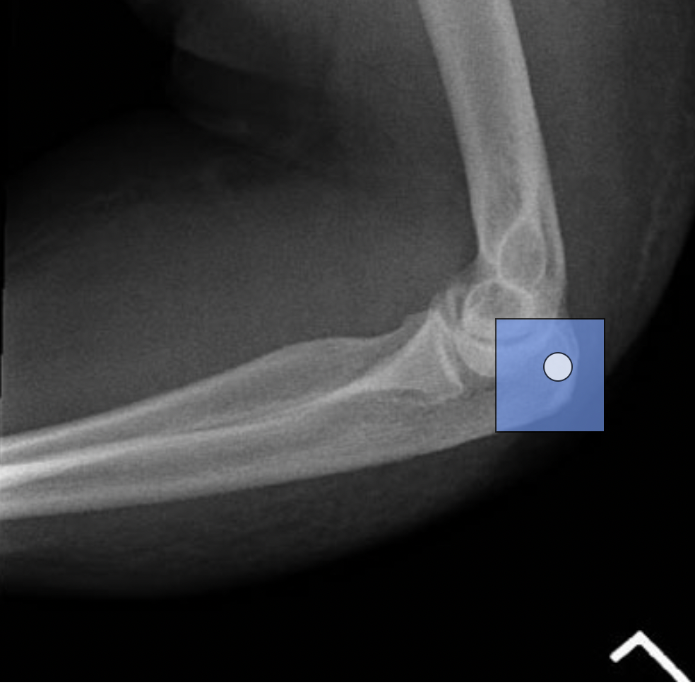
Place the cursor at the location of the olecranon process and left click.
Which position/projection of the elbow best demonstrates the trochlear notch of the ulna?
a. Anteroposterior (AP)
b. Anteroposterior (AP), internal oblique
c. Anteroposterior (AP), external oblique
d. Lateral, lateromedial projection
d. Lateral, lateromedial projection
Which structure is demonstrated without superimposition on a properly positioned medial oblique foot image?
a. Cuboid
b. First cuneiform
c. Navicular
d. First metatarsal
a. Cuboid
Which central ray (CR) angle is recommended to demonstrate the coronoid process of the elbow using the Coyle method?
a. 45 degrees toward the shoulder
b. 45 degrees away from the shoulder
c. 35 degrees toward the shoulder
d. 35 degrees away from the shoulder
b. 45 degrees away from the shoulder
Which wrist projection best demonstrates a Colles' fracture?
a. Lateral wrist
b. Posteroanterior (PA) with ulnar deviation
c. Posteroanterior (PA) oblique
d. Anteroposterior (AP)
a. Lateral wrist
The Alexander method is performed to demonstrate a dislocation of which joint?
a. Radioulnar
b. Glenohumeral
c. Acromioclavicular
d. Patellofemoral
c. Acromioclavicular
An anteroposterior (AP) shoulder image acquired with the epicondyles perpendicular to the image receptor (IR) will demonstrate which radiographic appearance?
a. The glenoid cavity is seen in profile, free of superimposition
b. The humeral head is seen in profile, free of superimposition
c. The greater tubercle is seen in profile laterally
d. The lesser tubercle is seen in profile medially
d. The lesser tubercle is seen in profile medially
Which appearance is demonstrated by a lateral projection of the shoulder obtained using the Lawrence method?
a. The intertubercular groove is seen in profile
b. The greater tubercle is projected onto the head of the humerus
c. The glenoid cavity is seen in profile
d. The proximal humerus is projected through the lung field
d. The proximal humerus is projected through the lung field
Which radiographic appearance results when the anterior surface of the patient's elbow is rotated 45 degrees medially for an anteroposterior (AP) elbow projection?
a. The olecranon process is seen in profile
b. The radial head and neck are free of superimposition
c. The coronoid process is seen in profile
d. The medial and lateral epicondyles are superimposed
c. The coronoid process is seen in profile
How are the patient's hand and fingers positioned for a posteroanterior (PA) wrist projection?
a. Hand pronated with fingers extended
b. Hand supinated with fingers extended
c. Hand pronated with fingers flexed
d. Hand supinated with fingers flexed
c. Hand pronated with fingers flexed
Which is the correct central ray (CR) location for a posteroanterior (PA) hand projection?
a. Third proximal interphalangeal (IP) joint
b. Third metacarpophalangeal (MCP) joint
c. Midpoint of the third metacarpal
d. Midpoint of the third carpometacarpal (CMC) joint
b. Third metacarpophalangeal (MCP) joint
The Holmblad method is performed to demonstrate which radiographic appearance?
a. Open intercondylar fossa
b. Open tibiofemoral joint space
c. Superimposed medial and lateral condyles
d. Patella in profile
a. Open intercondylar fossa
Which is the correct central ray (CR) location for an anteroposterior (AP) knee projection?
a. 1/2 inch below the apex of the patella
b. 1/2 inch above the apex of the patella
c. 1/2 inch below the base of the patella
d. 1/2 inch above the base of the patella
a. 1/2 inch below the apex of the patella
In which position is the knee placed in order to demonstrate the head of the fibula without superimposition?
a. 3-5 degrees lateral rotation
b. 3-5 degrees medial rotation
c. 45 degrees lateral rotation
d. 45 degrees medial rotation
d. 45 degrees medial rotation
Which bones make up the ankle mortise?
a. Tibia, fibula, and talus
b. Tibia, fibula, and calcaneus
c. Talus, calcaneus, and medial cuneiform
d. Talus, calcaneus, and navicular
a. Tibia, fibula, and talus
Which tarsal bone is best seen when the plantar surface of the foot is positioned perpendicular to the image receptor (IR), and a 40-degree cephalic central ray (CR) is directed to the base of the third metatarsal?
a. Cuboid
b. Talus
c. Lateral cuneiform
d. Calcaneus
d. Calcaneus
Which x-ray tube orientation is used to create a 90-degree angle between the central ray (CR) and the metatarsals for an anteroposterior (AP) axial projection of the foot?
a. Perpendicular to the image receptor (IR)
b. 10 degrees posteriorly
c. 15 degrees posteriorly
d. Parallel to the metatarsophalangeal (MTP) joint spaces
b. 10 degrees posteriorly
How should the radiographer rotate the patient's lower leg and foot for an anteroposterior (AP) oblique projection of the second toe?
a. 10 - 25 degrees medially
b. 10 - 25 degrees laterally
c. 30 - 45 degrees medially
d. 30 - 45 degrees laterally
c. 30 - 45 degrees medially
A posteroanterior (PA) axial projection of the coccyx is performed because the patient is unable to tolerate the supine position due to injuries sustained after falling backward. How should the central ray (CR) be adjusted for this projection?
a. 10 degrees caudad
b. 15 degrees caudad
c. 10 degrees cephalad
d. 15 degrees cephalad
c. 10 degrees cephalad
Which anatomical structure is the most medial?
a. Radial head
b. Radial notch
c. Coronoid fossa
d. Coronoid tubercle
d. Coronoid tubercle
Which anatomical structure is the most distal?
a. Radial tuberosity
b. Coronoid fossa
c. Radial head
d. Trochlea
a. Radial tuberosity
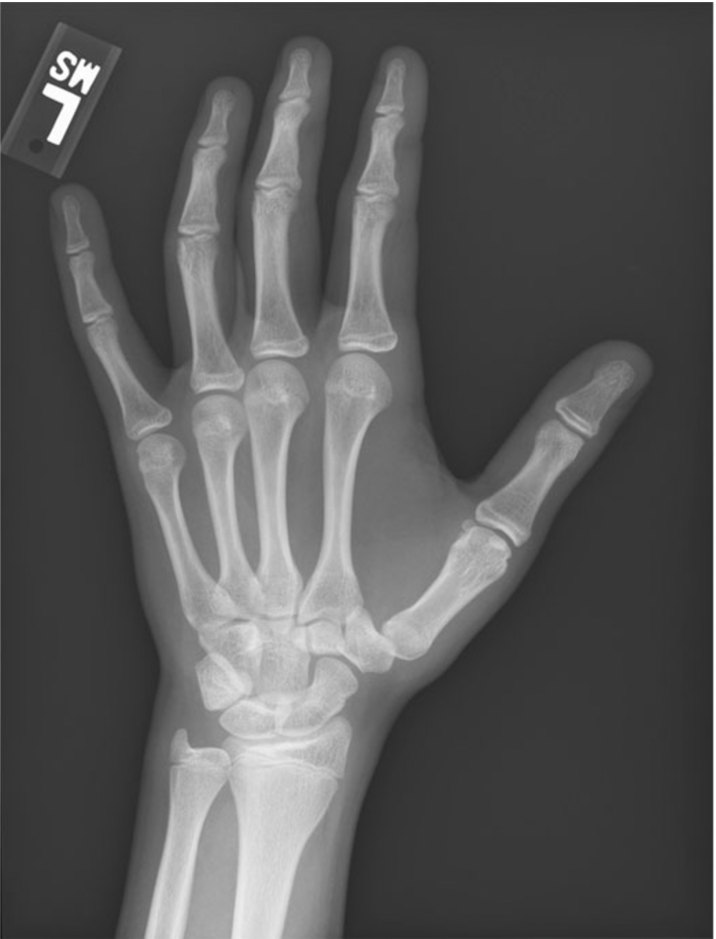
Click on the proximal phalanx of the first digit in the oblique image of the hand. (Click the chosen location. To change, click on the new location.)