Unit 3 - Thorax
1/31
There's no tags or description
Looks like no tags are added yet.
Name | Mastery | Learn | Test | Matching | Spaced | Call with Kai |
|---|
No analytics yet
Send a link to your students to track their progress
32 Terms
Thorax
From base of neck (thoracic inlet) to diaphragm muscle
Protects heart and lungs
Site for attachment of muscles for:
Upper limb
Back
Respiration
Abdomen
From diaphragm muscle to pelvic inlet
Contains major viscera of digestive system
No bony structure; musculature holds viscera in place
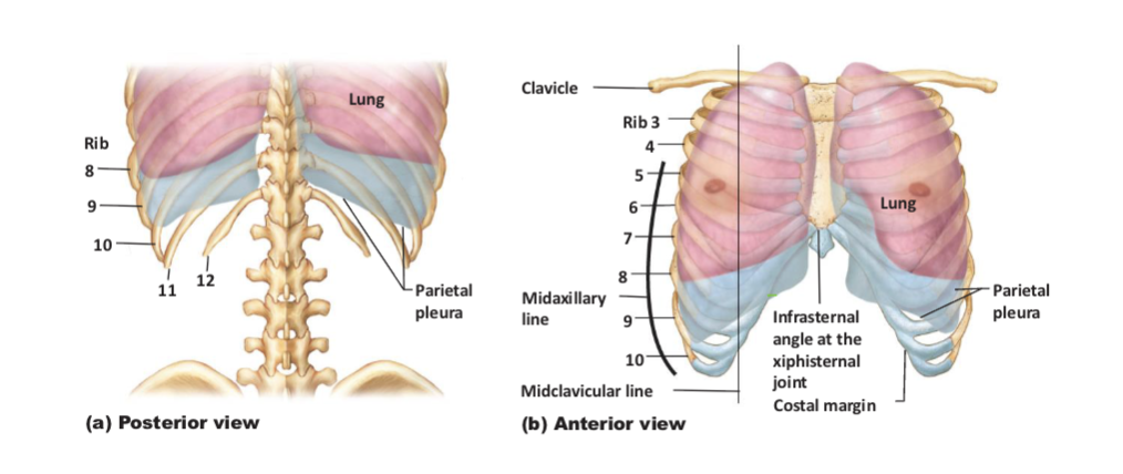
Describe the borders of the thorax
Superior border: thoracic inlet or superior thoracic
aperture
kidney-shaped opening formed by T1
vertebra, 1st pair ribs & sup. margin of
manubriumincludes:
• apex of lungs
• common carotid artery
• internal jugular vein
• subclavian artery & vein
• esophagus & trachea
• brachial plexus
• (clavicles close, but not part of it)
Inferior boarder: wide opening formed by T12
vertebra, 12th pair ribs, & costal arch
Anterior boarder: sternum, ribs & costal cartilages
Posterior boarder: thoracic vertebrae & ribs
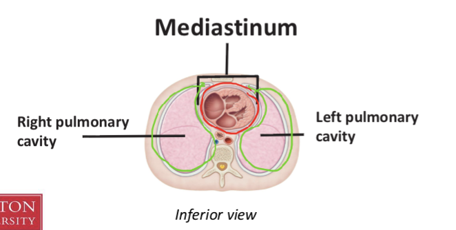
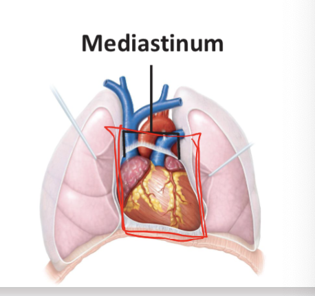
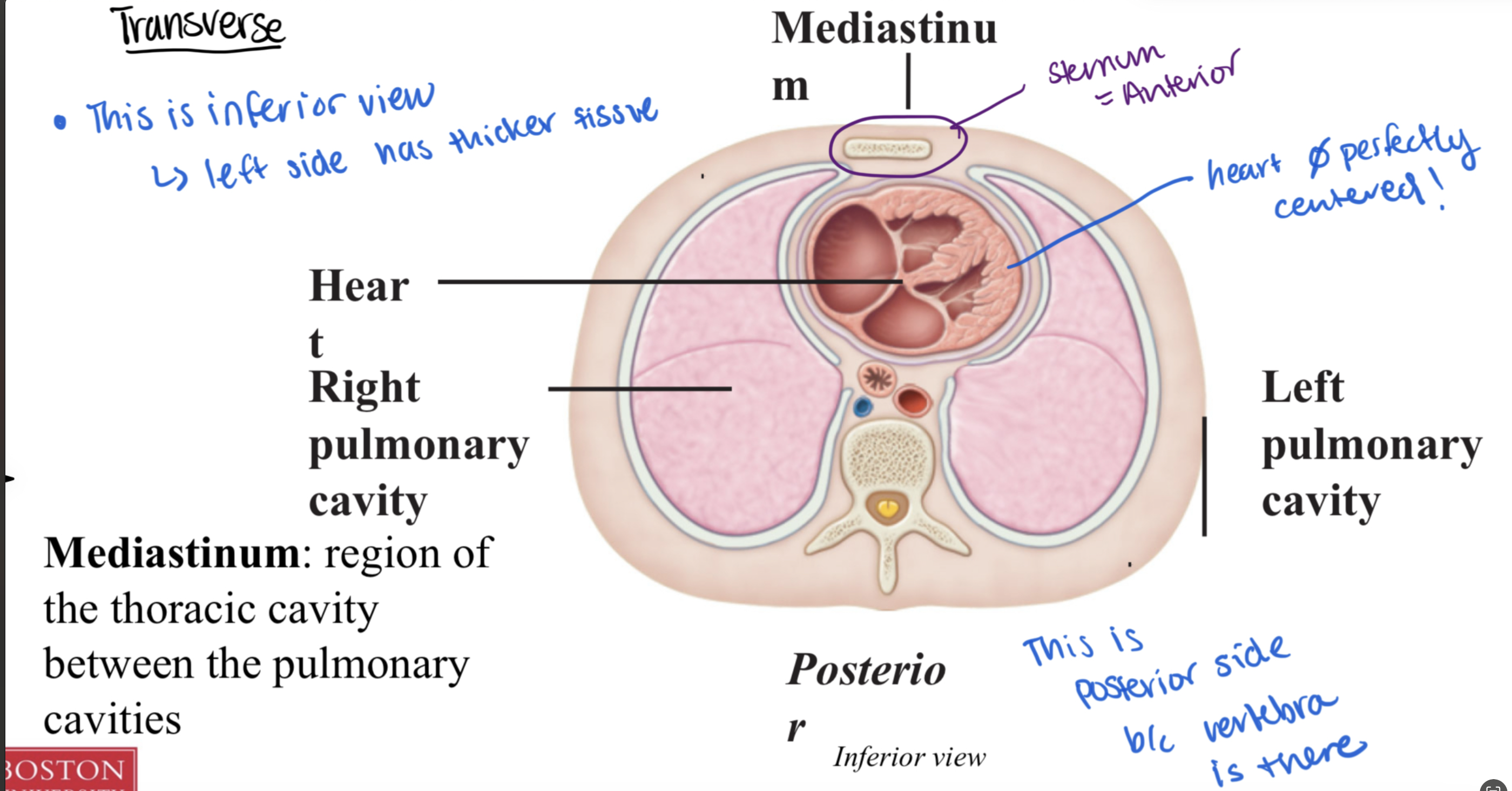
Define and describe the borders of the three thoracic spaces
mediastinum and two pleural cavities
Mediastinum:
in center and for the heart
region of the thoracic cavity between the pulmonary
cavities
Left and Right pleural cavities:
envelope the lungs
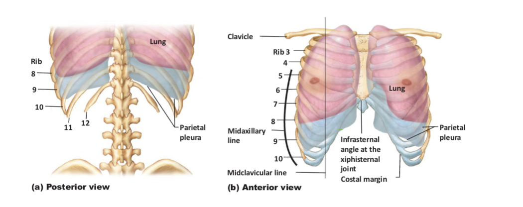
explain this image
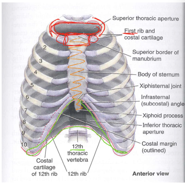
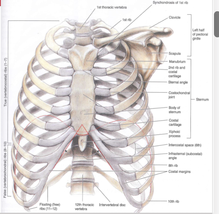
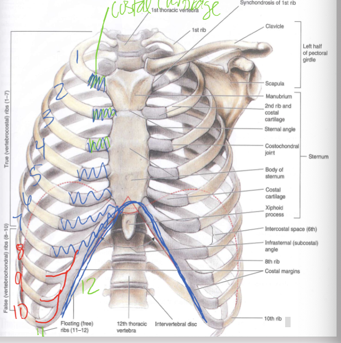
Thoracic Osteology (part 1)
12 thoracic vertebrae
• Sternum
ManubriumBody
• Xiphoid process
• Jugular notch
• Sternal angle
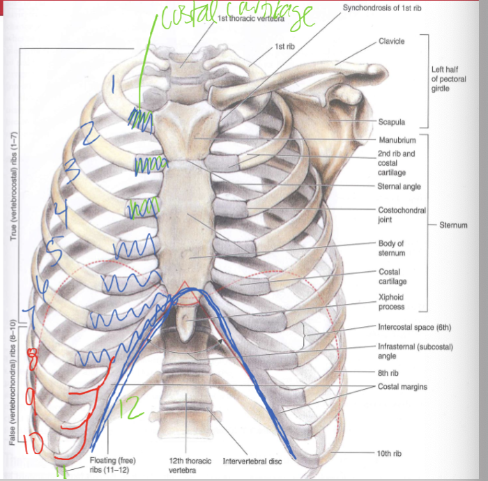
Compare and contrast true ribs, false ribs and floating ribs
12 ribs & their costal cartilages
True ribs (vertebral costal) attach to the
sternum —→ ribs 1-7
False ribs (vertebral chondral) attach to the
cartilage ——→ ribs 8-10
Floating ribs (free) no anterior attachment ——→ ribs 11-12
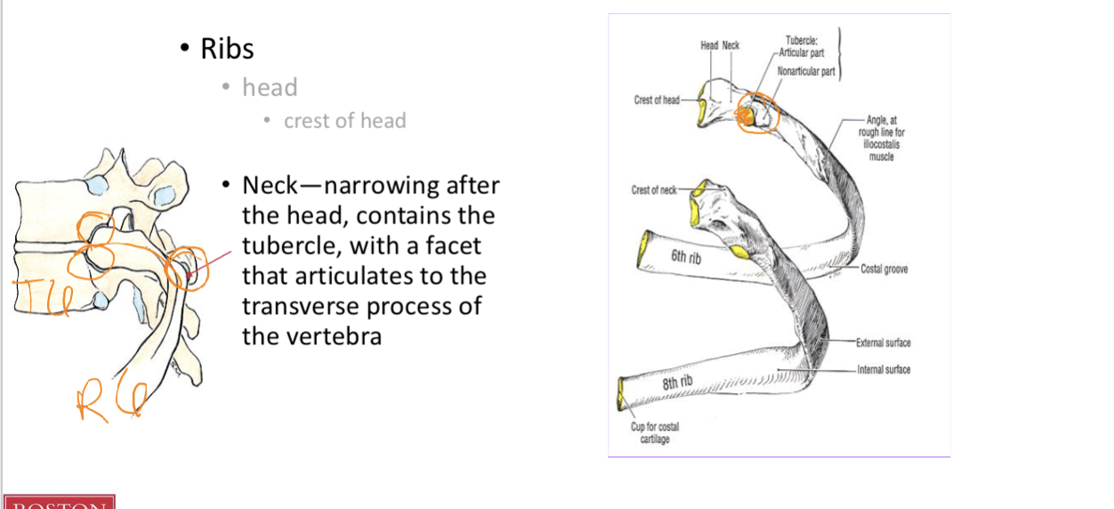
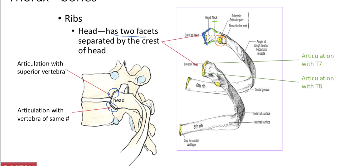
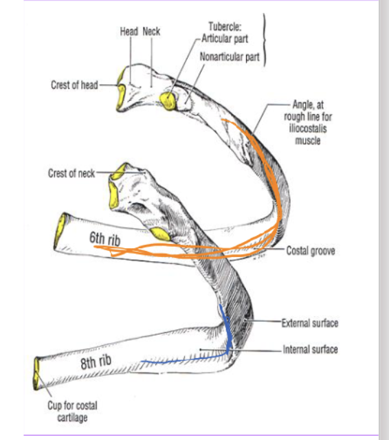
Identify the bony features of an individual rib (Neck and Head)
Head
has two facets separated by the crest of head
has 2 facets - 1 that articulates with T7 and another that articulates with T8
is posterior and medial b/c it articulates w/ verterbra
Neck
narrowing after the head, contains the tubercle, with a facet
that articulates to the transverse process of the vertebra
R6 articulates w/ T5
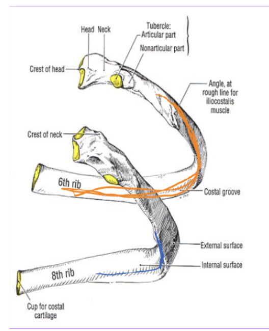
Identify the bony features of an individual rib (Body)
Body
is the rest of the rib
Has the costal angle, which is a the sharpest curve (site of attachment for
iliocostalis) and is lateral to rib’s tuberclehas the costal groove, is an indentation on costal angle where the spinal nerve and vessels passes
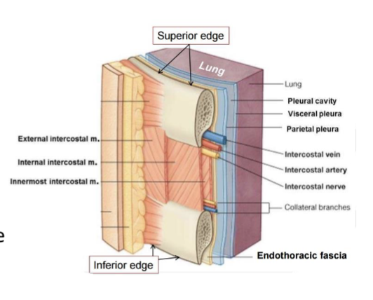
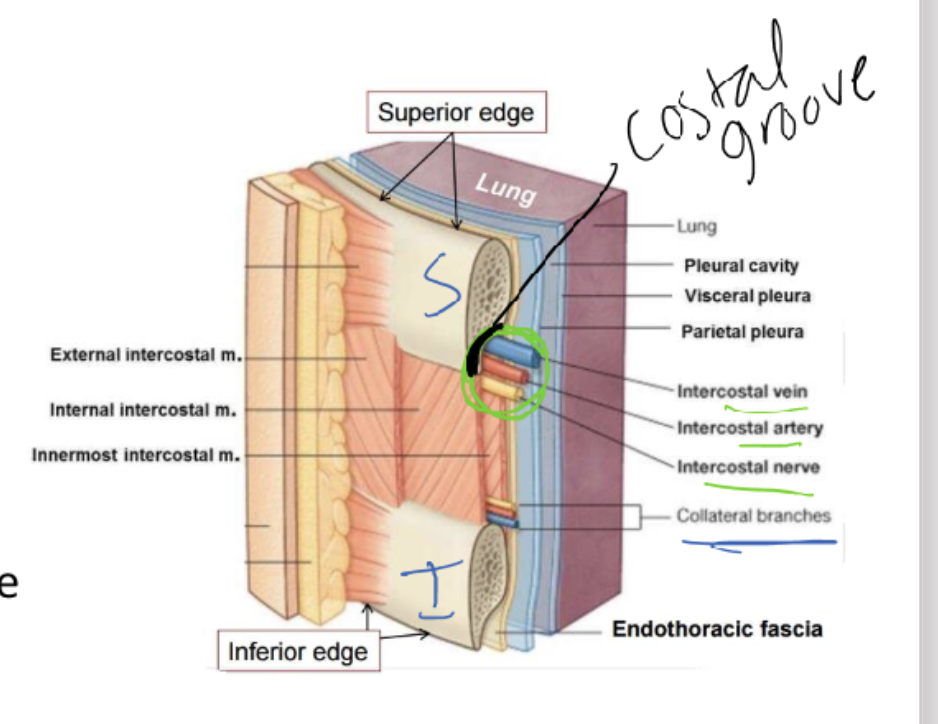
Identify the individual components of the intercostal spaces and understand their positions relative to each other
Between the ribs are intercostal spaces
named for the superior ribInside are 2 nerves and sets of blood vessels:
Main intercostal nerve, artery and vein are found in the
costal groove of the superior ribCollateral branches are found just above the inferior rib
Space below the 12th rib is the subcostal space
Costal groove is on inferior aspect of rib, which gives notch space for vein, artery, and nerve
Thoracic muscles & fascia
Thoracic wall is covered by muscles that belong to upper limb or move vertebral
column: (People Study To Learn Efficiently)
Pectoral muscles
Serratus anterior
Trapezius
Latissimus dorsi
Erector spinae
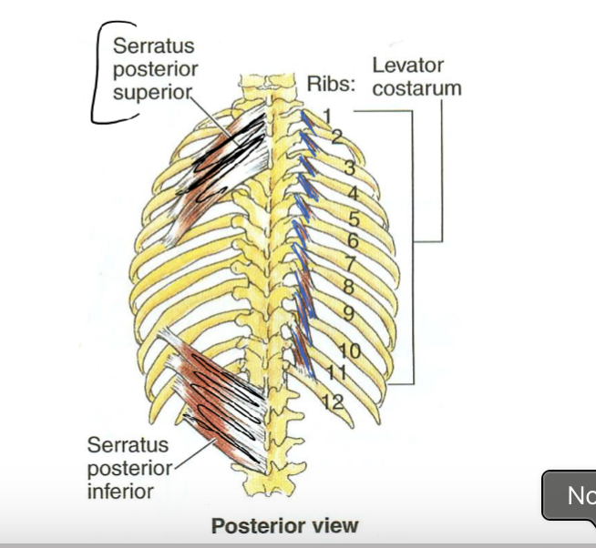
Thoracic muscles concerned exclusively w/ ribs for respiration: (DILS)
Diaphragm
Intercostals
Levator costarum
Serratus posterior
***Any muscle w/ attachment to ribs has potential to assist respiration under stress
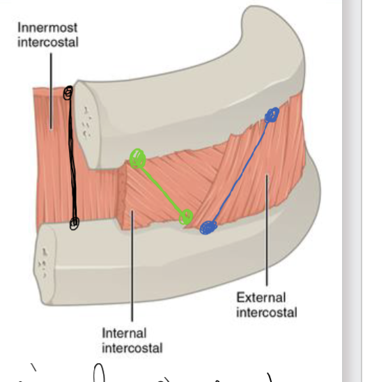
Identify the three layers of intercostal muscles
External
most superficial
oblique with inferior edge/border = medial and Superior edge/border = lat.
Internal
oblique with inferior edge/border = lat and Superior edge/border = med
Innermost
most deep
vertical ( meaning superior/inferior ) and NO oblique
helps in respiration
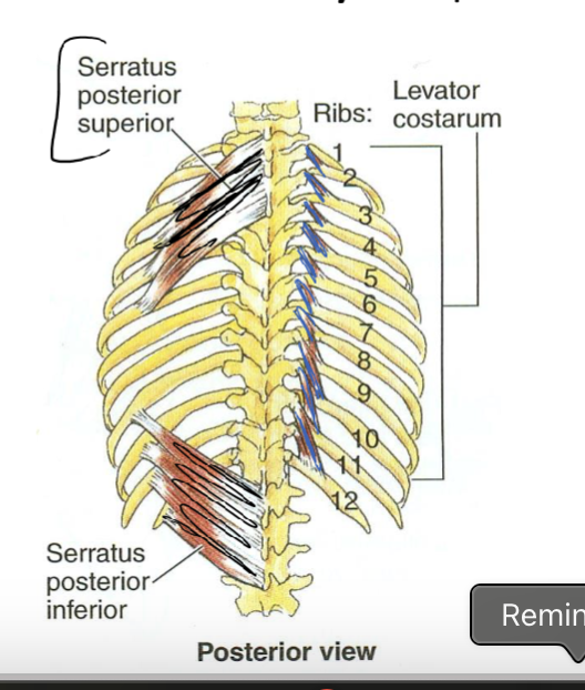
Additional muscles with only respiration functions
Serratus posterior
serrated like blade
Levator costarum
pulls ribs up for respiration
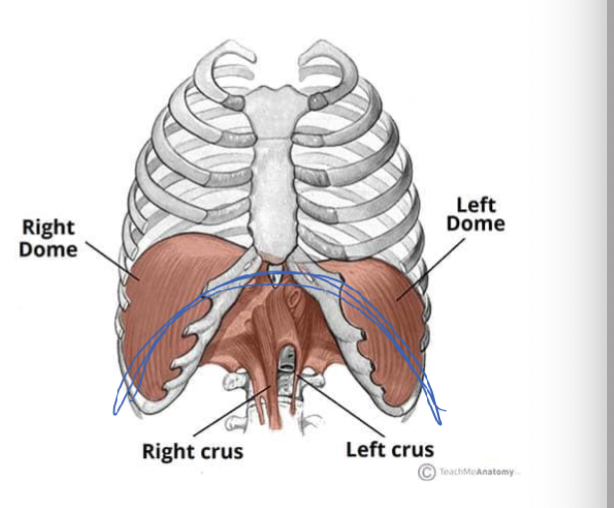
Diaphragm’s Origin, Insertion, Action,
Origin:
Xiphoid process
Lower costal cartilages
Upper Lumbar vertebrae
Medial and lateral arcuate ligaments
Insertion:
Central tendon of diaphragm
Action:
Expands thorax during inspiration
More about the diaphragm
it is on the inferior aspect of the thorax
this helps increase vertical space in thorax to allow air to enter
When flat = contracted
when in upside down U-shape = relaxed, which decreases V. space to expel air
REQUIRED FOR LIFE OR DEATH OCCURS
What does being on ventilator do to your diaphragm
it makes the diaphragm atrophy due to no usage of it, to the point when your off the ventilator it will make it hard to breathe
Thorax— 10 visceral contents
Heart
Esophagus
Pulmonary artery
Pulmonary veins
Aortic arch
Descending thoracic aorta
Superior vena cava
Inferior vena cava
Lungs
Trachea
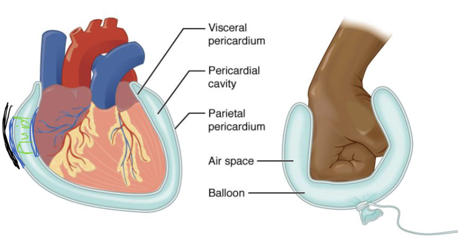
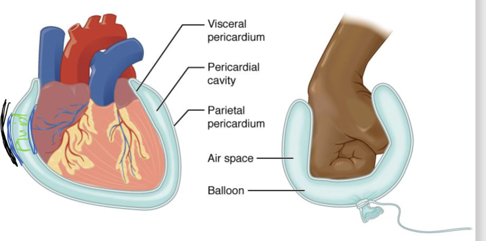
Compare and contrast serous and fibrous membranes
Most thoracic and abdominal organs are surrounded by a serous membrane and many have fibrous membrane
Serous membrane:
Double-sided and Fluid-filled membrane that allows for movement
reduces friction w/ lubricating fluids
Fibrous membrane:
exterior (superficial) to the serous membrane
contains movement
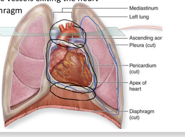
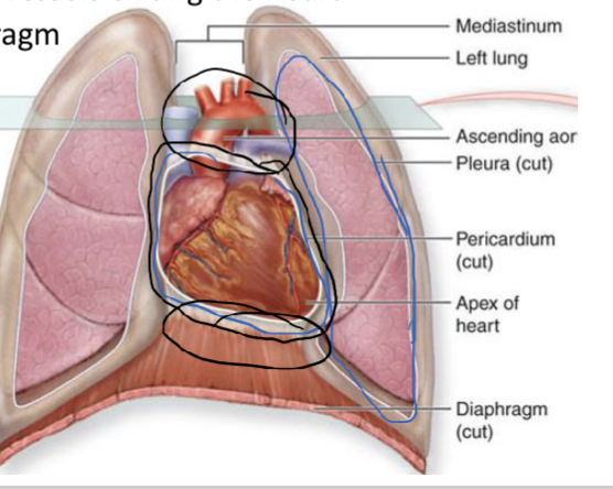
Fibrous pericardium
Main function: prevent overfilling of the heart
Location (black outline):
Superiorly to the large vessels exiting the heart
Inferiorly to the diaphragm
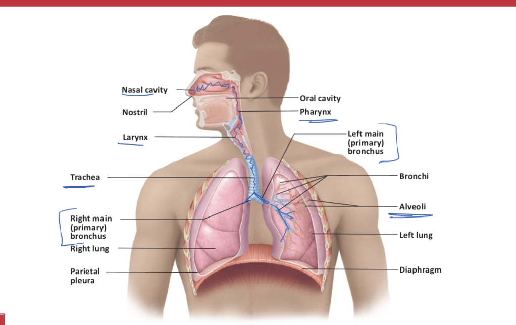
Compare and contrast the general locations and functions of the conducting and respiratory portions of the respiratory tract
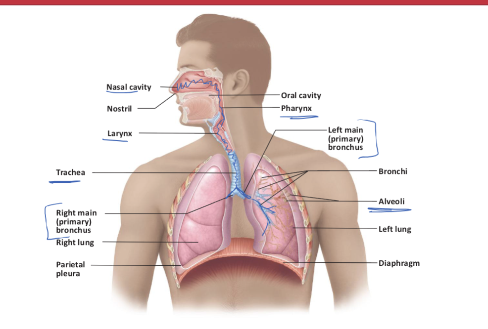
List, in order, the respiratory structures that air passes through during inhalation and exhalation.
IN 10 steps
Inhalation:
air passes via Nasal cavity
air goes down pharynx
air goes down larynx
air goes down trachea
air goes to primary bronchi
air goes to secondary bronchi
air goes to tertiary bronchi
air goes to terminal bronchioles
air goes to respiratory bronchioles
air goes to alveolar duct
air goes to alveolar sacs (bunch of grapes)
air goes to alveoli (single grape)
****exhalation is the reverse of the inhalation***
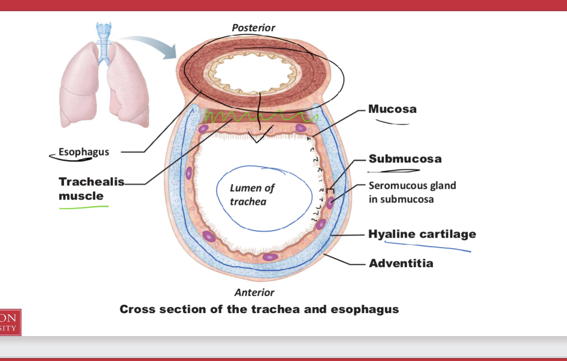
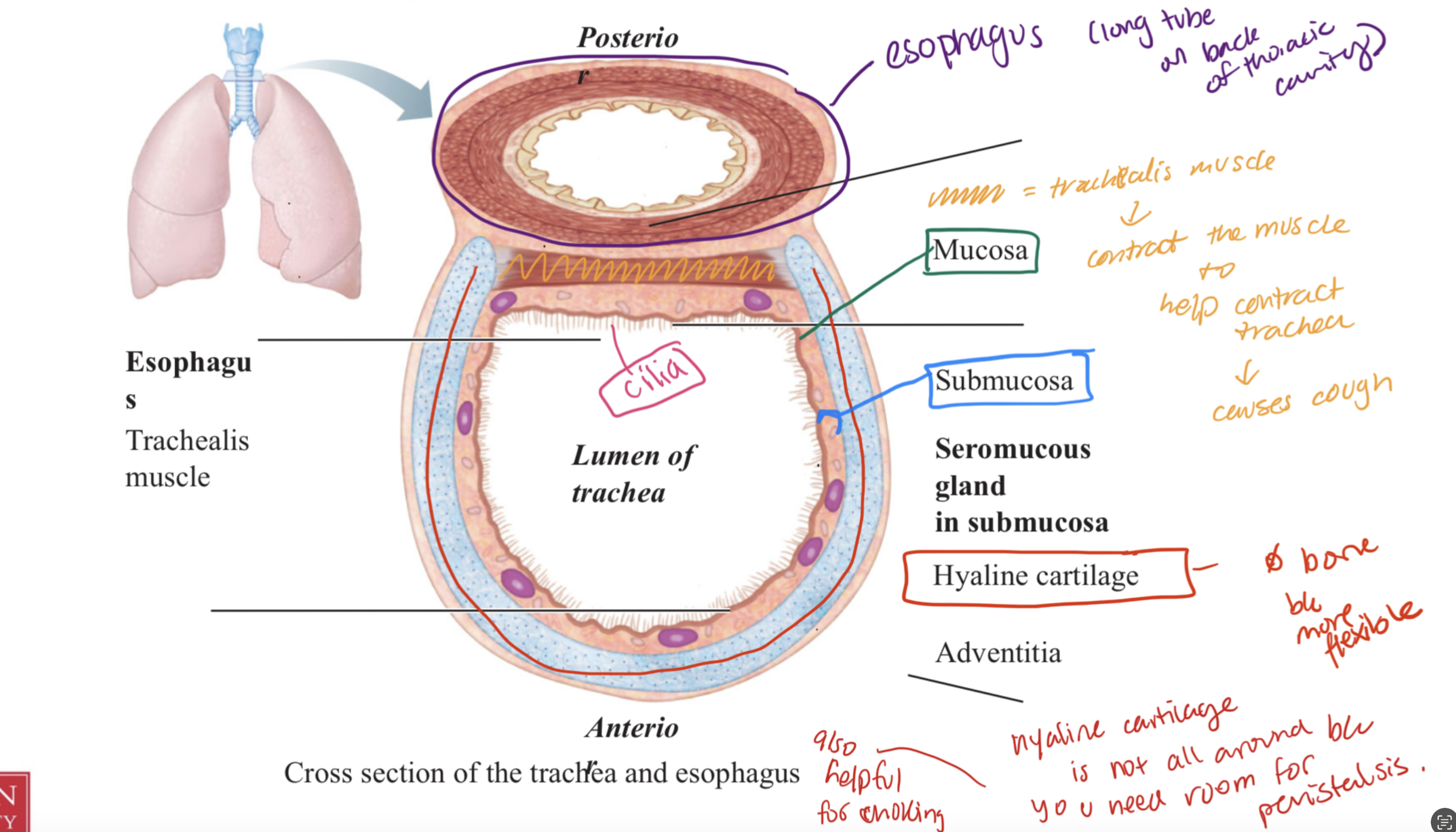
Describe the gross anatomical features of the trachea, including its positioning with respect to the esophagus
Trachea
made of hyaline cartilage b/c it provides flexibility to stretch without breaking
there is cilia for moving food
anterior to the esophagus
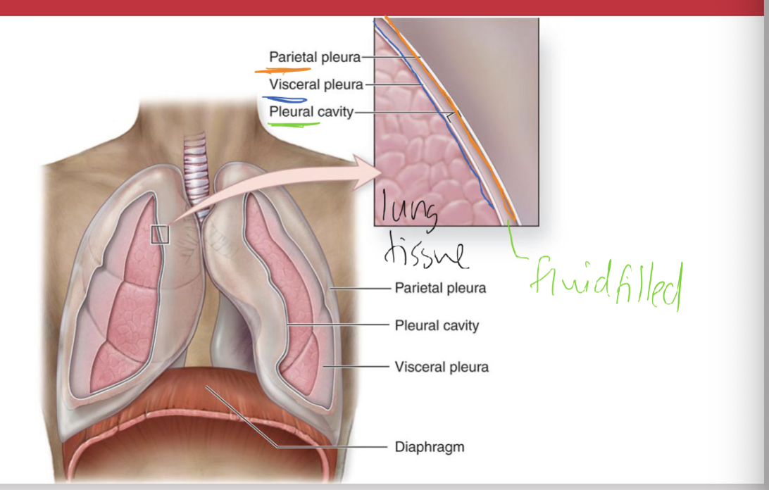
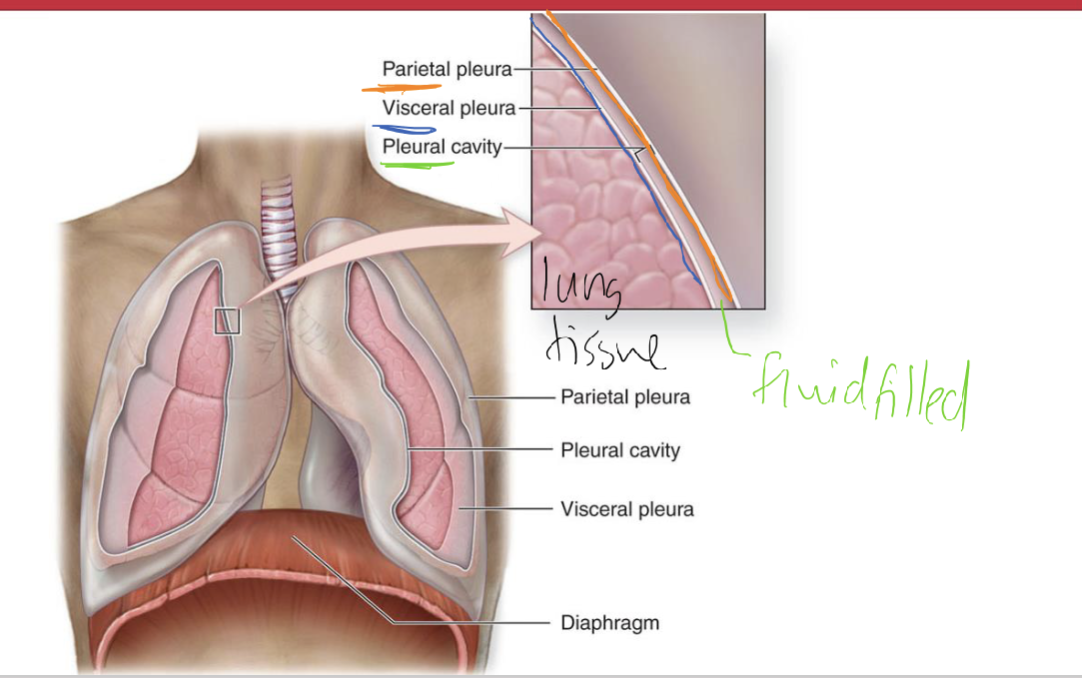
Explain the parietal and visceral pleurae and pleura cavity concept
parietal pleura
most superficial and congruent/inseparable to rib cage (and wall)
Pleural cavity
fluid filled in btw V and P pleurae
Visceral pleura
inner lining visceral membrane
The purpose:
pleural cavity’s fluid acts like glue (surface tension) b/c when parietal pleura moves due to inspiration so does the pleural cavity and visceral pleura and the lungs
this allows increasing of thoracic cavity to allow air to enter
Collasped lung
something enters pleural cavity and lung recoils —> NO air can enter
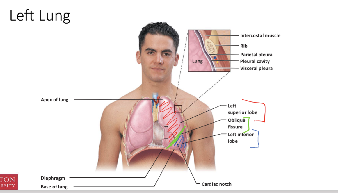
Parts of the Left lung
Red = left superior lobe
Green = oblique fissure
Blue = Left inferior lobe
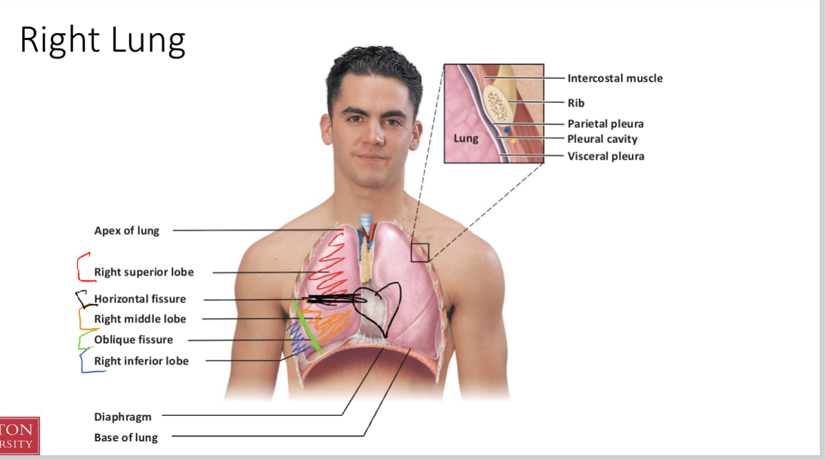
Parts of the Right lung
bigger than left and has more lobes b/c heart is placed more to the left
Red = Right superior lobe
Black = Horizontal fissure
Orange = Right middle lobe
Green = Oblique fissure
Blue = Right inferior lobe
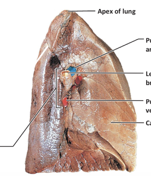
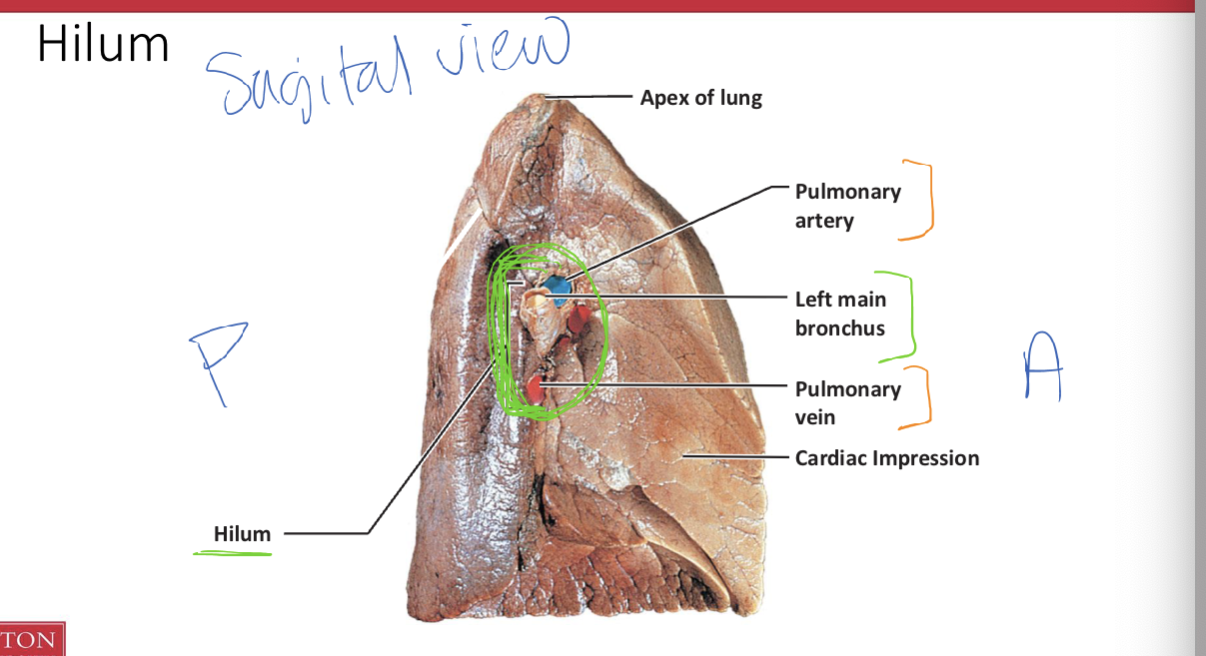
Hilum
seen in sagittal view
Pulmonary vein
pulmonary artery
left main bronchus
For posterior
it is darker lavitity: blood moves down b/c of gravity
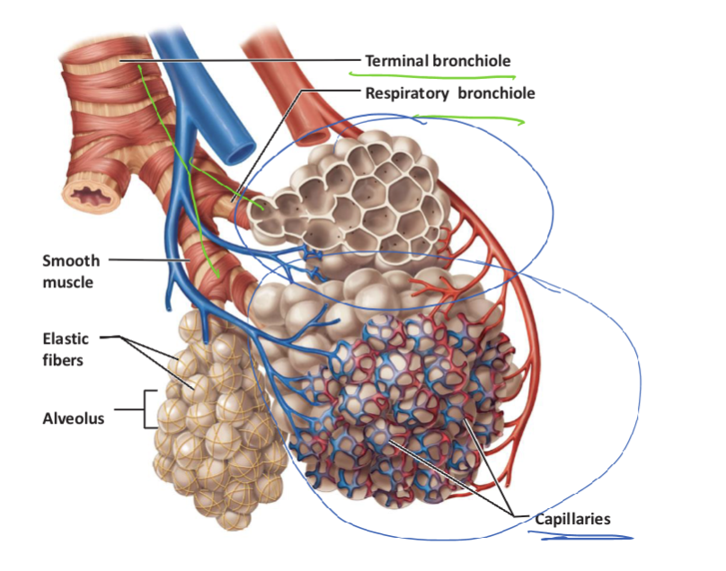
Identify and describe the gross anatomic features of the bronchial tree (e.g., bronchi, bronchioles, alveolar ducts, alveolar sacs and alveoli).
bronchi:
have cartilage b/c the trachea splits into bronchi
bronchioles:
have smooth muscles in their walls for constriction and dilation
alveolar ducts
extend from respiratory bronchioles and their walls are lined w/ alveoli
alveolar sacs
clusters of alveoli @ ends of alveolar ducts
alveoli
very small sacs
Capillaries
surround alveoli
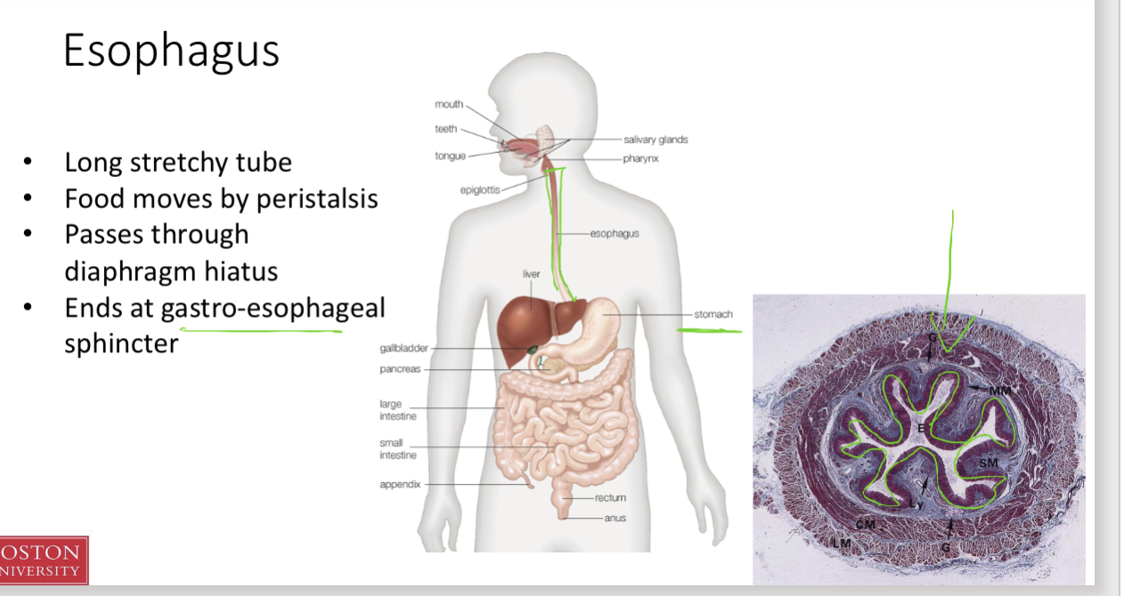
Identify the esophagus and describe its location relative to other body structures.
Long stretchy tube
Food moves by peristalsis
Passes through diaphragm hiatus
Ends at gastro-esophageal
sphincterposterior to trachea
Parietal vs Visceral
Parietal: body wall
Visceral: organ wall
Pink = exhaled lung size
grey = inhale expansion