lecture 5 CBNS108
1/41
There's no tags or description
Looks like no tags are added yet.
Name | Mastery | Learn | Test | Matching | Spaced | Call with Kai |
|---|
No analytics yet
Send a link to your students to track their progress
42 Terms
What forms at the beginning of gastrulation and marks the dorsal side?
➤ The dorsal lip of the blastopore
What does the dorsal lip do?
➤ It is part of the Spemann Organizer, which directs body axis formation
How is the dorsal-ventral axis established?
➤ Via cortical rotation after sperm entry → β-catenin builds up on one side → dorsal side forms
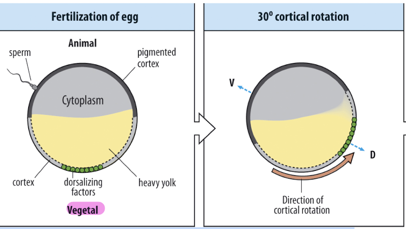
What triggers the breaking of radial symmetry in the frog embryo?
➤ Sperm entry
What does cortical rotation do? (right after sperm entry)
➤ Rotates cortex away from sperm entry → moves dorsalizing factors
The dorsal side is opposite to where the sperm entered
maternal dorsalizing factors
Wnt proteins, which turn on the β-catenin pathway on the future dorsal side
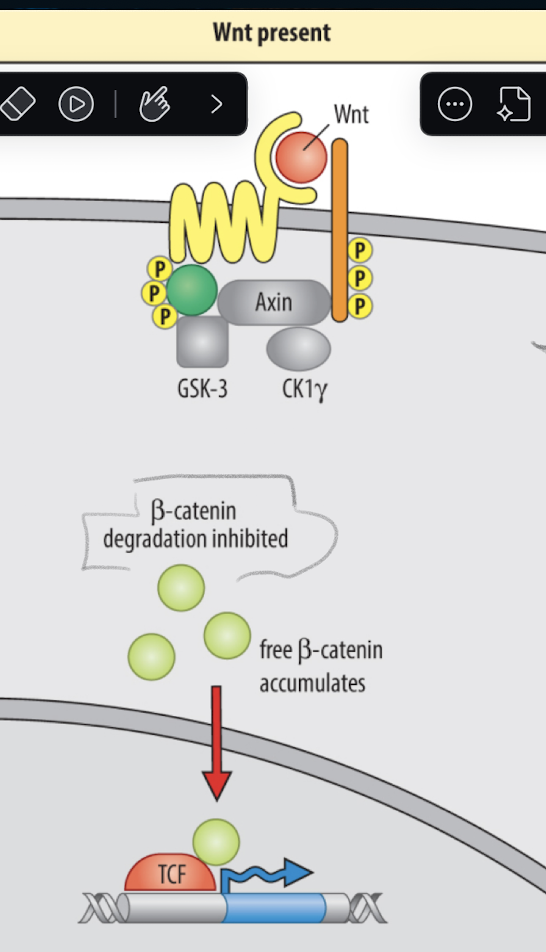
wnt pathway
1. Wnt ligand binds Frizzled receptor
2. Phosphorylation of β-catenin is blocked
Normally, GSK-3 adds phosphates to β-catenin → marks it for degradation.
But Wnt signaling inhibits GSK-3, so β-catenin is not phosphorylated.
This prevents it from getting broken down
3. β-catenin becomes stable and enters the nucleus
Now stable, β-catenin builds up in the cytoplasm.
It moves into the nucleus and partners with a transcription factor (TCF) to turn on dorsal-specific genes.
What happens to β-catenin when Wnt is NOT present?
➤ It gets phosphorylated by GSK-3 and degraded
in ventral cells
What initiates A-P axis patterning in frog embryos?
➤ Signals from the involuting mesoderm during gastrulation
What type of cells involute at the dorsal lip?
mesodermal cells
How does involution timing relate to axis patterning?
what axis is set up first?
➤ Early mesoderm induces anterior structures; later mesoderm induces posterior
➤ D-V axis forms first → helps guide A-P patterning
What germ layer is patterned by these signals?
➤ Ectoderm (specifically the neural ectoderm)
Hilde Mangold & Hans Spemann identified what?
A special region on the dorsal side of the embryo
They transplanted this region to another embryo, which caused the host to grow a 2nd body axis
This showed this region had organizing power (Spemann organizer)
Spemann organizer overview
Located at the dorsal lip of the blastopore.
It acts as a signaling center — it sends out molecular instructions to:
Start forming the central nervous system
Help define both the D-V and A-P axes
What does the organizer do molecularly?
➤ Sends out signals that induce the nervous system and pattern both axes
gray cresent
contains critical dorsalizing factors that set up the body plan.
🧬 It’s where the Spemann Organizer will form.
What happens if both blastomeres get gray crescent?
➤ Twin, fully developed embryos form
What happens if only one side gets the gray crescent?
➤ That side becomes a normal embryo; the other becomes a “belly piece” (no back, no nervous system).
How is the gray crescent formed?
➤ After sperm entry triggers cortical rotation, shifting dorsal determinants to the opposite side
What did the transplant experiment show? (Hilde transplanted DBL cells from one embryo to the opposite side of another)
➤ DBL cells can induce the formation of an entire second axis (including CNS) second spinal cord, notochord, etc.
Big Picture Summary: spemann organizer
The gray crescent is the region of the egg that gives rise to the
DBL (dorsal blastopore lip), which contains the
Spemann Organizer, a signaling center that patterns the embryo
What does the Spemann-Mangold experiment produce when DBL is grafted?
Where did most of the neural tube in the secondary axis come from?
➤ A twinned embryo with two heads and two nervous systems
(also created second organizer/secondary invagination/ new dorsal lip)
➤ From the host — the organizer induced host cells to form neural tissue
What are the three functions of the Spemann Organizer?
maintain, induce, organize
On the dorsal side,what enhances nodal signaling even more?
B-catenin, which is active here → Spemann Organizer forms there
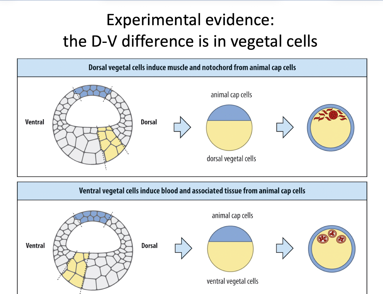
Dorsal vegetal cells Vs. Ventral vegetal cells
➤ They induce muscle and notochord (dorsal mesoderm)
➤ Blood and associated tissues (ventral mesoderm)
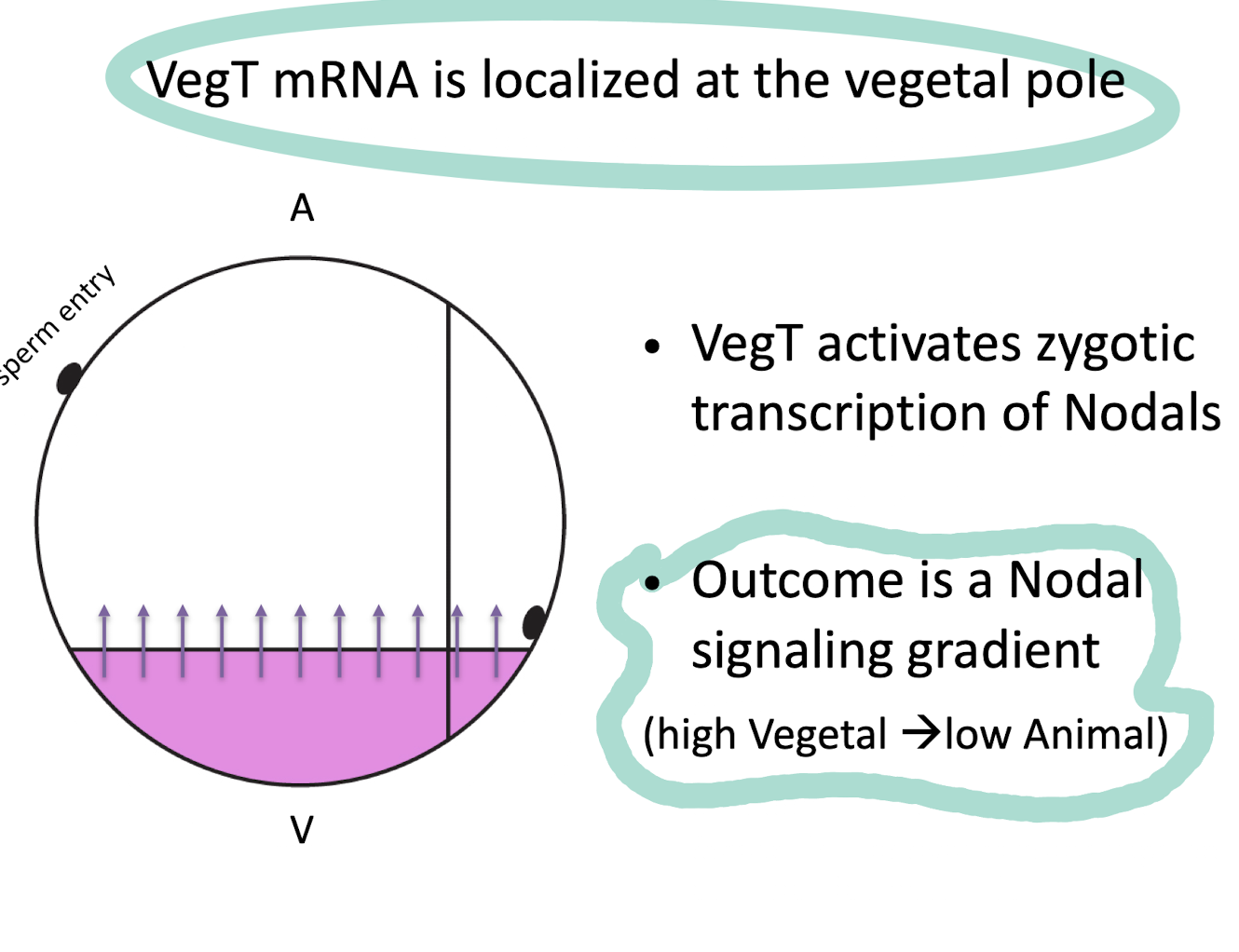
What is the result of VegT activity?
➤ A Nodal signaling gradient is formed: high at the vegetal pole, low at the animal pole
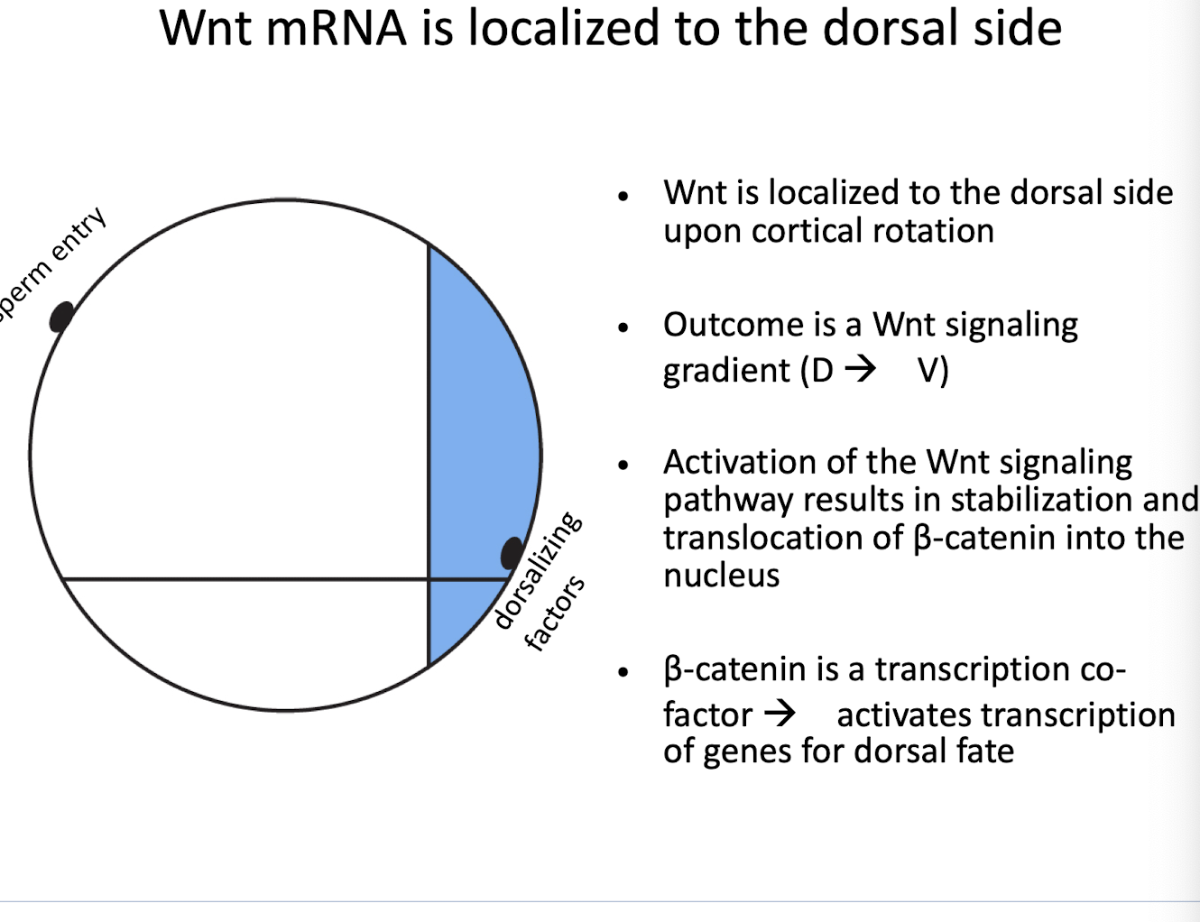
What happens when Wnt signaling is active?
➤ β-catenin is stabilized, enters the nucleus, and turns on dorsal genes
➤ On the dorsal side only
VegT mRNA is localized at the
vegetal pole
wnt mRNA is localized to the
dorsal side
Wnt is localized to the dorsal side → creates a Wnt signaling gradient (D ➝ V)
Neiuwkoop center
🟣 Purple = region with both VegT and β-catenin
dorsal-most vegetal cells
express Siamois, which is a key marker of this center
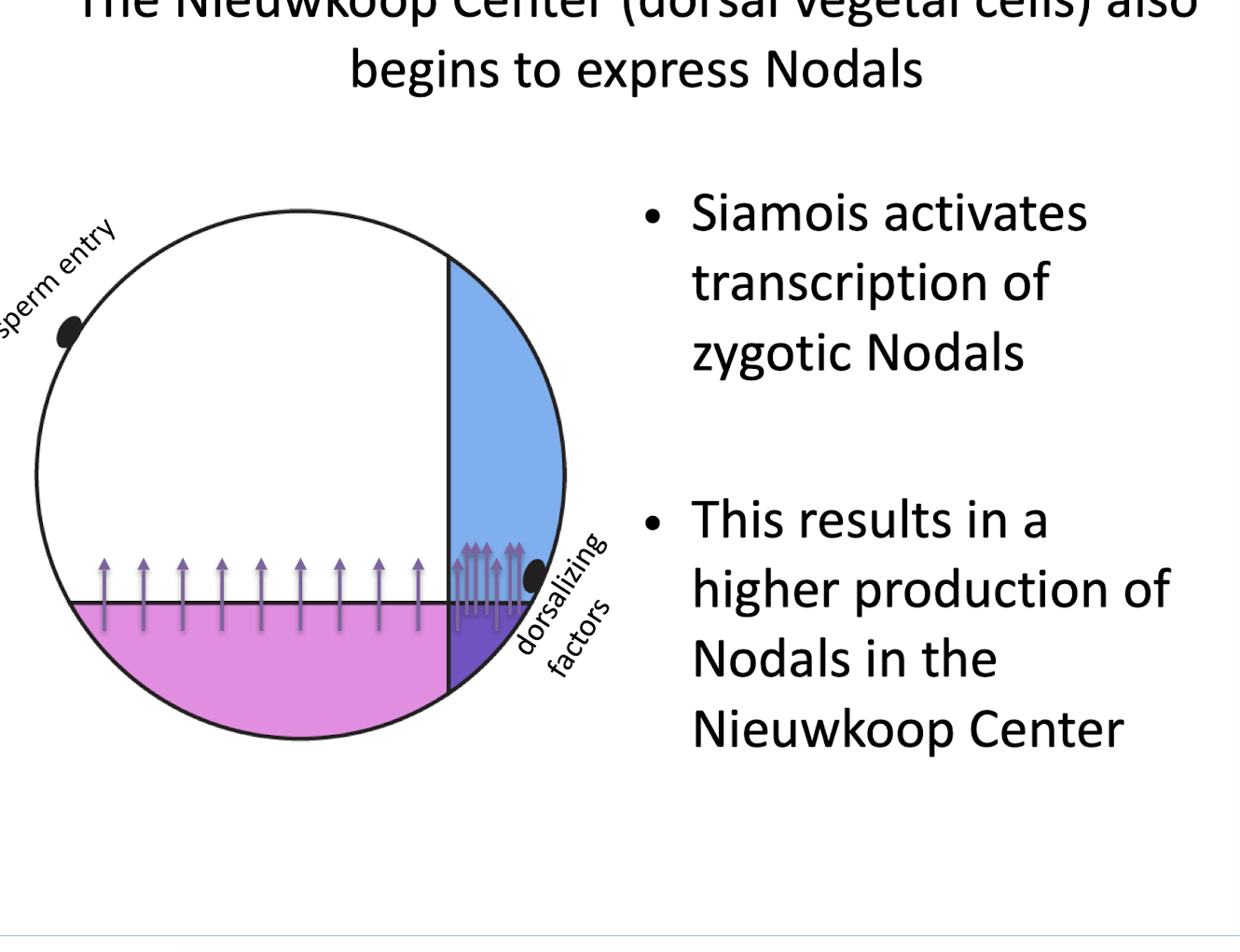
What is the function of the Nieuwkoop Center?
➤ It induces the Spemann Organizer in the overlying dorsal marginal zone
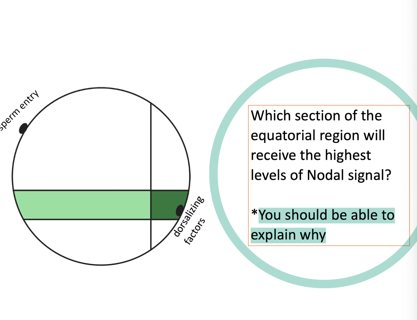
Where Is Nodal Strongest?
The dorsal equatorial (marginal) region — the dark green stripe near the dorsal side.
Why?
It lies directly above the Nieuwkoop Center, which produces the most Nodal.
These cells get both VegT-driven baseline Nodal and boosted Nodal due to Siamois activity from β-catenin presence.
This region becomes the Spemann Organizer (first site of involution at the dorsal lip).
How do we know that different concentrations of Nodal induce different mesoderm fates?
✅ Experiment:
Take animal cap cells (which normally become ectoderm).
Expose them to varying concentrations of Nodal (or Activin, a similar TGF-β ligand).
Observe what genes get turned on.
✅ Expected Outcome:
If the hypothesis is correct:
Low Nodal → activates low-threshold genes (e.g., brachyury, for general mesoderm like muscle or blood)
High Nodal → activates high-threshold genes (e.g., goosecoid, for organizer/notochord fate)
How do we know Nodal acts as a morphogen?
➤ Because different concentrations activate different genes, and it’s diffusible
Cell fate = signal strength
Low Nodal → lateral/ventral mesoderm
High Nodal → dorsal mesoderm + organizer
What do you expect if you transplant the Nieuwkoop Center to the ventral side?
➤ A secondary organizer forms, and possibly a partial or full twinned axis
Organizers dorsalizing functions
1. 🧬 Self-differentiates into dorsal mesoderm
➤ It becomes structures like the notochord (the backbone precursor).
2. 🧱 Dorsalizes surrounding mesoderm
➤ It turns nearby lateral mesoderm into somites (which become muscle, vertebrae).
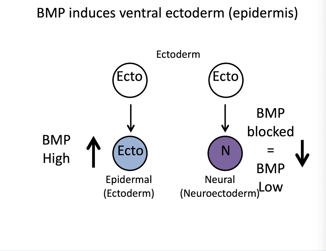
3. 🧠 Dorsalizes ectoderm → forms neural tissue
➤ Organizer molecules block BMP → ectoderm forms neural plate/tube (brain + spinal cord) instead of skin.
4. 🧬 Dorsalizes endoderm
➤ Influences deeper tissues, ensuring correct gut and organ development on dorsal side.
5. 🔄 Initiates movements of gastrulation
➤ Organizer is the starting point for involution and convergent extension.
What does an antagonist do?
It inhibits or blocks a signaling pathway.
Instead of activating a receptor or downstream genes, antagonists prevent the signal from working.
In the case of the organizer:
It blocks BMP signals that would otherwise ventralize the embryo.
It also blocks Wnt, which helps fine-tune anterior-posterior patterning.
What would happen if these antagonists were missing?
➤ Embryos would be ventralized (e.g., no nervous system, no notochord).
How is position along the D-V axis determined?
➤ By BMP concentration — cells read the gradient and adopt fates accordingl
Why are gradients critical for axis formation?
💡 Cells closer to the source (higher morphogen) = one fate
⚖ Middle cells (intermediate morphogen) = another fate
🧍 Farther away (low morphogen) = yet another fate
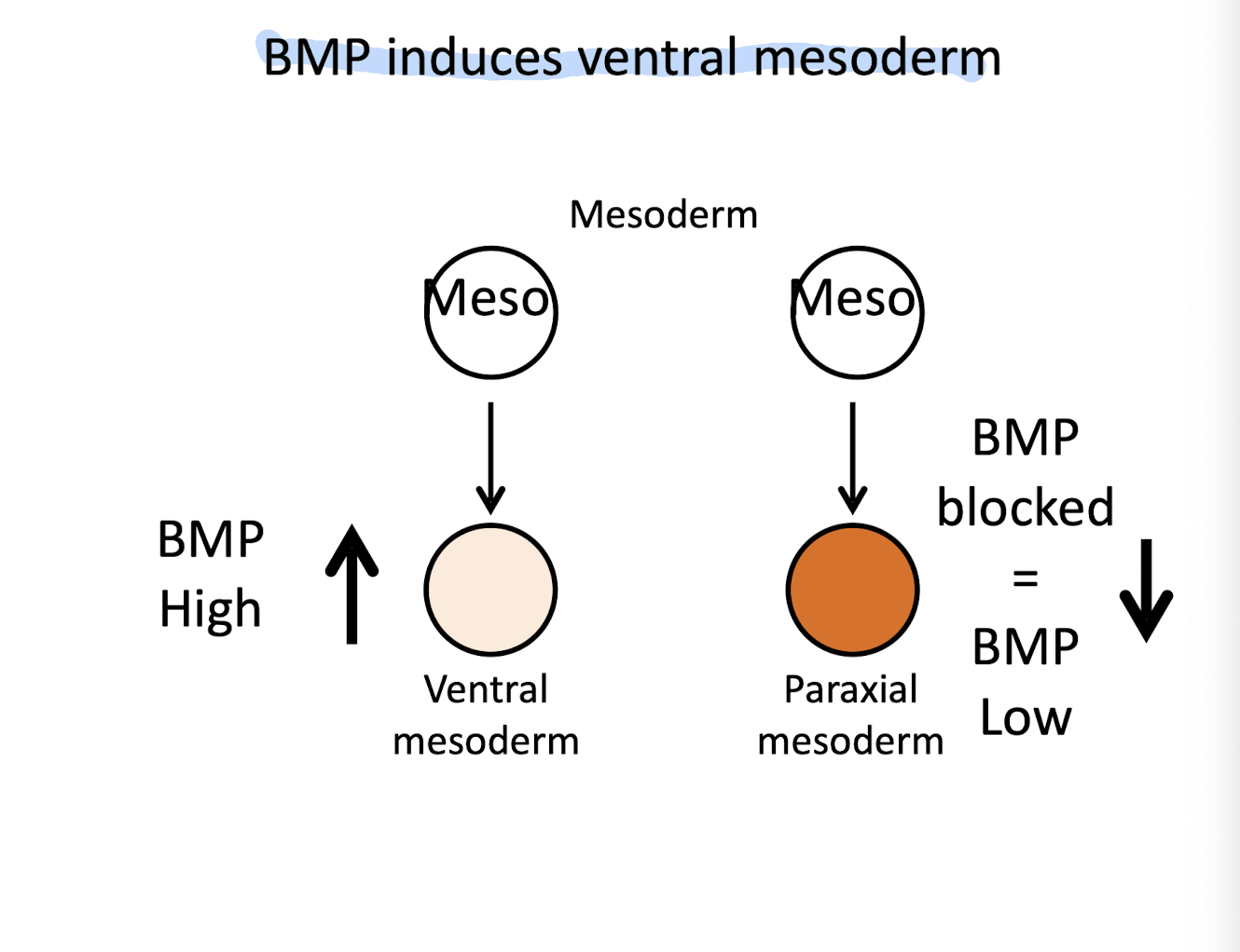
BMP and Mesoderm Fate
High BMP → mesoderm becomes ventral mesoderm (blood, kidney)
Low BMP → mesoderm becomes paraxial mesoderm (somites like muscle + vertebrae)
This is how BMP gradients shape the dorsal (muscle) vs. ventral (blood) mesoderm!
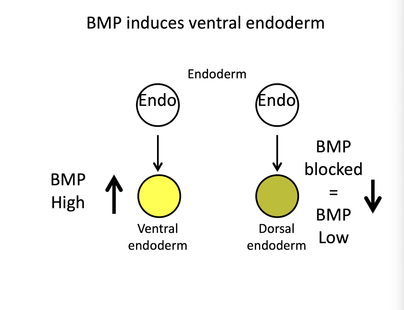
BMP and Endoderm Fate
High BMP → endoderm becomes ventral endoderm
Low BMP → endoderm becomes dorsal endoderm (gut-associated structures)