CH 11- Functional Organization of Nervous Tissue Pt 2.
1/94
There's no tags or description
Looks like no tags are added yet.
Name | Mastery | Learn | Test | Matching | Spaced | Call with Kai |
|---|
No analytics yet
Send a link to your students to track their progress
95 Terms
Gray matter
contains neuron cell bodies, dendrites
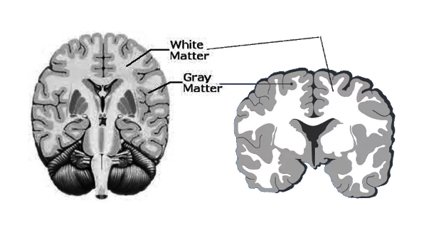
Gray matter CNS
Cortex- surface of the brain
Nuclei- clusters deep within the brain
Gray matter PNS
Ganglia- neuron cell bodies
White matter
bundles of myelinated axons

White matter CNS
Nerve tracts- carry action potentials from one area of the CNS to another
White matter PNS
Nerves- bundles of axons and their connective tissue coverings
Action Potential
electrical signals produced by the nervous system
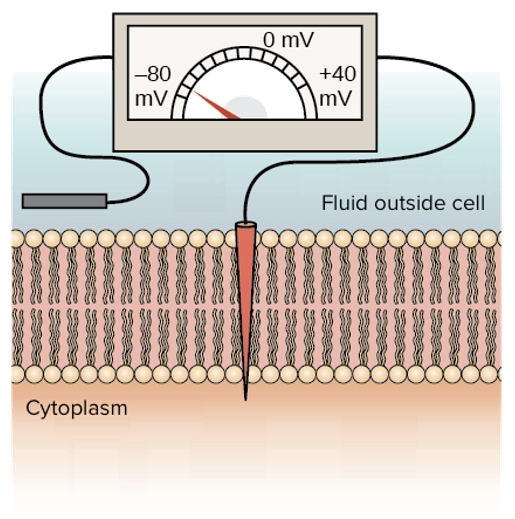
Membrane potential
measure of electrical properties of the plasma membrane due to
ionic concentration differences across the plasma membrane
permeability characteristics of the plasma membrane
Permeability of the plasma membrane
determined by the ion channels and pumps
sodium-potassium pump
help to maintain the difference in cytoplasmic and extracellular concentrations of ions
leak channels
gated channels
sodium-potassium pump
help to maintain the difference in cytoplasmic and extracellular concentrations of ions
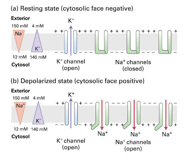
Leak channels
always open
responsible for permeability of the plasma membrane when it is at rest
determine permeability of resting membrane
more permeable to K+ and Cl- than to Na+
specific for one type of ion
Gated Ion Channels
open and close due to a specific signal
ligand-gated ion channels
voltage gated ion channels
mechanically-gated ion channels
thermoreceptors
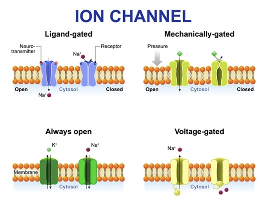
Ligand-gated ion channels
opened by binding of a specific molecule (ligand) on the extracellular side
channel crosses the membrane
Voltage-gated ion channels
open and close in response to specific voltage changes across the plasma membrane
required for action potentials
Mechanically- gated ion channels
open in response to mechanical stimulation
Thermoreceptors
respond to temperature changes
Establishing resting membrane potential
cytoplasm and extracellular fluid are electrically neutral
charge difference across the plasma membrane
is the plasma membrane polarized or unpolarized?
polarized
Potential difference
electrical charge difference across the plasma membrane
Resting membrane potential
potential difference in a resting cell
changes in resting membrane potential
Ions diffuse down their concentration gradients
Movement results in electrical current and changes in resting membrane potential
Two types of changes:
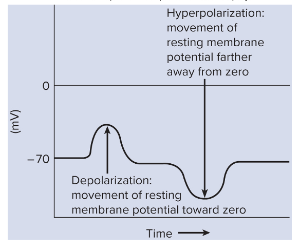
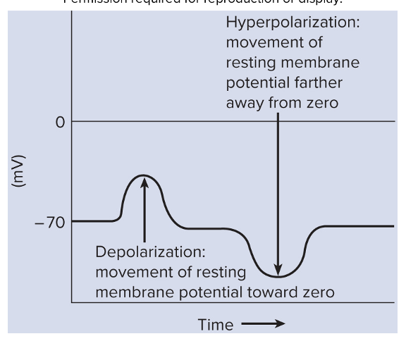
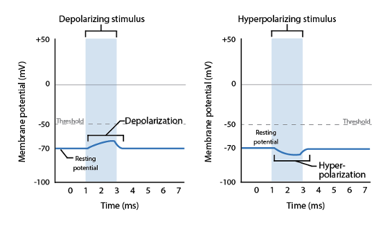
Depolarization
Hyperpolarization
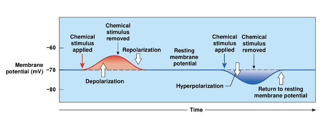
Depolarization
inside the cell becomes more positive
excitatory
several factors leading to depolarization
Depolarization- excitatory state
always moves the membrnae potential closer to the point of action potential generation
Factors that can lead to depolarization of neurons
Na+ entry
Ca2+ entry
Changes in extracellular K+ concentration
Sodium Ions
Na+ entry is the most common cause of depolarization
Limited Na+ leak channels
Entry of Na+ is typically regulated
Ligand-gated Na+ or voltage-gated Na+ channels
Calcium Ions
Calcium enters the cell which causes depolarization
important for some cardiac muscle cells to generate action potentials
Plays significant role in action potentials
regulates the voltage-gated sodium channels
regulation of neurotransmitter secretion at the presynaptic terminal
Hypocalcemia- lower levels of Ca2+ in the blood
Hypocalcemia
Lowers levels of Ca2+ in the blood
Symptoms include nervousness and uncontrolled skeletal muscle contraction
Caused by lack of dietary Ca2+ or vitamin D or insufficient PTH
Potassium Ions
Changes in extracellular K+ concentration can affect resting membrane potential
Increases can cause cytoplasmic K+ to stay inside the cell
When K+ stays inside the cell it can cause depolarization
Hyperpolarization
Inside of the cell becomes even more negative
Inhibitory-
makes the cell less likely to create an action potential
Two major ways to hyperlarize neurons
K+ exits
Cl- enters
Potassium Ions
Exit of K+ is primary cause of hyperpolarization after action potential
Voltage-gated K+ channels
Ligand-gated K+ channels
mechanism for some inhibitory neurotransmitters
Hypokalemia
lower blood K+ concentration
Hypokalemia
Lowers potassium concentration in the blood
DECREASE in extracellular potassium can cause more potassium to exit the cell through leak channels
Symptoms include muscular weakness, abnormal heart function, sluggish reflexes
Cause by starvation, alkalosis, and some kidney diseases
Chloride ions
Cl- concentration is higher outside the cell
Opening of ligand-gated Cl- channels allows Cl- to diffuse into the cell
Some inhibitory neurotransmitters use this mechanism
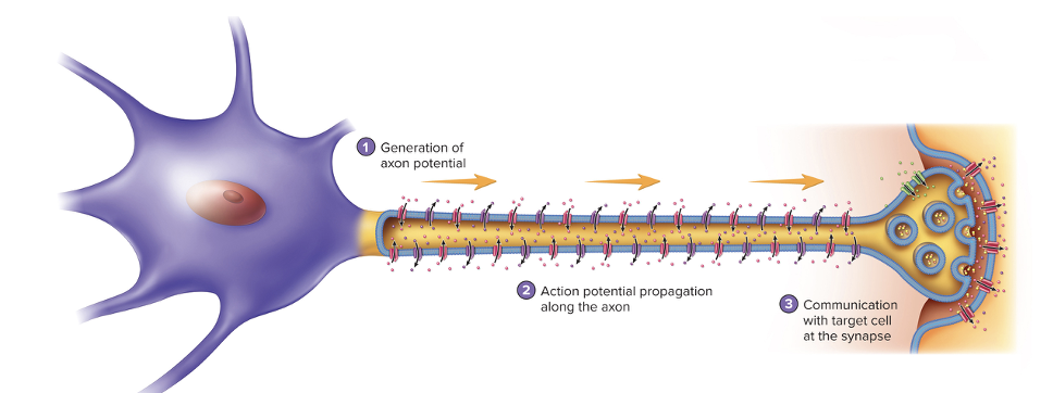
Neuron Communication
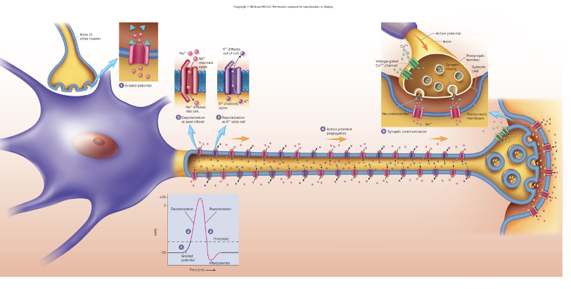
Generation of action potentials
Action potential propagation along the axon
Communication with a target cell at the synapse
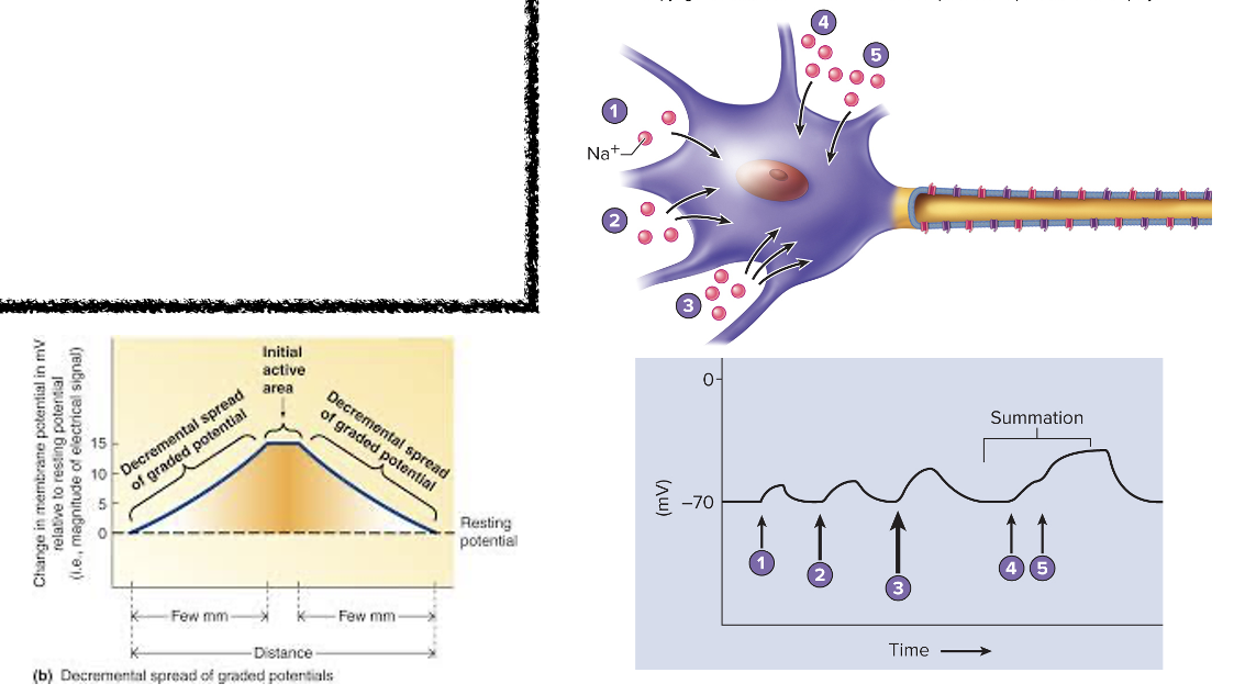
Graded Potentials
Relatively small change in membrane potential localized to one area of the plasma membrane
vary in size depending on strength of the stimulus
Caused by several types of stimuli
chemicals binding to ligand-gated ion channels
changes in voltage triggering opening or closing of voltage-gated ion channels
mechanical stimuli opening mechanically gated ion channels
temperature changes affecting specific temperature receptors
Graded potentials can be…..
Hyperpolarizing- inhibitory
Depolarizing-excitatory
Summation- Graded Potentials
Combination/adding graded potentials
Large enough (reaches threshold) will result in an action potential
Spread in decremental fashion
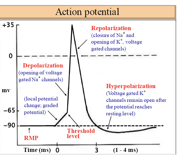
Action Potentials
Used by neurons for communication
Result from summation of graded potentials
LARGE change in membrane potential
Spreads (travels) without changing in magnitude over long distances
Comes in phases
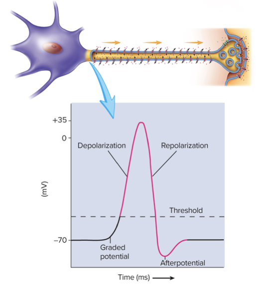
Phases of an action potential
Depolarization phase
Repolarization phase
Afterpotential
Return to resting membrane potential
All-or-None Principle
If graded potential reaches threshold, action potential is generated
voltage-gated channels open altering membrane permeability
If graded potential doe snot reach threshold, action potential is not generated
membrane potential returns to resting potential
Voltage-Gated Ion Channels & Action Potentials
Required for generation of action potentials
Comes in phases
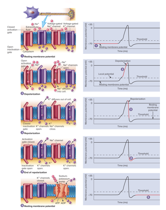
Phases of an action potential in Voltage-Gated Ion Channels & Action Potentials
Depolarization phase
Repolarization phase
After potential
Return to resting membrane potential
Refractory Period
Plasma membrane becomes less sensitive to further stimulation
Absolute refractory period
Relative refractory period
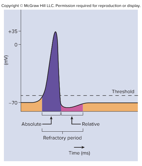
Absolute refractory period
First part of the refractory period
Membrane is completely insensitive to stimulus
From beginning of action potential until near the end of repolarization
Lets depolarization and repolarization phases to be ompleted before another action potential can begin
Prevents strong stimulus from causing prolonged depolarization of plasma membrane
Relative refractory period
Stronger than threshold stimulus needed to start another action potential
Membrane is more permeable to K+
Ends when voltage-gated K+ channels close and membrane potential returns to rest
Action Potential Frequency
Number of action potentials per unit of time in response to a stimulus
Directly proportional to stimulus strength and to the size of the graded potential
Subthreshold stimulus
Threshold stimulus
Submaxial stimulus
Maximal stimulus
Supramaximal stimulus
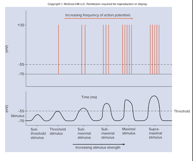
Subthreshold stimulus
stimulus not strong enough to reach threshold, does not generate an action potential
Threshold stimulus
graded potential just reaches threshold and causes a single action potential
Submaxial stimulus
stimuli between threshold and maximal stimulus strength
Maximal stimulus
strong enough to produce a maximum frequency of action potential
Supramaximal stimulus
stimulus stronger than maximal stimulus, does not increase action potential frequency
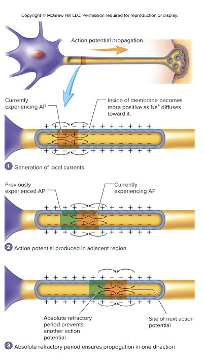
Propagation of Action Potentials
Involves the generation of a new action potential in adjacent region of the plasma membrane
Action potentials are generated in the trigger zone and travel in one direction down the axon
Types of action potentials
Continuous conduction
Saltatory conduction
Continuous Conduction
Happens in unmyelinated axons
Generates an action potential in each section of the plasma membrane
An action potential in one section of membrane allows for Na+ to diffuse to adjacent areas (local current) causing depolarization
new identical action potential is generated in response to the depolarization
Saltatory conduction
Happens in myelinated axons
Action potential is conducted from one node of Ranvier to the next
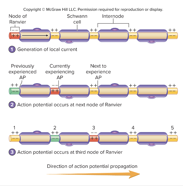
Speed propagation depends on:
Myelination
Thickness of myelin sheath
Diameter of the axon
Synapse is composed of
Presyneptic cell
Posynaptic cell
Types of Synapse
Electrical synapses
Chemical synapses
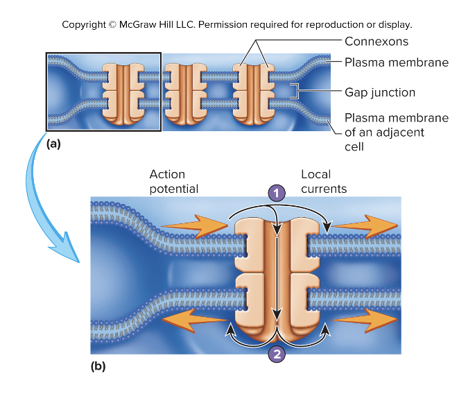
Electrical Synapses
Happens between cells connected by gap junctions
Allows ions flow from one cell to the next
Composed of connexons
6 tubular proteins (connexin)
Not common in nervous system
Found in cardiac muscle and some smooth muscle
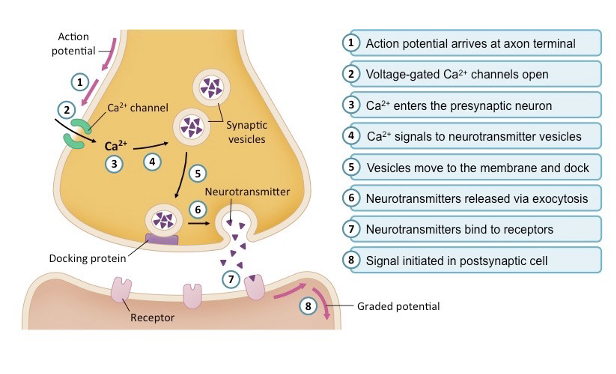
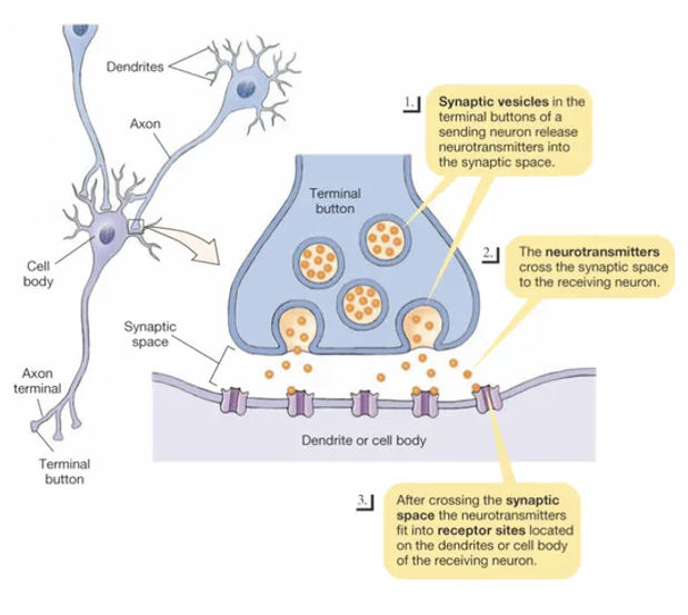
Chemical Synapses
Chemical messenger (neurotransmitter) is used to communicate between the cells
Release of neurotransmitter occurs due to action potential in the presynaptic terminal
Voltage-gated Ca2+ channels open ad Ca2+ entering the axon terminal triggers exocytosis of the neurotransmitter
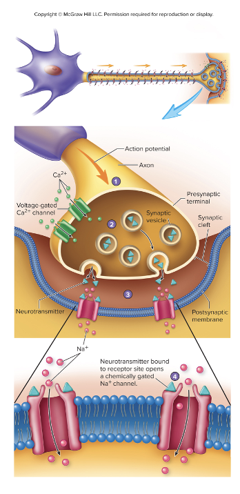
Chemical synapses are composed of
Presynaptic terminal-
axon terminal of the presynaptic cell that houses synaptic vesicles containing neurotransmitters
Synaptic cleft-
space separating the cells
Postsynaptic membrane-
membrane of the post synaptic cell (neuron, muscle cell, gland cell)
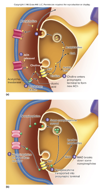
Neurotransmitter Removal
Neurotransmitter and receptor equilibrium:
High concentration of neurotransmitter in synaptic cleft results in more receptor binding
Rapid removal or destruction of neurotransmitter results in short term effects of neurotransmitter
Receptors in Synapses
Located on the postsynaptic cell
can also be found on some presynaptic cells
Highly specific
Determine the affect the neurotransmitter has on a cell
neurotransmitter can stimulate some cells and inhibit other
Neuron Communication
Graded potential
Action potential
depolarization
repolarization
Action potential propagation
Synaptic communication
Neurotransmitters
Chemical messengers released from neurons
Some neurons can secrete more than one type of neurotransmitter
Characteristics of neurotransmitters
Must be synthesized by the neurons and stored in synaptic vesicles sin presynaptic terminal
Action potential must stimulate its exocytosis into synaptic cleft
Must bind to a specific receptor on. the post synaptic membrane
Must evoke a response in the postsynaptic cell
Neurotransmitters are classified based on
chemical structure
effect on postsynaptic membrane
mechanism of action at their target
Chemical classification of neurotransmitters
Acetylcholine
Biogenic amines
catecholamines
indoleamines
Amino acids
Purines
Neuropeptides
Gases and lipids
Acetylcholine
Synthesized from precursors acetic acid choline
Biogenic amines
Catecholamines
derived from amino acid tyrosine, includes dopamine, norepinephrine, epinephrine
Indoleamines
derived from histidine and tryptophan, includes histamine and serotonin
Amino acids
Includes GABA, glycines, glutamate
Purines
Nitrogen containing compounds
Includes adenosine and ATP
Neuropeptides
10-40 amino acids
Includes substance P and endorphins
Gases and lipids
Gases (gasotransmitters)- nitric oxide (NO) and carbon monoxide (CO)
Lipids- endocannabinoids
Effect of Neurotransmitter on Postsynaptic Cells
Excitatory
causes depolarization
makes cell more likely to generate an action potential
Ex: glutamate, norepinephrine, dopamine
Inhibitory
causes hyperpolarization
Makes cell less likely to generate an action potential
Ex: GABA, serotonin, dopamine
Neurotransmitters mechanisms of action
Ionotropic effect
Metabotropic effect
Ionotropic effect
binding to ion channels
Metabotropic effect
binding to G protein-linked receptors
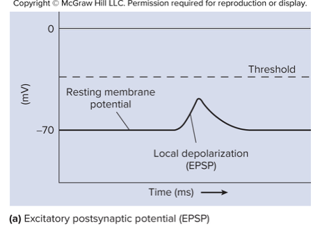
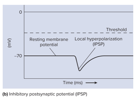
Postsynaptic Potentials
Excitatory postsynaptic potential (EPSP)
Inhibitory postsynaptic potential (IPSP)
Excitatory postsynaptic potential (EPSP)
Depolarization
Could generate an action potential
Typically results from increase permeability of membrane to Na+
Inhibitory postsynaptic potential (IPSP)
Hyperpolarization
Do not generate action potentials
Typically results from increase in the plasma membranes premeability to Cl- or K+
Neuromodulators
Substance released by neurons that influence the likelihood of an action potential being generated in the postsynaptic cell
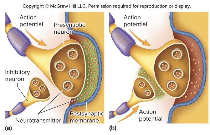
Axoaxonic synapses-
axon of neuron synapses on the presynaptic terminal (axon) of another
allows the release of neuromodulator to influence the action of another neuron
Neuromodulation
Presynaptic inhibition
Presynaptic facilitation
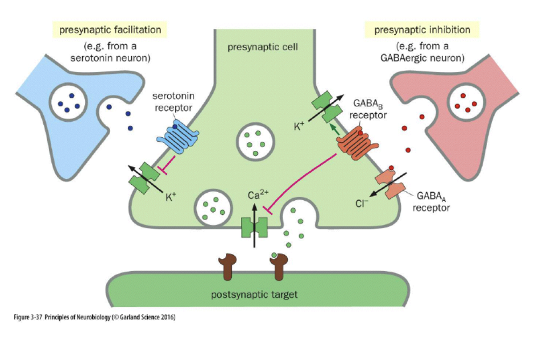
Presynaptic inhibition
Amount of neurotransmitter released from presynaptic terminal is reduced
Enkephalins and endorphins released by inhibitory axoaxonic synapses to reduce or eliminate pain sensation by blocking release of neurotransmitter from sensory neurons
Presynaptic facilitation
Amount of neurotransmitter released from presynaptic terminal is elevated
Serotonin released from axoaxonic synapses increases release of neurotransmitters
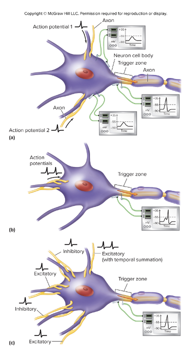
Summation of Graded Potential
Generation of an action potential is determined by the sum of all graded potentials generated by stimulation of the neuron
IPSP’s
EPSP’s
Spatial summation
multiple action potentials get at the same time from separate neurons
Temporal summation
two or more action potentials arrive very close together from the same neuron
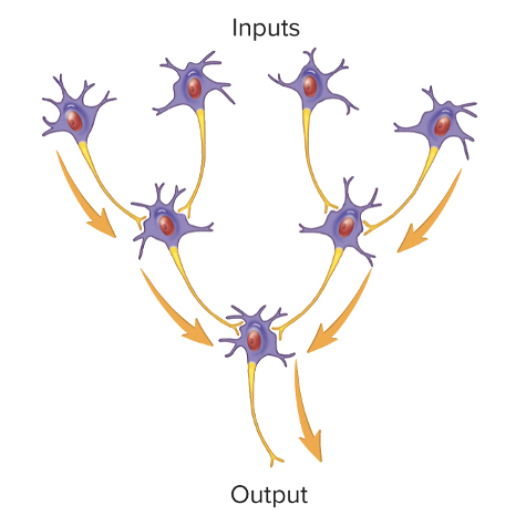
Neural Pathways and Circuits
Serial pathway
Parallel pathway
Serial pathway
simples organization
input travels along only one pathway
Parallel pathway
most pathways
more complex
input travels along several pathwyas
comes in different patterns
Patterns of parallel pathways
Convergent pathways
Divergent pathways
Reverberating circuits
Parallel after-discharge circuits
Convergent Pathways
Multiple neurons converge upon and synapse with smaller number of neurons
Allows different parts of the nervous system to activate or inhibit the activity of neurons
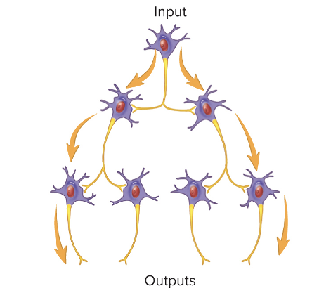
Divergent Pathways
Smaller number of presynaptic neurons synapse with a larger number of postsynaptic neurons
Allows information transmitted in one neuronal pathway to diverge into two or more pathways
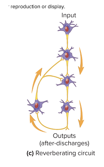
Reverberating Circuits
Chain of neurons with synapses with previous neurons in the chain
Makes positive-feedback loop
Lets action potentials entering the circuit to cause a neuron farther along in the circuit to produce an action potential more than once (after-discharge) to prolong response to stimulus
Circuit will continue to discharge until the synapses are fatigued or inhibited by other neurons
Control rhythmic activities
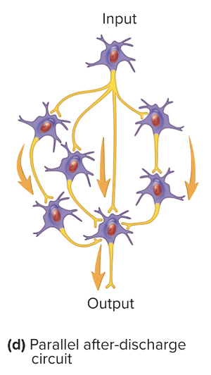
Parallel After-Discharge Circuits
Neurons that stimulate neurons in parallel organization
All converge upon a common output cell
Involved in complex neuronal processes