Male Carnivore INTERNAL Genitalia (Mod. 5)
1/18
There's no tags or description
Looks like no tags are added yet.
Name | Mastery | Learn | Test | Matching | Spaced |
|---|
No study sessions yet.
19 Terms
Describe the “outer capsule” of the testes… what is it also known as?
Tunica albuginea
Predominantly connective tissue
Large vessels can be seen on its surface… pattern of vessels is characteristic between species
Describe the parenchyma of the testes… what is it? What is it made of? What are other notable features? (think of the central groove…)
The parenchyma is the tissue of the testes
Made primarily of interstitium; comprised of Leydig cells and has a connective tissue/vascular framework
Supports seminiferous tubules
IS LOBED… lobules formed by invaginations of capsule
Notable features:
Central groove running through testicle, called the Mediastinum Testis
This contains the Rete Testis, a space where the seminiferous tubules drain into
Now knowing what you do about the parenchyma and the structures it holds, describe the path of sperm…
Sperm produced in seminiferous tubules → drains into mediastinum testis → drains to rete testes → small efferent ductules carry sperm to head of epididymis
Describe the epididymis… what is its function? What are the three parts it is composed of, and what are their functions?
A firm, palpable ridge that lies dorsally to the testes
Function: Allows sperm storage and maturation
Travels from head → body → tail → ductus deferens
Head:
Firmly attached to testes… receives the efferent ductules, which COMBINE to form the convoluted epididymal duct
Body:
Loosely attached to the surface of the testes… creates a space between it and testicle called the testicular bursa
Tail:
Firmly attached to testes as well… epididymal duct exits here to form the ductus deferens / vas deferens
What are the 3 main testicular attachments / ligaments? Describe them. What is their significance clinically?
1) Proper ligament of testis - Attaches TAIL of epididymis to testes
2) Ligament of tail of epididymis - attaches TAIL to PARIETAL VAGINAL TUNIC
3) Scrotal ligament - attaches vaginal tunic to scrotum
When castrating, will need to break these down, EXCEPT FOR PROPER LIGAMENT!!!!! Can be left alone
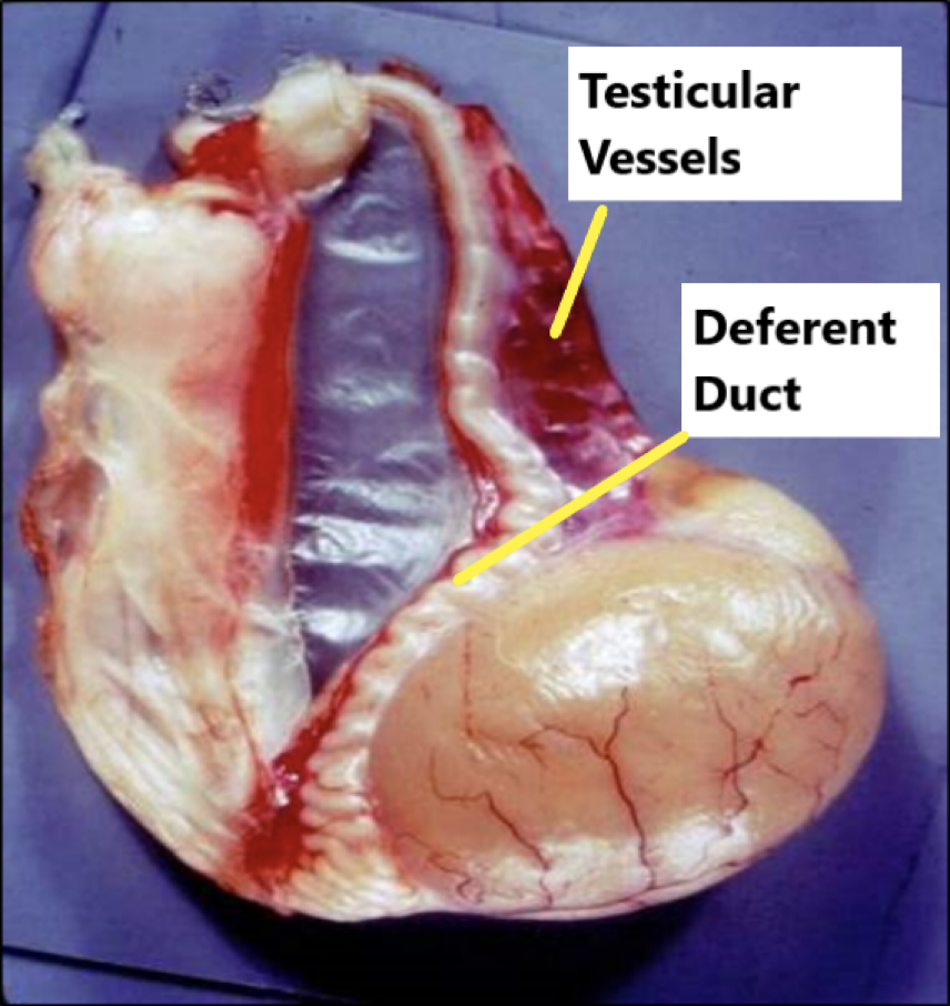
What 5 structures are found within the spermatic cord?
1) Deferent duct (vas deferens)
2) Deferent Artery + Vein - Small blood vessels; supply vas deferens
3) Large Testicular Artery + Vein
4) Lymphatic structures
5) Testicular nerves - predominantly SYMPATHETIC
What structure is ALSO associated with the spermatic cord, but is OUTSIDE of it?
Cremaster muscle
Sits outside vaginal tunic
Describe the deferent duct (ductus deferens, vas deferens, same thing); what is its function? Where does it lead into?
Function: Carries sperm to urethra
Travels through spermatic cord
Runs through inguinal ring, then crosses over the dorsal neck of bladder
Enters the PROSTATE and joins with the urethra
What is the pampiniform plexus of the testicles? What is its function?
An elaborate complex of veins that is arranged right at the base of the spermatic cord
Wraps around the distal convoluted testicular artery
Function: heat exchange mechanism + venous drainage
Arterial blood coming down to the testes is cooled as it passes through the pampiniform plexus, which is draining slightly cooler blood from the testicles
This helps reduce the temperature of the blood entering the testicles, keeping the sperm safe
At the same time, the arterial blood warms the cooler blood being drained as it goes up the spermatic cord and back into the body
What is the cremaster muscle, and what is its function?
Is a strip of muscle originating from the internal abdominal oblique
Caudal to dep inguinal ring (so further into the body)
Runs along vaginal tunic; inserts onto tunic as well
Function: when contracted, brings testis closer to body to increase testicular heat
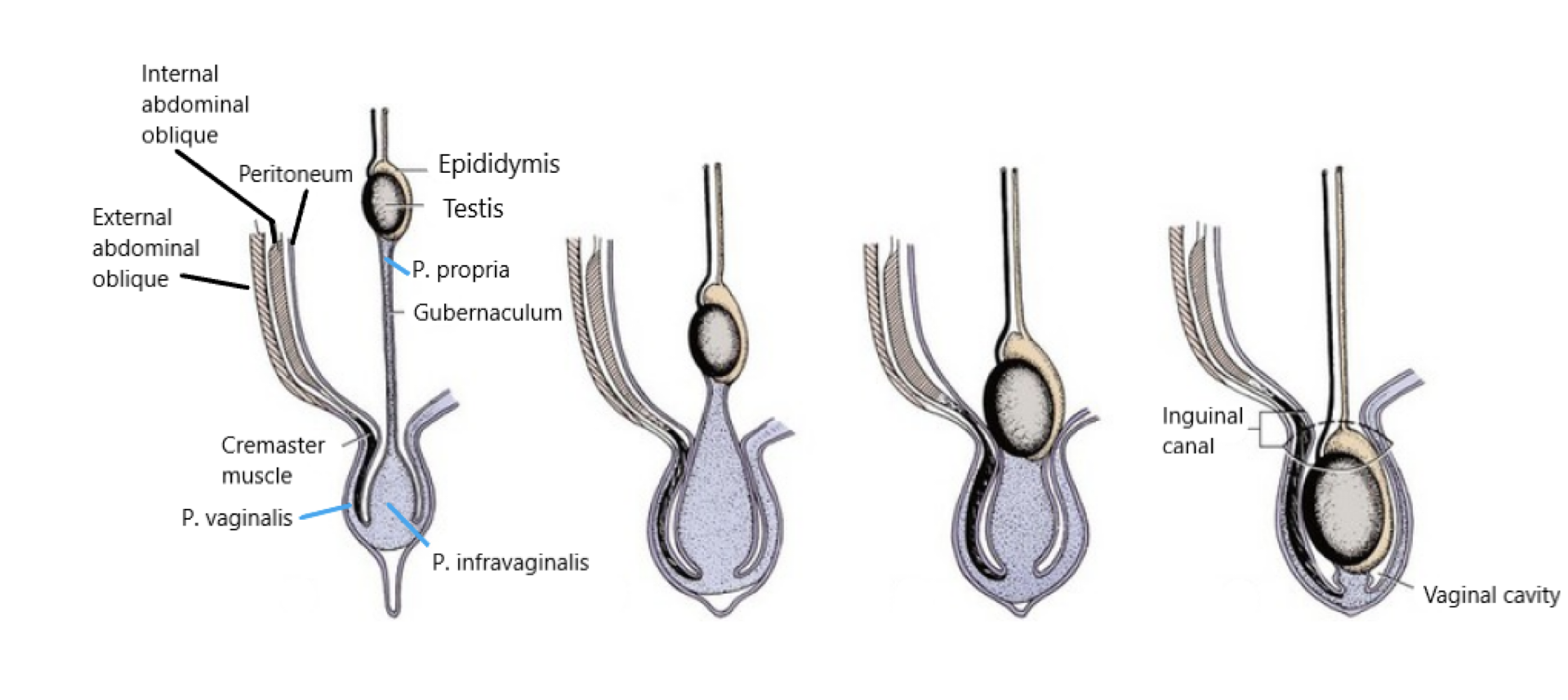
What is the structure that REGULATES TESTICULAR DESCENT?? (hint: funny name)
Describe it and its structure, including its 3 main layers.
Gubernaculum testis
A mesenchymal structure that extends from abdominal testes in immature males… runs through inguinal canal and opens into scrotum
Forms the vaginal tunic when it makes balls drop
Gubernaculum swells and shortens, which opens the inguinal canal wide enough to draw testes down
Three main layers:
1) Pars propria - visceral layer; extends up to catch the testes
2) Pars vaginalis - parietal layer; lines the INSIDE of the SCROTUM
3) Pars infravaginalis - distal to invagination, but is the junction between the other two layers (see image)
What are the two main INTERNAL reproductive organs?
1) Urethra
2) Accessory Reproductive Glands
Ampullary glands (NOT IN CATS)
Prostate
Bulbourethral glands (NOT IN DOGS)
Describe the male urethra… what are its two “regions” its divided into?
Urethra runs from bladder to tip of penis
Its two regions:
1) Pelvic urethra - portion that runs through pelvic canal
2) Penile urethra - runs through penis
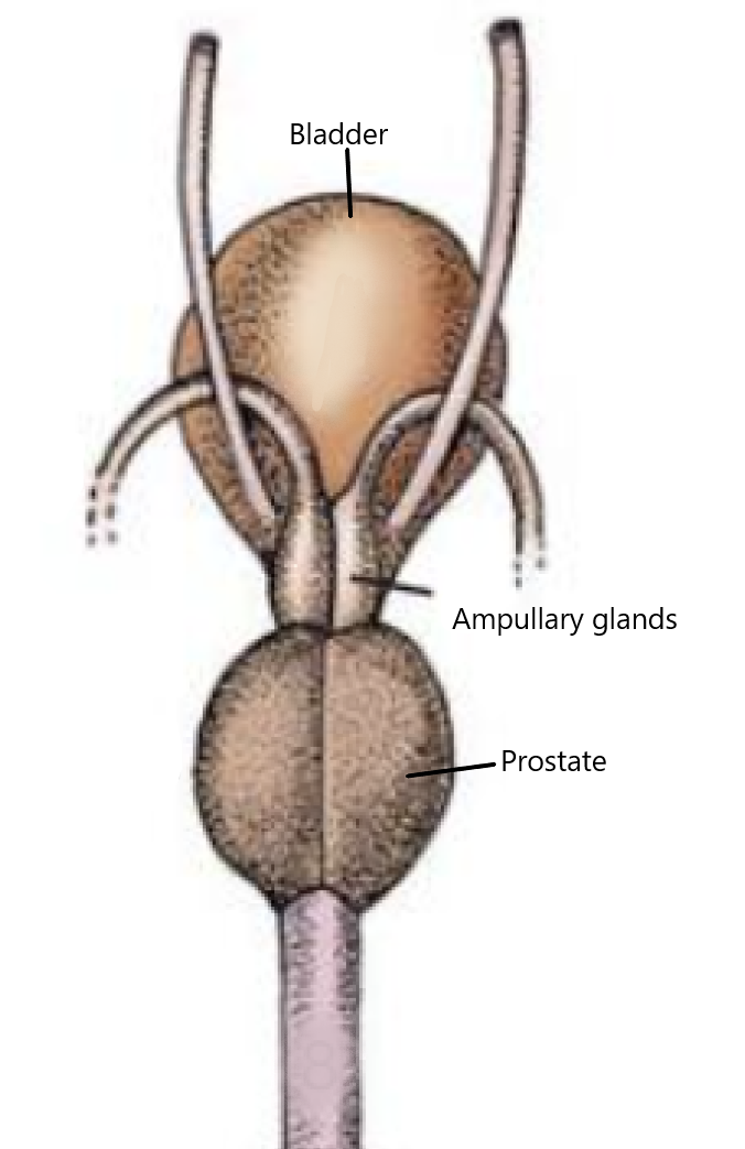
Describe the pelvic urethra… where does it insert? What is the “dorsal ridge”? Where is it oriented in the body?
Starts at the bladder, but is contained in the prostate initially
Has a “dorsal ridge” called the seminal colliculus
Projects into the lumen
This is where the VAS DEFERENS and PROSTATIC DUCTS open up into to release sperm + semen
Lies along the pelvic floor (see image) with a muscular sleeve called the urethralis
Describe the penile urethra (very simple)… where does it run through?
Runs through penis between cavernous tissues
THEN runs through ventral groove of OS Penis
What are the ampullary glands?
Dilations of the terminal vas deferens before entering the prostate (see image)
Lies dorsal to the bladder
Function: Lined with glandular tissue → contributes to a small amount of ejaculate
Has a capsule and a layer of septa w/ smooth muscle… assists in the expulsion of fluid (small structure, so not a lot of force)
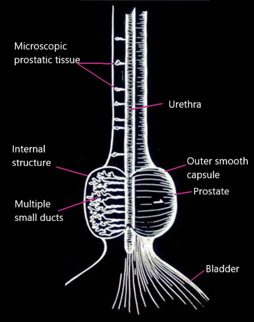
Describe the prostate… what is its function? Why is it so important? What are its two main components?
Function: Produces the bulk of seminal fluid
Has two parts:
1) Vestigial disseminated part WITHIN URETHRAL MUSCOSA
So tissue within the wall of the urethra; “microscopic prostatic tissue”
2) The large compact part around the proximal urethra
Is bi-lobed (see image)
Describe the structure of the larger portion of the prostate gland:
Divided into left and right lobes by a dorsal groove and internal septum
Is located at the neck of the bladder; surrounds the proximal urethra
In dogs, urethra completely surrounded
In cats, ventral area of urethra free
INTERNALLY, there are multiple small ducts that drain into the urethra by the seminal colliculus (dorsal groove on pelvic urethra)
What is the most common cause of prostatic enlargement in dogs? What are some other possible causes?
Most common cause: Benign Prostatic Hyperplasia
Hormonally induced
Seen in almost every dog that is still intact around 5-6 years old
May compress the rectum or urethra
Causing constipation
Other causes:
Neoplasia (cancer)
Inflammation (prostatitis)
Cystic disease
***** RARELY SEEN IN CATS