PIE- How to take a radiograph of a dogs shoulder
1/26
There's no tags or description
Looks like no tags are added yet.
Name | Mastery | Learn | Test | Matching | Spaced | Call with Kai |
|---|
No analytics yet
Send a link to your students to track their progress
27 Terms
What two views are used to take a radiograph of a dogs shoulder?
-Lateral view
Technically this is a medio-lateral view
-Caudo-cranial view
dog will need to be anaesthetised for caudo-cranial view
where does the xray beam enter and exit in lateral view
x ray beam will enter the through the
medial aspect of the shoulder joint and exit through the
lateral aspect to to the table
where does the xray beam enter and exit in caudo cranial view
the x ray beam enters through the
shoulder joint, it enters effectively from Caudally and exits cranial.
Describe the limbs and neck in lateral view
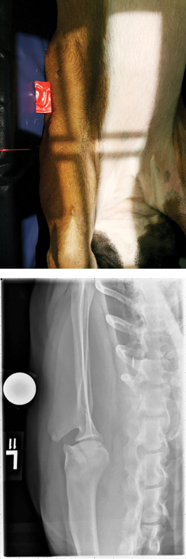
The dog is in lateral recumbency with the leg to be imaged against the table
The limb to be radiographed is pulled into extension using a rope tie
this also pulls the shoulder joint away from the neck soft tissues
The upper limb is moved caudally out of the way with a rope tie
The neck is flexed dorsally and secured using a sandbag
this moves the cervical spine so it does not superimpose over the shoulder joint

What are the steps to taking a lateral view to radiograph the shoulder?
Place a foam pad under the chest to ensure the dog is parallel to the table and assist shoulder extension
The exposed area should include the distal 50% of the scapula and proximal 50% of the humerus
Use L / R label
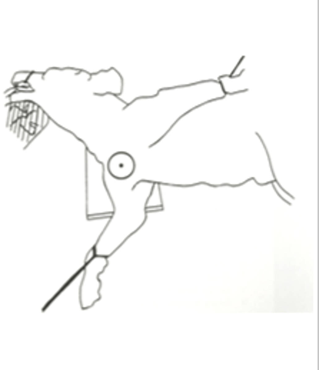
how can we detect the acromion
just by sliding our fingers underneath the dog and palpating.
where centre for lateral view
The centre of collimation should be the shoulder joint (just distal to the acromion)
where collimate for lateral view
Collimate to include soft tissues cranial and caudal to shoulder
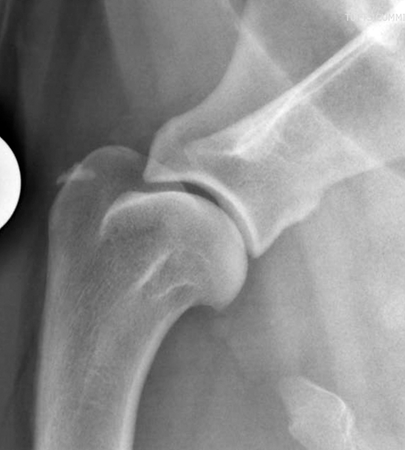
What should be seen in a well positioned lateral view radiograph of the shoulder?
In a well positioned radiograph the joint space should be well visualised and there should be no overlying tissue
Some radiographers deliberately try to overlie the shoulder joint with the trachea to increase contrast
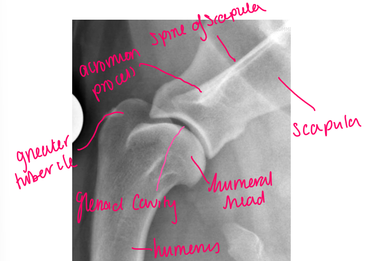
label
acromion process
spine of scapula
scapula
humeral head
humerus
glenoid cavity
greater tubercle
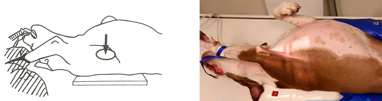
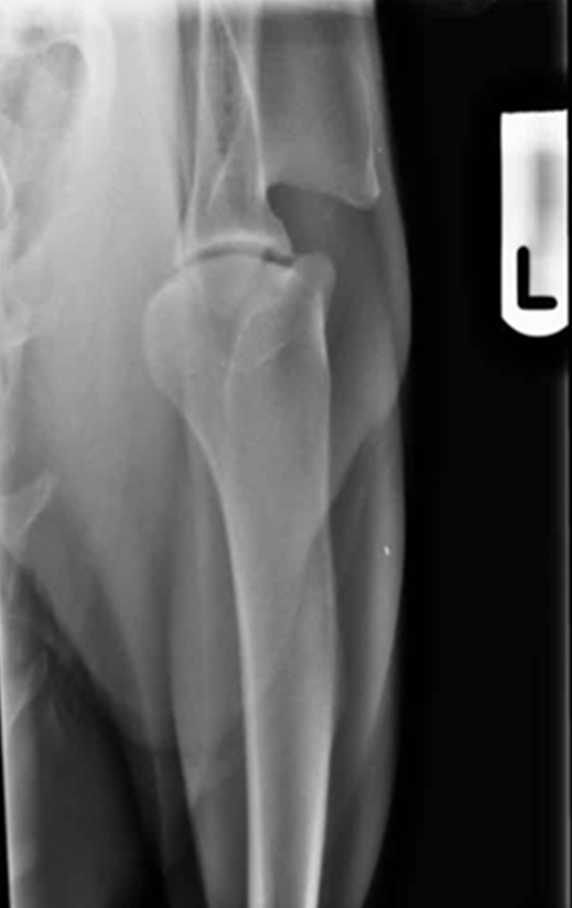
Describe the position of dog in caudo-cranial view for shoulder radiograph
The dog is put in dorsal recumbency with the limb to be radiographed pulled into extension (X-rays enter caudal aspect and exits cranial aspect of the shoulder)
The limb is pulled slightly away from the midline (approx. 5 degrees)
A sandbag may be use to push the head and neck slightly away from the limb to reduce superimposition
Use L/R label
how should the long axis of scapula and humerus be in the caudo cranial view
Aim to hold the long axis of scapula and humerus in line with each other
see the joint space nice and prominently demonstrating the radiograph is suitably positioned.
how centre caudo cranial
Centre at the middle of the shoulder joint (just distal to the acromion)
how collimate in caudo cranial view
Collimate to include the distal 50% of the scapula and proximal 50% of the humerus
Collimate to include the lateral and medial soft tissues
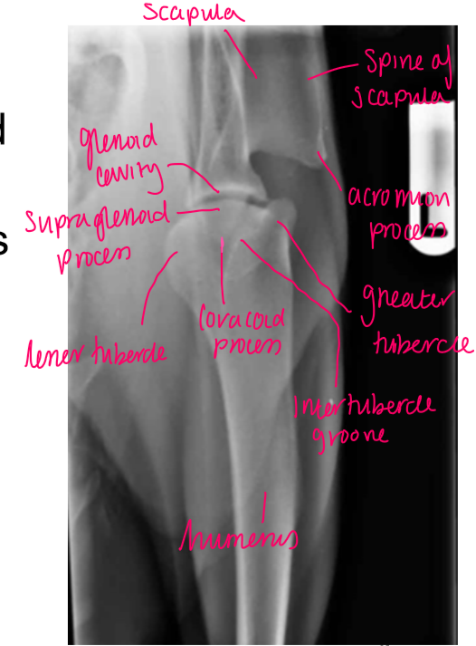
label
scapula
spine of scapula
acromion process
greater tubercle
intertubercle groove
coracoid process
lesser tubercle
supraglenoid process
glenoid cavity
humerus
What should be seen in a well positioned caudo-cranial view of a shoulder radiograph?
§A well positioned radiograph has the scapula and humerus in line with each other
§Centering on the middle of the joint means there is good visualisation of the joint space
Problems that can occur with a cranio-caudal (not caudo-cranial) view of a shoulder...
The cranio-caudal view can result in magnification distortion as it is difficult to get the shoulder close to the table
This technique would be common in the standing horse but not in dogs
what common pathology of the shoulder is there
medial shoulder luxation
osteochondrosis
chronic shoulder arthrosis
medial shoulder luxation
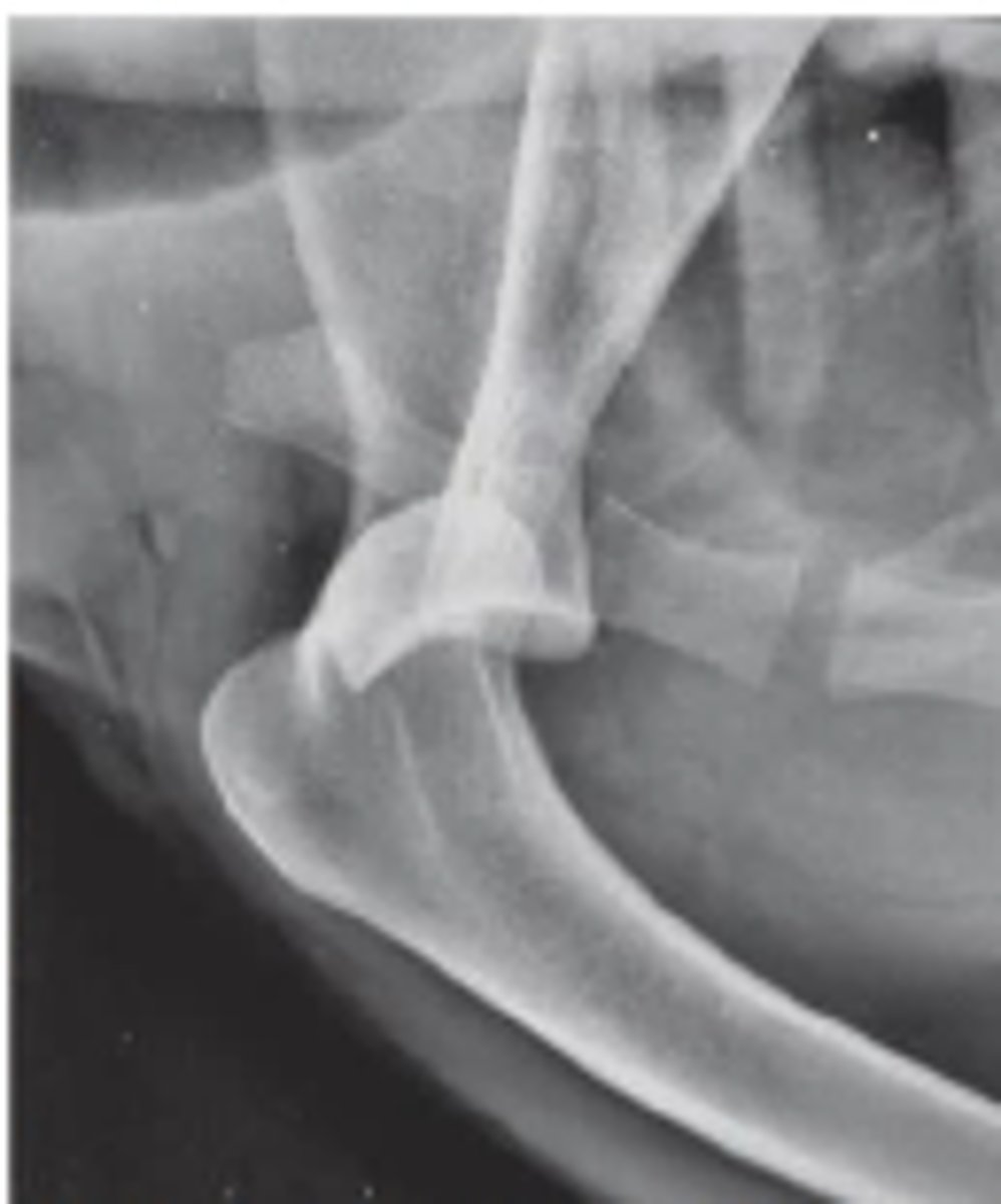
Describe a medial shoulder luxation due to trauma
unusual position of humeral head compared to glenoid cavity of the scapula
-Humeral head has been displaced medially
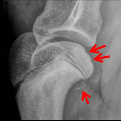
Describe osteochrondrosis
-Cartilage defect associated with an underlying bone problem
-Causes flattened appearance of humeral head
-Multiple disc like depressions of humeral head
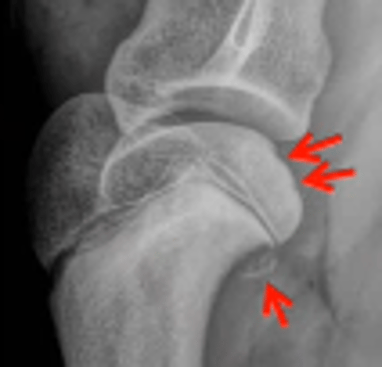
Osteochondrosis
-Red arrows show disc like depressions
What will osteochondrosis (developmental) present as?
Loss of rounded contour of caudal surface of humeral head
Radiolucent defect
May have surrounding sclerosis
May have calcified free body in joint
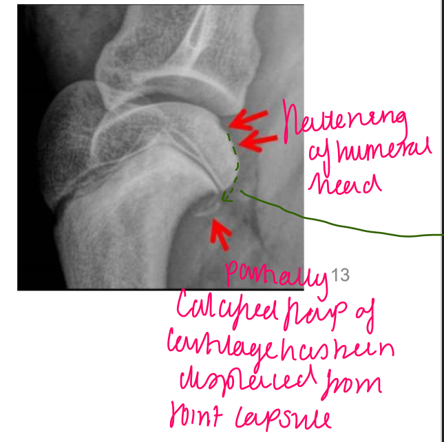
label
flattened/ indent of the humeral head
partially calcified flap of cartilage → displaced from joint capsule
What will be seen in chronic shoulder arthrosis?
Lots of new bone (osteophytes)
Underlying cause often not evident
label the normal
sclerosis and osteophytes
sclerosis, uneven joint space, osteophytes (New bone)
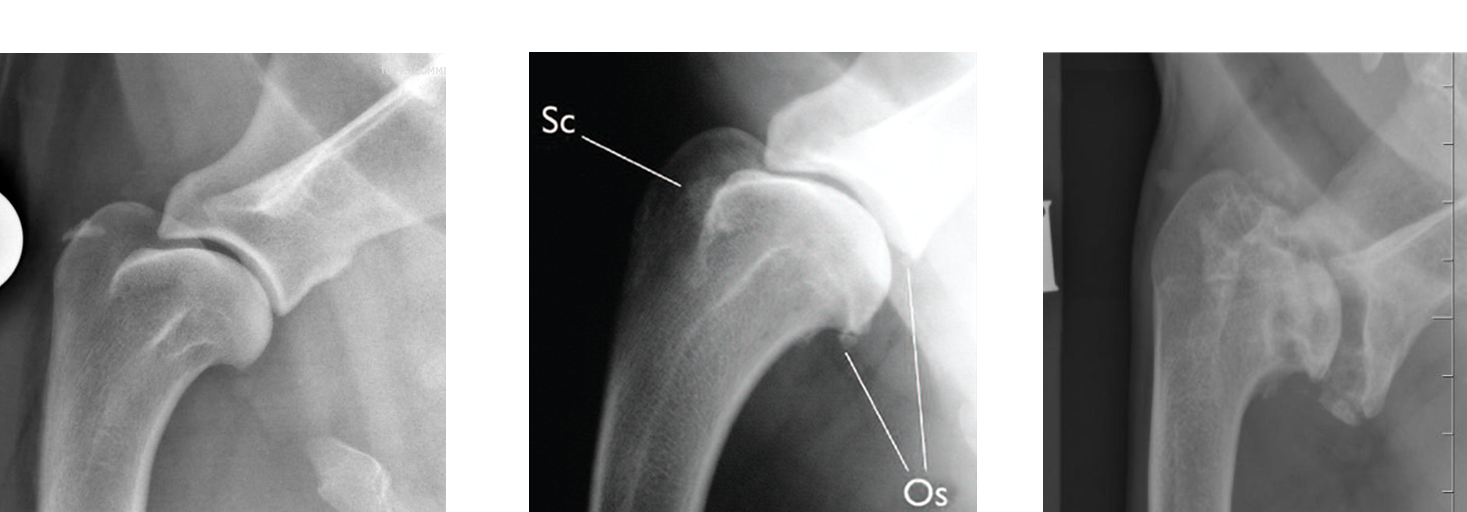
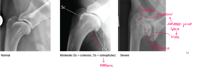
WHAT do we expect to see at normal joint
nicely rounded and smooth margin to the
head of the humerus and look at the margins of
the distal part of the scapula spine and the joint
space here.
So nice and smooth and nice.