Currents by Name: Variations of the basic currents
1/33
There's no tags or description
Looks like no tags are added yet.
Name | Mastery | Learn | Test | Matching | Spaced | Call with Kai |
|---|
No analytics yet
Send a link to your students to track their progress
34 Terms
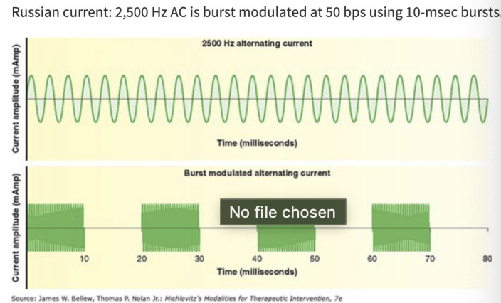
Russian Current
variation of AC
activates skeletal muscle
burst* modulation - defining characteristic
2,500 Hz (carrier frequency) alternating sinusoidal current that is interrupted & delivered in short bursts
50 bps using 10-msec bursts
10/50/10
actual duty cycle = 16.7%
relative duty cycle = 50%
current is flowing half of the time
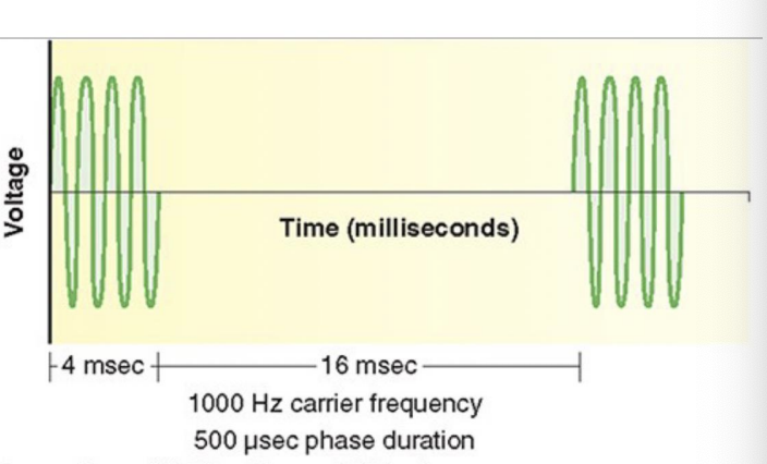
Aussie Current/Burst Modulated AC
created from 1,000 Hz burst modulated AC
delivered in 4-msec bursts
greater torque production, efficiency, and decreased rate of muscle fatigue
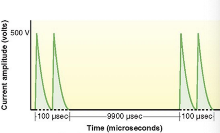
High Volt Pulsed Current
twin-peaked monophasic pulsed current waveform
peak voltage ~150-500 V
Pulse duration short ~50 to 100 usec
Frequency of 1-100 Hz*
low relative duty cycle <1%
low average treatment voltage
indications: wound healing*, pain
High Volt vs. Low Volt Pulsed Current
refers to the magnitude of the voltage used to drive the current
High volt — voltage: 150-500 V
Low volt — voltage: <150 V
Low intensity DC/Microcurrent
microcurrent
DC or monophasic pulsed current
pulse duration is .5sec — longer than other pulsed currents
frequency 1 Hz (up to 1,000 Hz)
facilitates tissue healing
Microcurrent
any current with an amplitude <1 mA
Symmetrical & Asymmetrical Biphasic Pulsed Currents
represents a group of waveforms widely used for muscle stimulation and pain modulation
square, rectangular, triangular
vary in duration & amplitude
little data for use of asymmetrical over symmetrical
Electrical Stimulation Indications
strengthening of skeletal muscle
re-education of skeletal muscle
stimulate denervated skeletal muscle
pain modulation
preventing & reducing edema
increasing blood flow
tissue healing
delivery of medication
Indications (Scorebuilders)
Bell’s Palsy
decreased ROM
facial neuropathy
fracture
Idiopathic scoliosis
joint effusion
labor and delivery
muscle atrophy
muscle spasm
muscle weakness
open wound/ulcer
pain
stress incontinence
should subluxation
Contraindications
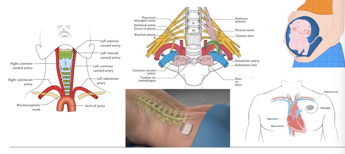
Electrodes should NOT be placed over:
the trunk or heart in patients with pacemakers or defibrillators
the pelvic, abdominal lumbar, or hip region in pregnant individuals
carotid bodies (between STM and trachea)
over urinary bladder stimulators
areas of known peripheral vascular disease
the phrenic nerve, eyes or gonads
areas of active osteomyelitis
areas of hemorrhage
Anatomical Landmarks
Contraindications (scorebuilders)
cardiac arrhythmia
cardiac pacemaker
malignancy
osteomyelitis
over a pregnant uterus
over carotid sinus
patient with a bladder stimulator
phlebitis
seizure disorder
Precautions
Electrotherapy should be used with caution in patients:
without intact sensation
who are unable to communicate
with compromised mental ability or lack of cognition
with cardiac dysfunction
with epilepsy or seizure disorders
over active or previous neoplasms
over compromised skin
over tissues vulnerable to hemorrhage or hematoma
in the cervical or craniofacial region in patients with hx of CVA or seizures
Do not use ES device within 5 yards of diathermy units or other sources of electromagnetic radiation
Therapeutic Effects (scorebuilders)
decreased edema
decreased pain
eliminate disuse atrophy
facilitate bone repair
facilitate wound healing
improved ROM
increased local circulation
muscle re-education
muscle strengthening
relaxation of muscle spasm
Knowledge Check 1: Decreased sensation
Which of the following is not an absolute contraindication to use of electrical stimulation?
A. osteomyelitis
b. over the carotid sinus
c. over the eyes
d. decreased sensation
Dials, Buttons, & Touchscreens
make sure estim unit has working batteries
connect electrodes to the lead wires and connect wires to unit
place electrodes on the skin
pull the lid cover down to access controls
to turn on: turn the amplitude knob of at least one channel
mode: synchronous, constant, alternate
set: rate, width (Hz) (microseconds), ramp (s), on time (s), off time (s), time (min)
up/down arrows
turn the channel knob to adjust intensity
Electrodes
the device that relays current between the electrical stimulation device and the patient
two classes of electrodes:
surface/transcutaneous
invasive/indwelling
carbonized silicon rubber
variety of shapes and sizes
Current density, impedance, current flow
small electrodes
large electrodes
distance apart
Applying electrodes
evaluate skin surface
shaving recommended 1 day prior
consider use of water to improve adhesiveness
full contact with skin, avoiding bumps in pad or pads touching one another
if pads won’t stick, do not attach with tape or elastic
check plastic sheet, usually “ON”
Electrode Placement
base off what the treatment is trying to accomplish
ex: motor nerves = motor points (muscle belly or proximal 1/3)
interelectrode distance - distance between electrodes
wider placements = greater depth of penetration/more motor unit or sensory fiber activation
Electrode Size
Small Electrodes:
increased current density
increased impedance
decreased current flow
Large Electrodes:
decreased current density
decreased impedance
increased current flow
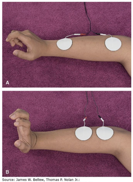
Electrode Configuration: Monopolar
active electrode positioned over the treatment area while another electrode is placed over nearby nontreatment area
active electrode vs. reference/inactive electrode
cathode is active and produces cell depolarization, produces net charge
Electrode Configuration: Bipolar
all the electrodes are placed over the treatment area
parallel or perpendicular to the fiber direction of the muscle
electrodes alternate between + and - with no net charge produced
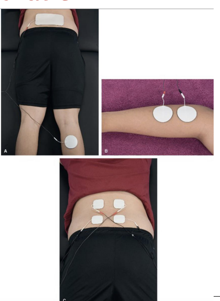
Electrode Configuration: Quadripolar
four electrodes from two separate circuits, placed so currents are intentionally crossed over target tissues
requires specific application of channels to achieve effect
Pain Modulation parameters
high frequency, low frequency, brief intense, hyperstimulation
Preventing & Reducing Edema (FYI)
acute (24-72 hours) or existing edema (Subacute or chronic)
Increasing Blood Flow (FYI)
vasospastic disorders and diminished arterial blood flow
Tissue healing (FYI)
epithelialization, autolysis, reaction of inflammatory process
promotion of granulation of wound
bacterial effect for infection
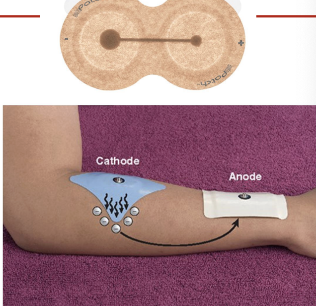
Iontophoresis
used to induce movement of ions across skin
delivers medicinal ion
typically uses DC current
risk for burns
soft tissue inflammatory conditions, neuralgia, edema, ischemic skin ulcers, hyperhidrosis, plantar warts, gouty arthritis, calciific tendonitis, scar tissue, Peyronie’s disease
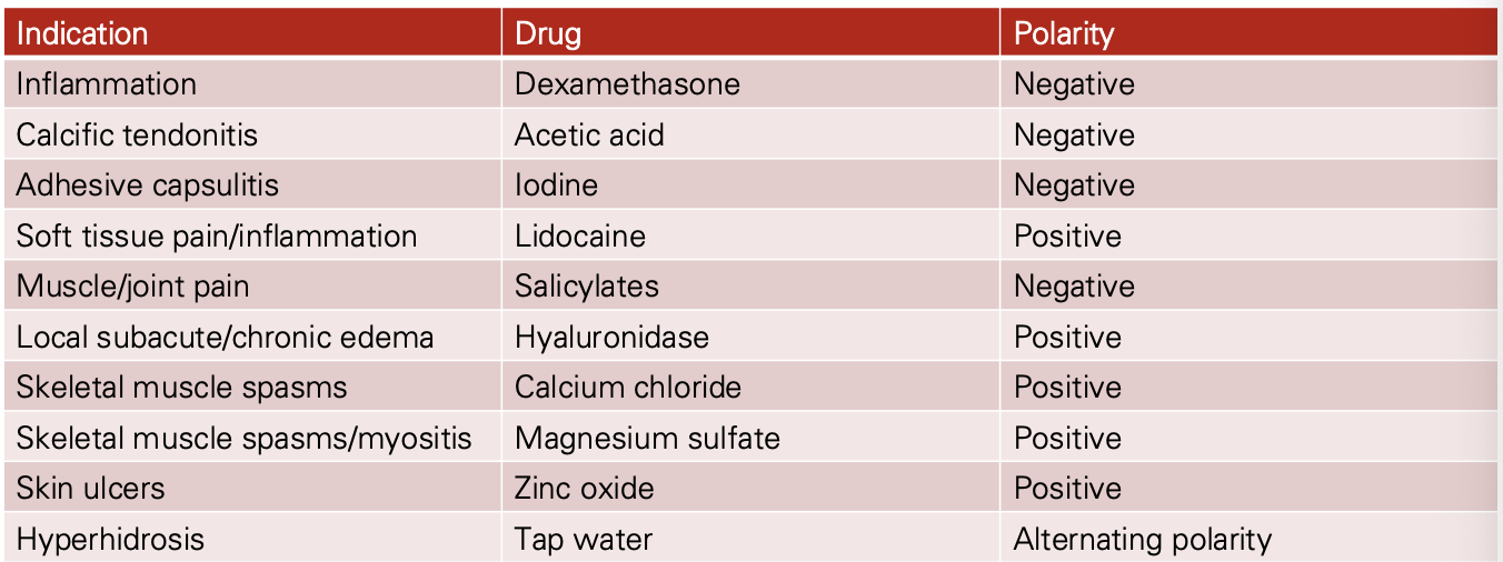
Iontophoresis Ions
Iontophoresis Dosage
Dosage (mA min) = Current (mA) x Duration (min)
Typical: 20-80 mA min
Amplitude: 0.5-4 mA
Ex: 4 mA at 10 minutes = 40 mA min
INCREASING time can INCREASE absorption
Documentation
screen for contraindications/precautions
skin integrity
modality type
waveform type
waveform parameters (pulse duration, frequency, amplitude, on/off time, ramp up/down, burst duration, beat frequency, sweep, scan, swing)
treatment duration
electrode size
electrode placement
patient position/body position
response to treatment, including skin integrity
First, DO NO HARM
effective use of estim is founded on two factors:
careful differentiation of patients who stand to benefit from the intervention from those who are not appropriate
careful, knowledgable, and safe application of the intervention
There are risk associated with most all treatment interventions
proper screening is critical
be aware of surroundings — inspect equipment and report problems
Key safety information
proper screening must be performed PRIOR TO use of estim
contraindications (required)
sensation is a precaution only — check if concerned
capillary refill/blanche is not required
Must inc. intensity SLOWLY
must provide patient with off switch/bell
must dec. intensity prior to shutting off
must inspect skin FOLLOWING application
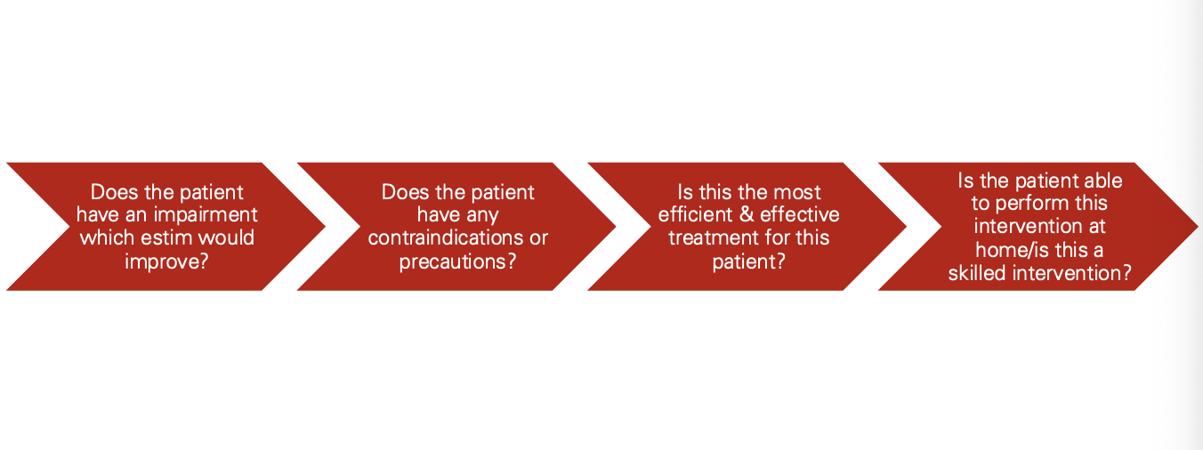
Clinical Decision Making