BIOL 1010 Exam 3
1/94
There's no tags or description
Looks like no tags are added yet.
Name | Mastery | Learn | Test | Matching | Spaced | Call with Kai |
|---|
No analytics yet
Send a link to your students to track their progress
95 Terms
When You are taking your blood pressure in your arm, you are measuring the force exerted on that blood by:
Contracting left ventricle
The pulmonary capillaries contain a high level of carbon dioxide. Where will that carbon dioxide go next?
The Alveoli
Maximum lung capacity in adult males:
+/- 5.7 Liters
Maximum lung capacity in adult females:
+/- 4.2 Liters
Vital Capacity
Amount of air that can move out of the lungs in one breath
Tidal Volume
Air flowing into and out of the lungs inn the respiratory cycle (+/- 0.5 liters)
Residual volume remains in lungs and does what?
Keeps the lungs inflated
Alvelous
Epithelial cells with membrane of outer surface
Pulmonary capillaries consist of what?
They consist of endothelial cells, basement membrane
Thin respiratory membrane =
Alveolar epithelium + Pulmonary endothelium + membranes
Oxygen flows into:
The pulmonary capillaries
Carbon Dioxide flows out into:
The alveoli
Extracelluar fluid is kept within tolerable ranges by the:
The Urinary System
Water and solutes are ADDED by:
Absorption, Metabolism, and Respiration
Water and solutes are LOST by:
Urinary excretion, evaporation from respiratory surfaces, sweat, and elimination in feces
Components of the Urinary System:
Kidneys, Urine, Ureter, Urinary bladder, Urethra
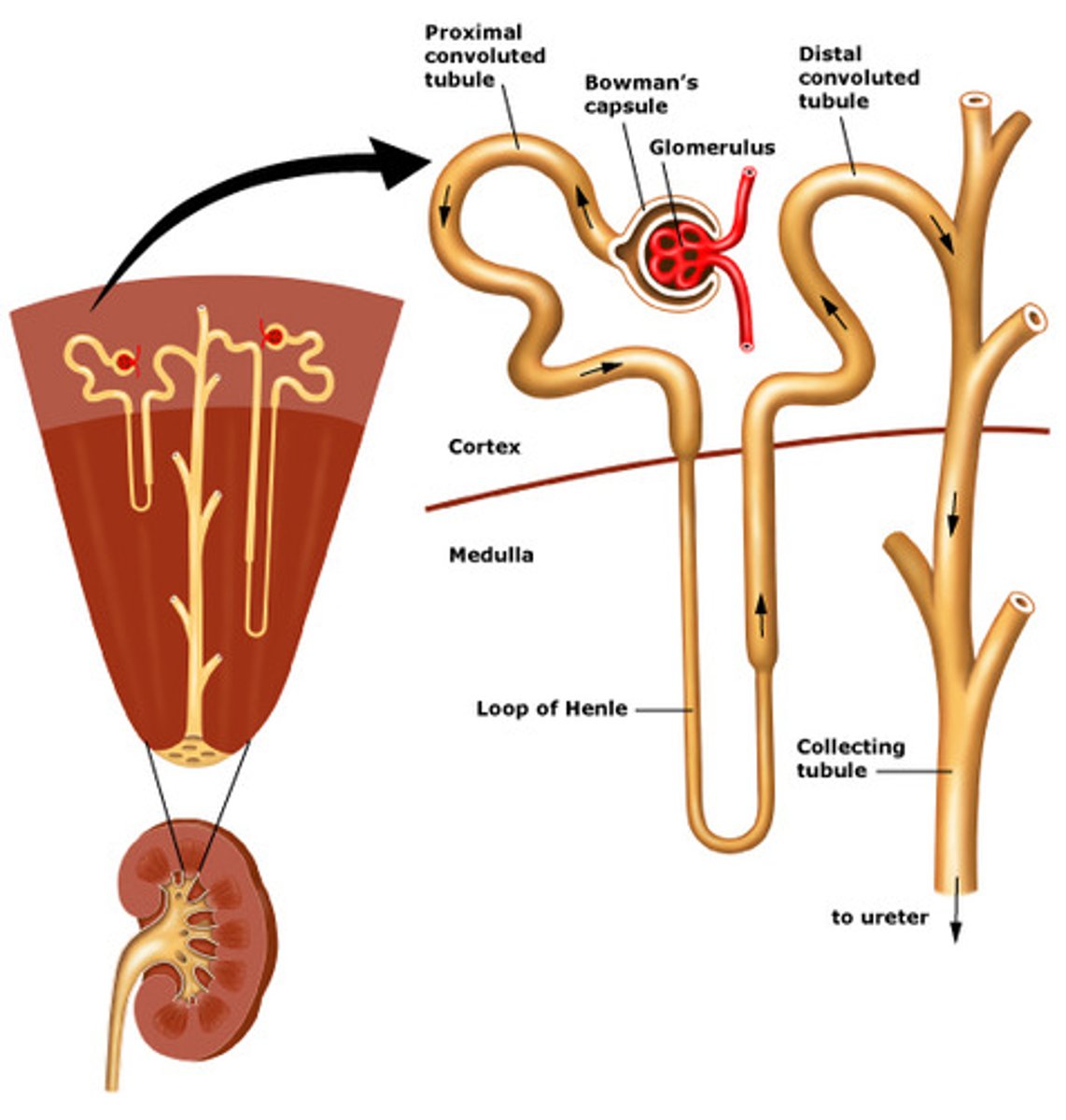
Kidneys
1. composed of a cortex (outer) and a medulla (Inner)
2. Filter water, mineral ions, organic wastes, and other substances from the blood
3. 99% returns to the blood; the 1% that doesn't is urine
Urine
Fluid that rids the body of water and solutes that are not needed
Urinary Bladder
Muscular sac that stores urine
Nephrons (basic function of the kidney)
1. Slender tubules that extend from the kidney cortex down through the medulla
2. Water and solutes are filtrated from blood (most will be reclaimed in the peritubular gap)
3. Each one starts at the Bowman's capsule. Inside the capsule is a blood filtering unit called the glomerulus
4. The filtrate leaves the Bowman's capsule and flows through the rest of it
Path of filtrate through the Nephron:
Proximal convoluted tubule - loop of Henle - distal convoluted tubule - collecting duct
Urine formed by three processes:
1. Filtration
2. Reabsorption
3. Secretion
Reabsorption
Solutes and water which move out of the nephron tubules are absorbed into the peritubular capillaries and return to general circulation
Secretion
Solutes from the peritubular capillaries are secreted into the nephron
Excretion
- Water and solutes that were not reabsorbed, or were secreted into the tubule, flow into the renal pelvis
- Eliminated from the body by the urinary tract
Path of filtrate through the Nephron (picture)
Filtration
Pressure filters blood by forcing water and solutes out of the glomerular capillaries
Sweat Glands (for excretion)
Release water and some solutes (urea)
Large Intestine (for excretion)
Remove salts and heavy metals (lead) from blood
Lungs (for excretion)
Remove carbon dioxide, water, and alcohol from the blood - exhaled
Adrenal Cortex
Secretes cortisol - raises blood glucose levels
Thyroid Gland
1. Located at the base of the trachea
2. Secretes thryoxine - regulates metabolism
3. Produces calcitonin which promotes the deposition of calcium into the bone
Hypothydroism
Low levels of blood in the thyroid can cause it; gain weight easily, sluggish, dry skinned, confused, depressed
Hyperthyroidism
If blood levels in the thyroid are too high; causes increased heart rate, elevated blood pressure, profuse sweating, and weight loss
Parathyroid Gland
1. 4 glands located on the posterior side of the thyroid
2. Parathyroid hormone (PTH)
- Raises blood calcium levels (goes from bone to blood)
Glucagon
Raises blood glucose levels
Insulin
Causes glucose uptake by the muscle and adipose cells from the blood (lowers blood glucose level)
Diabetes Melitus
Insulin deficiency which causes glucose to accumulate in the blood, then in urine
1. Urination becomes excessive and the body becomes dehydrated
2. Without glucose body cells start depleting their own fats and proteins as sources of energy
Type 1 Diabetes
Juvenile onset diabetes; lymphocytes destroy insulin secreting cells; insulin shots required
Type 2 Diabetes
Cells produce less insulin; usually emerges during middle ages; can often be controlled with diet and taking prescription drugs
Testosterone responsible for:
1. Development of secondary sex characteristics
2. Promotes development of sperm
3. Responsible for sex drive in males
Ovaries
Produce estrogen and progesterone
Estrogen
Produces secondary sex characteristics and maintains pregnancy
Progesterone
Maintains the uterine lining for pregnancy
Thymus
Located superior to the heart
1. Large during childhood, but absent in adults
2. Thymosine hormones - causes the maturation of lymphocytes
Pineal Gland
Produces melatonin
Melatonin:
1. Delays the onset of sexual maturity
2. Regulates biorhythms such as day and night activity cycles
Testes
Sperm production
1. Scrotum
2. 95 degrees
3. seminiferous tubules - coiled tubes
- Sperm production
Epididymis
Duct that sperm enters after leaving that testes - becomes mobile
Vas Deferens
Tube, carries sperm from epididymis to urethra
Urethra
Tube, interior of the penis
Semen
Formed when glandular secretions mix with sperm
Seminal Vesicle
Secrete fructose into semen, sperm use as an energy source
Prostate Gland
Secretions buffer the pH of the female reproductive tract
Bulbourethral Glands
2 glands, secretions thought to buffer pH of female reproductive tract
Cushing Syndrome
A condition that occurs from exposure to high cortisol levels for a long time (Adrenal Cortex)
Hormones
Products secreted into the bloodstream by endocrine glands; regulate processes such as growth, reproduction, and maintenance
Hypothalamus
In forebrain, synthesizes two hormones which are conveyed to the to the pituitary gland
Pituitary Gland (Master Gland)
At the base of the hypothalamus - anterior lobe, posterior lobe
Posterior Lobe
Secretes two hormones synthesized in the hypothalamus
Antidiuretic Hormone
Responsible for reabsorption of water from the nephron
Diabetes Insipidus
Excessive urination due to the lack of the antidiuretic hormone
Hormones produced by the Pituitary gland
Antidiuretic hormone, oxytocin, FSH, LH, TSH, ACTH, prolactin, somatropin
Hormones produced by the Posterior lobe (Pituitary gland)
Antidiuretic hormone, oxytocin
Hormones produced by the Anterior lobe (Pituitary gland)
FSH, LH, TSH, ACTH, prolactin, somatropin
Oxytocin
Females - responsible for labor contractions and milk letdown
FSH (Follicle stimulating hormone)
Females: causes follicle to mature
Male: Stimulates testes to produce sperm; starts spermatogenesis
LH (Luteinizing hormone)
Female: Ovulation, formation of corpus luteum (formed from remnants of the rupture on ovary)
Male: Promotes cells to secrete testosterone
TSH (Thyroid Stimulating hormone)
Stimulates the thyroid gland to produce hormones
ACTH (Adrenocorticotropin hormone)
Stimulates corext of adrenal glands to produce cortisol
Prolactin
Responsible for milk production in females
Somatropin
Growth hormone
Acromegaly
Overproduction of somatropin; age quickly
Hormones produced by the pineal gland
Melatonin
Hormones produced by the thyroid gland
Calcitonin
Calcitonin
Promotes the deposition of calcium into the bone
Hormones produced by the Parathyroid Gland
Parathyroid hormone (PTH)
Parathyroid Hormone (PTH) definition
Raises blood calcium levels (goes from bone to bone)
Adrenal Glands layers
Adrenal cortex and adrenal medulla
Hormones produced by the Adrenal cortex
Secretes cortisol
Hormones produces by the Adrenal Medulla
Epinephrine and norepinephrine
Hormones produced by the pancreas
Glucagon, Insulin
Hormone secreted by testes
Testosterone
Hormones secreted by ovaries
Estrogen and Progesterone
Spermatogenesis (picture)
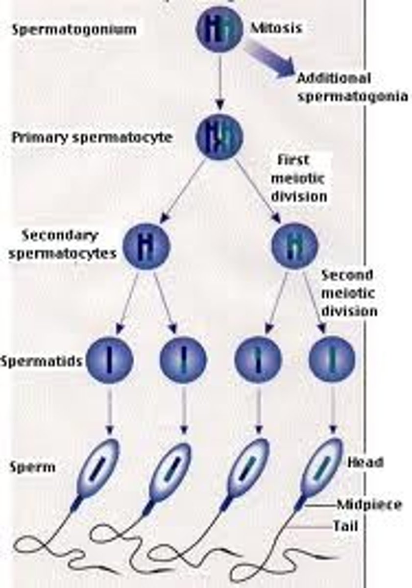
Spermatogonia
Cells in testes, undergo mitosis and produce primary spermatocytes
Hormones in Spermatogenesis:
LH, FSH, and testosterone
Ovaries produce:
Oocytes (immature eggs)
Oviducts (2)
Channel from the ovary to the uterus
Uterus
Hollow organ in which the embryo can grow and develop
Endometrium
Inner lining of the uterus wall - where the embryo implants
Cervix
Narrow portion of the uterus above the vagina
Vagina
Muscular tube - extends from the cervix to the surface of the body. Receives sperm and is part of the birth canal
Oogenesis (picture, one with two)
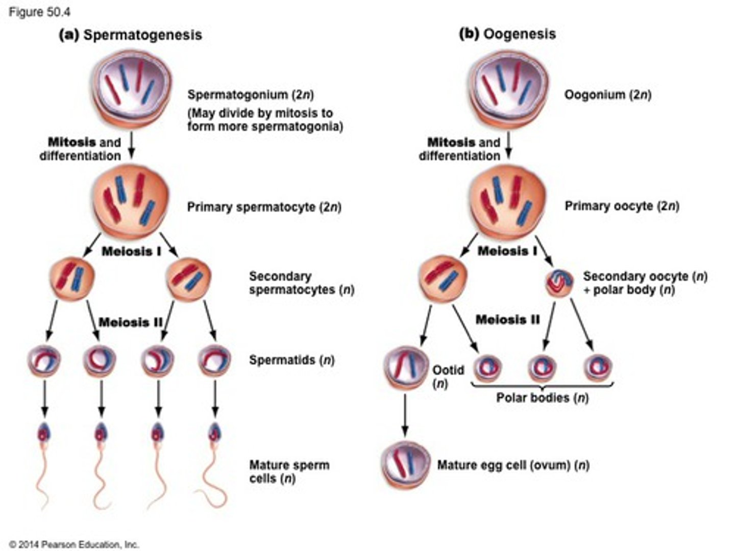
Follicle
The primary oocyte and the cell layer around it