PATH FINAL EXAM (everything)
1/174
There's no tags or description
Looks like no tags are added yet.
Name | Mastery | Learn | Test | Matching | Spaced |
|---|
No study sessions yet.
175 Terms
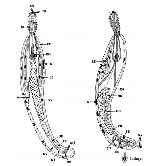
*PHYLUM ACANTHOCEPHALA
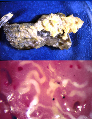
Macracanthorhynchus hirudinaceus
Polymorphus spp.
Piscine Acanthocephalans
-
MORPHOLOGY:
elongate, no digestive tract
praesoma - proboscis and lemnisci
metastoma (trunk) - sexual organs
armed proboscis with nerves that reach down main body
lemnisci - sensory nerve bundles from proboscis
giant nuclei with cuticle
MALE
central testes and cement gland
copulatory bursa
FEMALE
ventral ligament sac - eggs
uterine bell - sorts eggs in ventral ligament sac and ejects them into uterus
-
LIFE CYCLE:
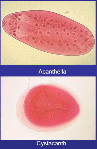
eggs with partially developed acanthors in feces > IH > hatch and penetrate gut wall, lodges > develops proboscis and loses hooks > acanthella
mature nonfunctional gonads > cystacanth > DH > sexual maturity
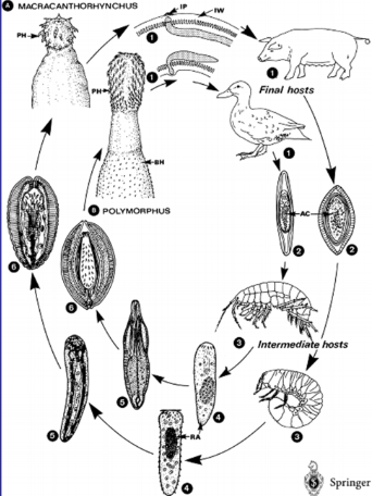
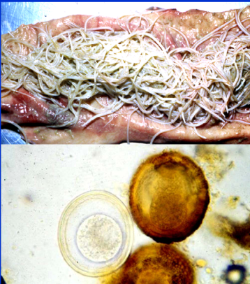
*Macracanthorhynchus hirudinaceus
PHYLUM ACANTHOCEPHALA
HOST: Swine, humans rarely (DH); beetle grub larvae (IH)
-
large, pink
acquired through ingestion of IH
typical lifecycle

*Polymorphus spp
PHYLUM ACANTHOCEPHALA
may be found in high numbers in duck, can be very pathogenic
proboscis causes local damage to mucosa > penetration and hooks
-
LIFE CYCLE:
typical life cycle
acanthella and cyctacanth develop in freshwater copepods
modify behaviour
*Piscine Acanthocephalans
PHYLUM ACANTHOCEPHALA
numerous species affecting fish
more than one IH
relatively common in freshwater and marine environments
*PHYLUM PLATYHELMINTHES (flatworms)
Class Monogena (ectoparasitic flatworms)
Class Trematoda (trematodes)- Aspidogastrea, Digenea
Class Cestoda (tapeworms)
*CLASS MONOGENA (ectoparasitic flatworms)
PHYLUM PLATYHELMINTHES
pest in caged fish farming and aquaria
-
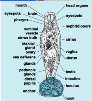
MORPHOLOGY:
hermaphrodites, male organs mature before female
distinct anchor with sucker and complex hook arrangement
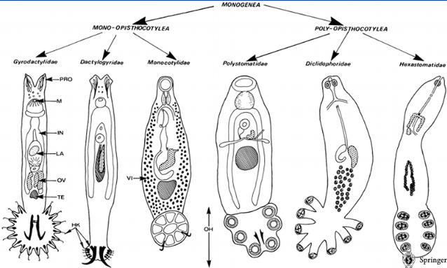
divided into 2 types
Mono-opistocotylea
Poly-opisthocotylea
-
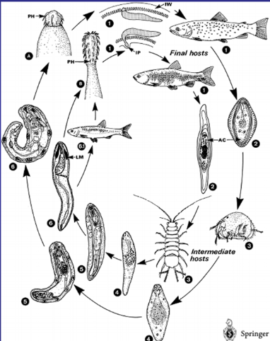
LIFE CYCLE (direct, cyclodevelopmental):
ectoparasites of fish (gills)
no asexual reproduction, no IH
some lay eggs > oncomiracidium (larval stage)
search out fish host by swimming with cilia
*CLASS TREMATODA (trematodes)
ORDER DIGENEA
intestinal: Alaria spp., Nanophyetus salmincola
blood: Schistosoma spp.
lung: Paragonimus spp.
liver: fasciola hepatica, fascioloides magna, opisthorchis sinensis
others: lancet liver fluke, green-banded broodsac, swimmer’s itch
-
ORDER ASPIDOGASTRIA
*ORDER DIGENEA
CLASS TREMATODA
-
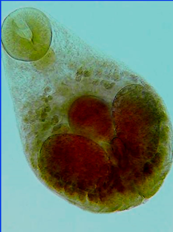
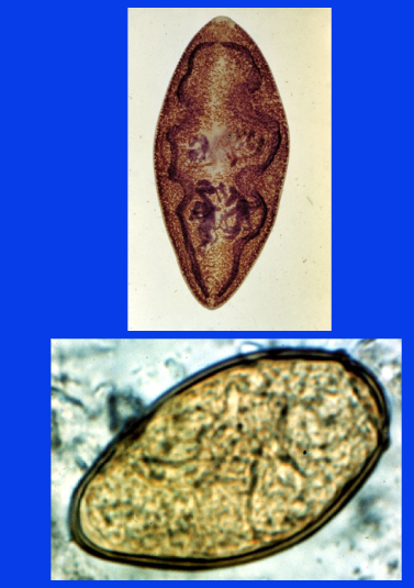
MORPHOLOGY:
dorso-ventrally flattened, spiny cuiticle, esophagus and blind ceca
ventral and oral suckers, shared genital pore
hermaphroditic (most)
female: ovary, uterus, vitellaria
male: paired testes
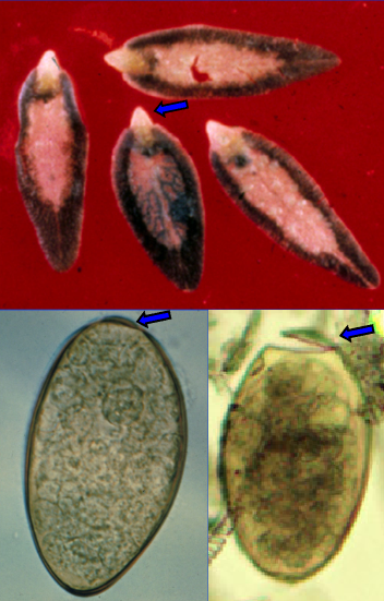
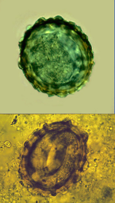
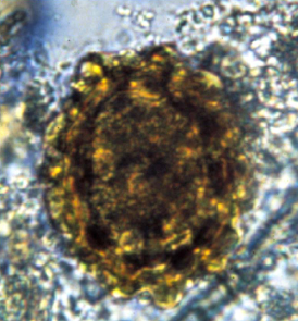
eggs: oval, operculate, brown-yellow, contents fill eggs completely
-
LIFE CYCLE (indirect):
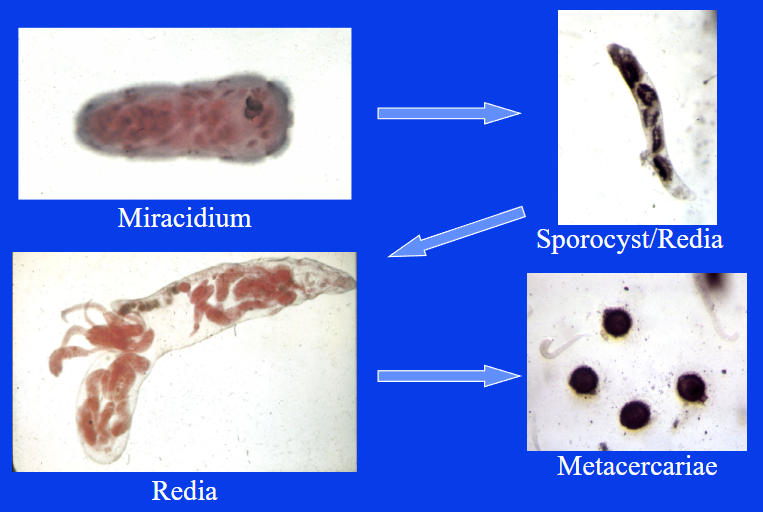
miracidium > sporocyst > redia > cercaria > (sometimes meta/mesocercaria)
most immature stages in IH, some in environment
HOST: specific snail (IH), varied vertebrate (DH)
geographic distribution of parasite follows snail host
miracidium > operculum > penetrate IH > asexual replication (sporocyst and redia) > cercaiae
cercariae
penetrate DH > mature
encyst on vegetation or in another IH > metacercaria
penetrate IH > mesocercaria

*Alaria spp. (intestinal fluke)
CLASS TREMATODA, CLASS DIGENEA
-
HOST: cats/dogs (DH), snails/tadpoles (IH), various (PH) - CAN INFECT HUMANS
-
MORPHOLOGY:
small, anterior flattened, cylindrical posterior
anterior region has oral and ventral suckers
eggs: yellow-brown, operculate, segmented embryo when egg in feces
-
LIFE CYCLE (indirect):
immature eggs in feces > miracidium hatch in water > IH > cercariae
IH/DH/PH > mesocercariae > lungs > metacercariae > respiratory tract and swallowed > mature in SI
Zoonotic threat: cercariae > tadpoles > mesocercariae
eating uncooked PH
only replicate in IH
-
PATHOGENESIS + DIAGNOSIS
tightly packed in clusters in SI
not normally pathogenic except in heavy infection
egg on fecal float
*Nanophyetus salmincola (Salmon Poisoning Fluke) (intestinal fluke)
CLASS TREMATODA, ORDER DIGENEA
-
HOST: dogs/cat/mink (DH), snail, fish (IH)
restricted to Pacific Northwest and Siberia
-
MORPHOLOGY:
small, creamy white
eggs: small, INDISTINCT operculum, yellow-brown
-
LIFE CYCLE (typical):
eggs in feces > miracidia in water > snail IH > cercariae > fish IH > metacercariae > DH > fluke > SI
PATHOGENESIS + TREATMENT
vector of Neorickettsia helminthoeca
not really pathogenic
treated with tetracyclines
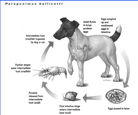
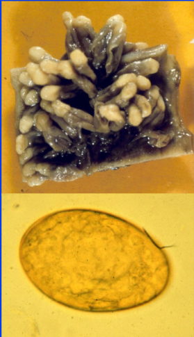
*Paragonimus spp. (lung fluke)
CLASS TREMATODA, ORDER DIGENEA
-
MORPHOLOGY:
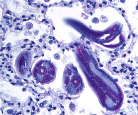
ovoid, large, in cysts in pairs, reddish-brown, spiny cuticle
egg: small, distinct operculum, yellow-brown
-
LIFE CYCLE (typical):
eggs in cyst > shed into bronchioles > swallowed > eggs in feces > miracidia > snail IH > cercariae > crayfish IH > metacercariae
DH > release fluke into SI > lungs and associate in pairs in lung parenchyma near bronchioles > fibrotic cyst
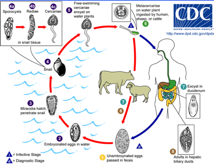
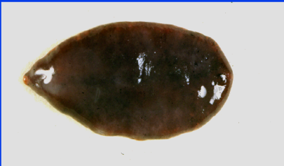
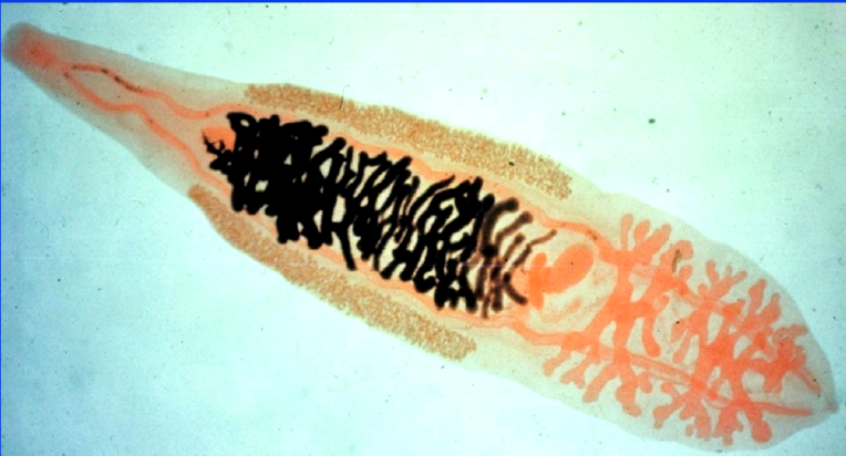
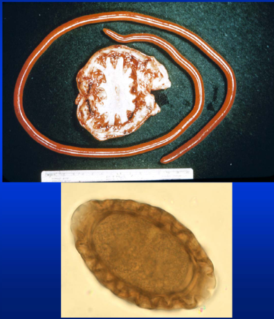
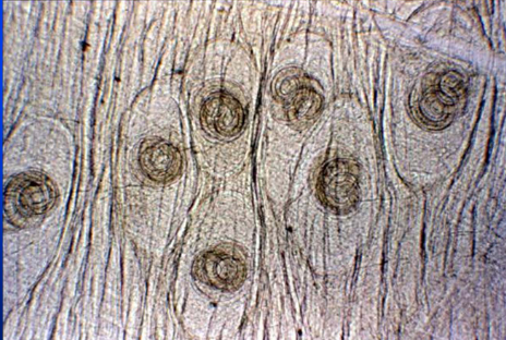
*Fasciola hepatica (liver fluke)
CLASS TREMATODA, ORDER DIGENEA
-
HOST: humans can be infected
usually imported animals, restricted geographically in Canada
MORPHOLOGY:
large, leaf-like, cephalic cone
eggs: indistinct operculum
-
LIFE CYCLE (typical):
eggs in feces > miracidium > IH > cercaria > vegetation > metacercaria
DH ruminant > fluke released > SI > liver capsule > migrates through parenchyma > bile duct > mature
-
PATHOGENESIS:
pipe-stem fibrosis of liver
within bile ducts, extensive fibrosis of duct wall

*Fascioloides magna (liver fluke)
CLASS TREMATODA, ORDER DIGENEA
-
HOST: wild cervids (DH, Canada)
widely distributed geographically
-
MORPHOLOGY:
large, leaf-like, no cephalic cone
egg: typical fluke
-
LIFE CYCLE (typical):
eggs pass through bile duct > eggs in feces > miracidium > IH > cercariae > vegetation > metacercaria
cervid DH > fluke released > SI > liver capsule > liver parenchyma > cyst in bule duct
-
PATHOGENESIS AND DIAGNOSIS:
diagnosed post-mortem in domestic animals, no patency
no clinical signs in wild and domestic cervids
fluke eggs seen on fecal float
CATTLE:
no clinical signs, no patency, accumulate
SHEEP:
no encysting, migrate constantly, mortality before patency
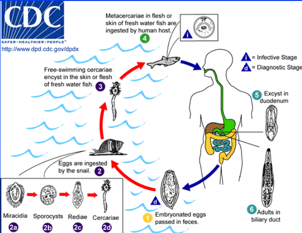
*Opisthorchis sinensis (liver fluke)
CLASS TREMATODA, ORDER DIGENEA
can infect humans
-
LIFE CYCLE:
eggs in feces > snail IH > cercariae > metacercariae in skin/flesh of fish
human DH > excyst in duodenum > mature in billiary duct
-
PATHOGENESIS + DIAGNOSIS:
inflammation, obstruction of biliary duct, acute pain, chronic infection
small, operculate egg in stool or duodenal aspirate
treatable
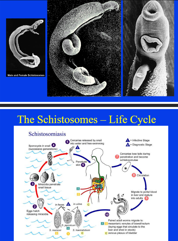
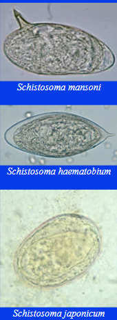
*Schistosoma spp. (blood fluke)
CLASS TREMATODA, ORDER DIGENEA
mansoni, japonicum, haematobium
can infect humans
-
LIFE CYCLE:
eggs in feces/urine > miracidia > snail IH > sporocysts > cercariae released
penetrate DH skin > cercariae lose tails > schistosomulae > portal blood in liver > mature > pair
can cause immune interactions during egg migration
-
PATHOGENESIS AND DIAGNOSIS:
acute (eosinophilia)
chronic: bloody diarrhea, portal hypertension, pulmonary hypertension
diagnose by egg morphology, intestinal species in feces/urine
treatable
*ORDER ASPIDOGASTREA
CLASS TREMATODA
-
MORPHOLOGY:
large ventral sucker, anterior mouth
posterior sucker modified into large adhesive disk/plate
eggs shed into enviro > hatch to larvae or ingested and hatch
-
LIFE CYCLE:
generally not host specific
Obligate: mollusk > fish (DH)
eggs > mollusk > form that is not sexually mature > larval form > fish host > SI > matures
facultative:
eggs/larvae > mollusk > mature adult
mollusk > fish/turtle host > establish in digestive tract > produce egg
-
PATHOGENESIS:
not associated with diseases
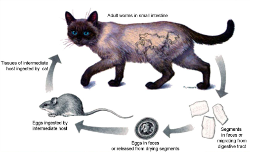
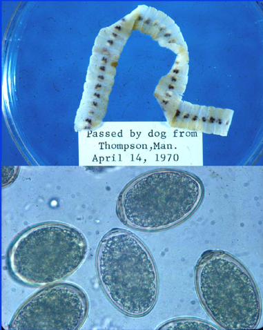
*Class Cestoda (tapeworms)
Cyclophyllidean: (long strobila, proglottids)
Taenia Pisiformis, Taenia Taeniaeformis, Taenia Crassiceps, Taenia Saginata, Taenia Solium
Echinococcus Granulosus, Echinococcus multilocularis
Arthopodan: Dipylidium Caninum, Anocephala perfoliata, Monezia spp.
-
Pseudophyllidean:
Crustacean: Diphyllobothrium latum
-
DH: host specific, do not replicate, not very pathogenic
IH: usually host specific, a prey species, may be pathogenic
-
MORPHOLOGY:
most are large (some small)
3 body regions
scolex, neck, strobila
no mouth/digestive tract, hermaphroditic
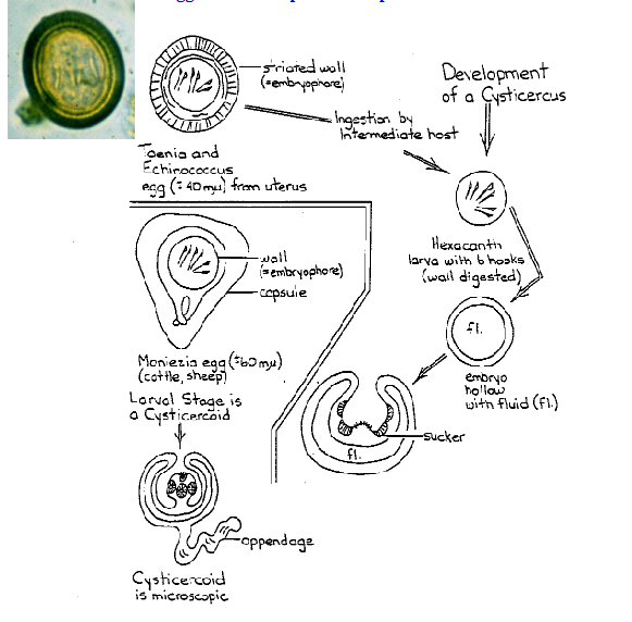
eggs: hexacanth embryo within embryophore, shed in segments/feces
larva: various stages
-
LIFE CYCLE (indirect):
eggs in feces/segments > hexacanth embryo > IH > migrates to final site of development > infective
IH eaten > scolex attaches to DH gut > matures
most species only have 1 IH, some aquatic use 2
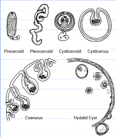
*LARVAL FORMS CLASS CESTODA
Cyclophyllidea:
Taenia spp: cysticercus, strobilocercus, coenurus
Echinococcus spp: hydatid cyst, alveolar hydatid cyst
Dipylidium, Noezia, Anoplocephala: cysticercoid
-
Pseudophyllidea:
Diphyllobothrium: procercoid, plerocercoid
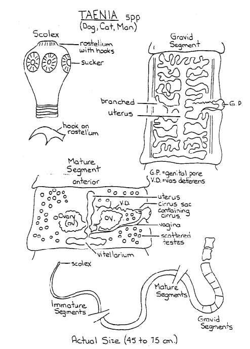
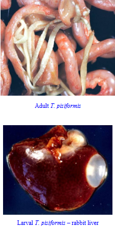
*Taenia pisiformis (rabbit tapeworm)
Class Cestoda, Cyclophyllidea
-
HOST: dogs (DH), rabbits (IH)
-
MORPHOLOGY:
shiny white strobila, rectangular segment
scolex 4 suckers, 2 rows of hooks on rostellum, single genital pore
Larva: cysticercus, fluid filled bladder
egg: typical taeniid, thick radially striated shell, found singly
-
LIFE CYCLE:
eggs from segments > IH > hexacanth larva hatches > peritoneal cavity/liver > cysticercus
DH > scolex in cysticercus evaginates > attaches to gut > strobila
-
PATHOGENESIS:
non-pathogenic, motile gravid segments
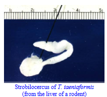
*Taenia taeniaeformis (rat tapeworm)
Class Cestoda, Cyclophyllidea
-
HOST: cats (DH), rodents (IH)
-
MORPHOLOGY:
scolex 4 suckers, 2 rows of hooks on rostellum
larva: strobilocercus
egg: typical taeniid
-
LIFE CYCLE:
similar to Taenia pisiformis
IH in liver, DH in small intestine
-
PATHOGENESIS:
not pathogenic, gravid segments in feces
*Taenia crassiceps
Class Cestoda, Cyclophyllidea
can infect humans if dog infected
-
HOST: foxes (DH), groundhogs (IH)
-
MORPHOLOGY:
larvae: budding cysticercus
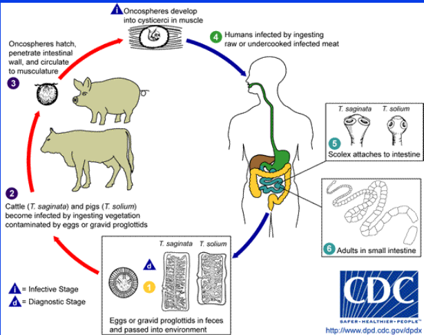
*Taenia saginata (beef tapeworm)
Class Cestoda, Cyclophyllidea
-
HOST: humans (DH), cattle (IH)
-
MORPHOLOGY:
unarmed rostellum, 4 suckers, segments with 1 genital pore irregularly alternating
larva: cysticercus
egg: typical taeniid
-
LIFE CYCLE:
segments in feces > eggs emerge > IH > hexacanth larvae > SI > muscles
hexacanth larvae > cysticercus (viable for long periods of time)
DH > uncooked meat > SI > matures
-
DIAGNOSIS + TREATMENT:
relatively benign, little damage
cysticercosis not possible in humans
improve sanitation
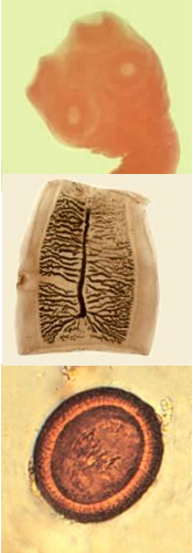
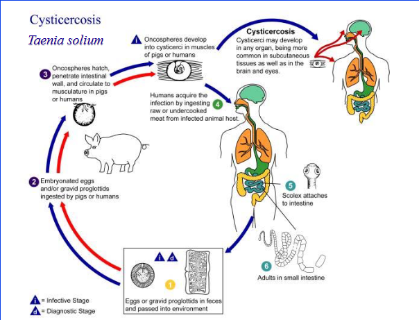
*Taenia solium (pork tapeworm)
Class Cestoda, Cyclophyllidea
-
HOST: humans (DH);swine, humans (IH)
-
MORPHOLOGY:
armed rostellum, 2 rows of hooks, irregular alternating genital pore
larvae: cysticercus
egg: typical taeniid
-
LIFE CYCLE: same as T. saginata
segments in feces > eggs emerge > IH > hexacanth larvae > SI > muscles
hexacanth larvae > cysticercus (viable for long periods of time)
DH > uncooked meat > SI > matures
-
PATHOGENESIS + DIAGNOSIS
cysticercosis possible in humans (self-infection > humans IH)
eggs shed fully mature and immediately infective
sanitation, determine source, avoid eggs
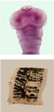
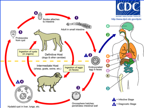
*Echinococcus granulosus
Class Cestoda, Cyclophyllidea
zoonotic threat
-
HOST: wolf (DH), moose (IH)
-
MORPHOLOGY:
smallest tapeworm of dogs, scolex 4 suckers, 2 rows of hooks, 3-4 segment strobila
larvae: hydatid cyst (asexual reproduction)
egg: typical taeniid
-
LIFE CYCLE:
eggs in segments > IH > hexacanth larvae > intestine > lungs > hydatid cyst grows
DH > scolex attach to SI mucosa
-
DIAGNOSIS + TREATMENT:
non pathogenic (DH), unaffected usually (IH)
space occupying lesions in humans
eggs in feces (cannot distinguish from Taenia), surgery
*Echinococcus multilocularis
Class Cestoda, Cyclophyllidea
zoonotic threat
-
HOST: fox (DH), rodents (IH) - Canada
cosmopolitan distribution
-
MORPHOLOGY:
smallest tapeworm of dogs, scolex 4 suckers 2 rows of hooks, strobila 3-4 segments long
larva: alveolar hydatid cyst (asexual replication, budding mass)
egg: typical taeniid
-
DIAGNOSIS + TREATMENT:
massive replication, budding and metastasis in IH (liver, other organs)
eggs in feces (cannot distinguish from Taenia), drugs, surgery
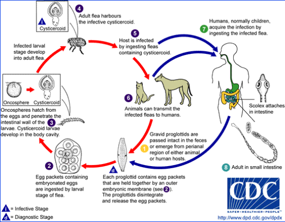
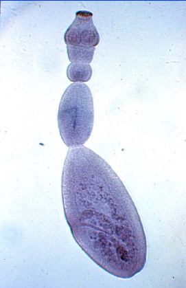
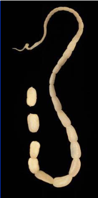
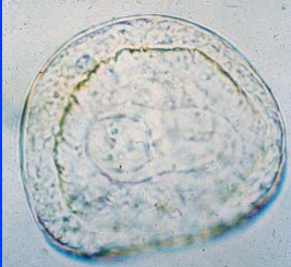
*Dipylidium caninum (Flea tapeworm)
Class Cestoda, Cyclophyllidea
-
HOST: dogs, cats, humans - rare (DH); fleas (IH)
-
MORPHOLOGY:
scolex with 4 suckers, rostellum with several rows of hooks, long strobila, barrel segments, pores each side
larvae: cysticercoid
eggs: in packets, thick unstriated embryophore
-
LIFE CYCLE:
eggs in segments (motile) > IH > hexacanth larvae > body cavity > cysticercoid
DH > scolex attaches to SI mucosa
-
DIAGNOSIS + TREATMENT:
non-pathogenic (vertebrates)
differentiate between taeniid
migrating segments in feces, rarely eggs in fecal floats, cestodicide
*Anocephala perfoliata
Class Cestoda, Cyclophyllidea
-
HOST: horse/ponies (DH), orbatid mites (IH)
role in stomach upsets
-
MORPHOLOGY:
lappets on scolex, in ileocecal junction
irregular eggs with pyriform apparatus
treatable
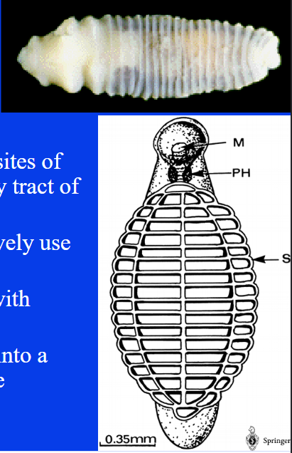
*Monezia spp.
Class Cestoda, Cyclophyllidea
-
HOST: cattle/sheep/goats (DH), orbatid mites (IH)
-
MORPHOLOGY:
in small intestine
irregular eggs on fecal with pyriform apparatus
-
DIAGNOSIS:
clincal signs only in heavily infested and young
treatable
*Diphyllobothrium latum
Class Cestoda, Pseudophyllidean
humans can get infected
HOST: fish-eating mammals (DH); 2 IH - copepod (procercoid) > fish (pleurocercoid)
MORPHOLOGY:
pair of bothria (grooves) on scolex, large strobila
eggs: operculate, light brown, ciliated hexacanth larva > coracidium
PATHOGENESIS + TREATMENT:
usually non-pathogenic, B12 deficiency
potentially treatable
*PHYLUM NEMATODA
Order Rhabditida (rhabditids, threadworms)
Order Strongylida (strongylids, hookworms, lungworms)
Order Oxyurida (pinworms, oxyurids)
Order Ascaridida (ascarids)
Order Spirurida (spirurids, filarids, Dracunculus)
Order Enoplida (trichurids/whipworms, Dioctophyme, capillarids, Trichin
-
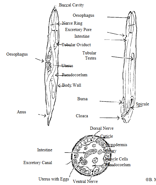
MORPHOLOGY:
elongate tubular bodies,
thick, resistant cuticle, muscles under
hydrostatic pressure to maintain shape and rigidity
simple alimentary tract, separate sexes
male: tube with testes, vas deferens, ejaculatory duct, copulatory bursa, cuticular spicules (sometimes)
female: pair of blind-ended ovaries to uteri, vagina, vulva, can store sperm
-
LIFE CYCLE:
egg > L1 > L2 > L3 (infective) > L4 > L5 > adult
life cycle can be divided among different hosts
*Infection and Migration in Nematodes
Infection:
oral ingestion (fecal-oral)
penetrate skin
transplacental, transmammary
predator-prey with IH or PH
vector borne
-
Migration:
tracheal: enter bloodstrem by penetrating gastrointestinal wall, end up in airway and swallowed
somatic: trachea migration but stay in bloodstream, distributed around body
mucosal: unrelated to tracheal and somatic, penetrate gastric pits/mucosa before returning to lumen
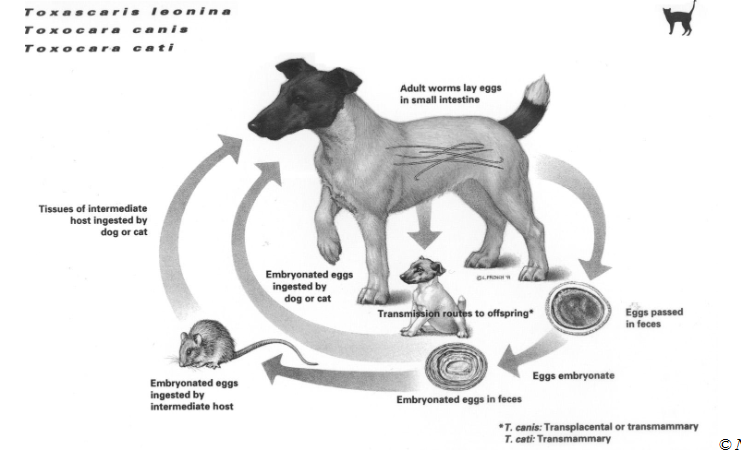
*Order Ascaridida (ascarids)
Phylum NEMATODA
Toxocara canis, Toxocara cati
Toxascaris leonina, Parascaris equorum
Ascaris lumbricoides, Ascaris suum
Ascaridia galli, Heterakis gallinarum, Baylisascaris procyonis
-
GENERAL:
generally robust, heavy bodied
infect SI (DH) - except Heterakis gallinarum
highly host specific, various life strategies
eggs often resistant to enviro (infectious for long times)
-
LIFE CYCLE (direct, may have PH):
prolific, many alternate migration routes
eggs mature with L3 in enviro (usually oral infection)
L3 penetrate SI > migration > SI > mature
-
PATHOGENESIS:
poor growth, potbelly, obstruction, minor lesions from migration
possible zoonoses (ocular/visceral larval migrations)

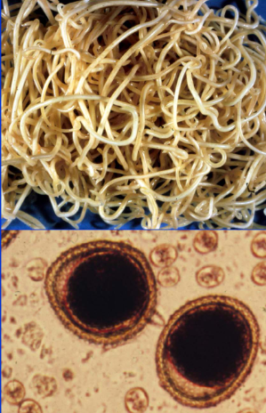
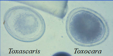
*Toxocara canis (canine roundworms)
ORDER ASCARIDIDA
can infect humans
-
HOST: dogs (specific)
-
MORPHOLOGY:
large, heavy bodied
egg: thick shelled, pitted, single cell in feces
-
LIFE CYCLE: PPP 4-5w, 3w
eggs in feces > L3 > oral transmission > gastrointestinal
migration route dependent on age
tracheal (<3mo), somatic (6+mo), mixed (3-6mo)
pups often born with parasite (transplacental, hypobiotic larvae)
partial tracheal migration
potential PH with larvae ingestion
short patency, no migration
-
PATHOGENESIS + TREATMENT:
mainly light infections, usually non-pathogenic
heavy infection: unthriftiness, stunted growth, dry skin, dull coat
potentially painful abdomen, death may occur before patency, eosinophilic gastroenteritis, lung issues, focal lesions
vomition = heavy infection
many treatments
some antihelmintics could potentially cause obstruction

*Toxocara cati (feline roundworms)
ORDER ASCARIDIDA
-
HOST: cats (specific)
-
MORPHOLOGY:
arrowhead cervical alae (cuticular extension), large
egg: thick shelled, pitted, single cell in feces
-
LIFE CYCLE: PPP 4w
eggs in feces > L3 > oral transmission > gastrointestinal
L3 penetrates stomach wall > tracheal (young) OR somatic (older)
transmammary infection possible
PH ingestion can cause infection (cats that can hunt)
-
PATHOGENESIS + TREATMENT:
not as pathogenic as T. canis
vomiting even in moderate, unthriftiness and diarrhea (unusual)
fewer treatment compounds
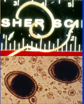
*Toxascaris leonina
ORDER ASCARIDIDA
-
HOST: dogs, cats (generalist)
not as common as Toxocara spp.
-
MORPHOLOGY:
same size as T. cati, no cervical alae
eggs: smooth shells, undulating membrane, single cell that does not fit shell
-
LIFE CYCLE: PPP 2mo
eggs feces > L3 > oral transmission > gastrointestinal
no tracheal migration > L3 intestinal wall > mucosal migration > lumen > mature
possible infection by PH ingestion
-
PATHOGENESIS + TREATMENT
not as pathogenic as Toxocara spp.
usually not heavy burdens
similar treatments as Toxocara spp.
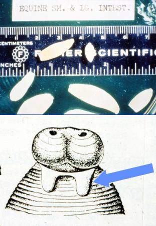
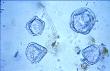
*Parascaris equorum (equine roundworm)
ORDER ASCARIDIDA
-
HOST: horses, ponies donkeys
-
MORPHOLOGY:
heavy bodied, white
eggs: sub-spherical, thick protein coat
-
LIFE CYCLE: PPP 10-12w
tracheal migration > liver and lung
-
PATHOGENESIS + TREATMENT
clinical signs in foals and weanlings
heavy burdens can impact colic
unthrifty, loss of appetite, hypoalbuminenia
typical eggs can be found in feces
can be treated
some compounds can cause obstruction

*Ascaris lumbricoides (human roundworm)
ORDER ASCARADIDA
-
HOST: humans, swine
-
MORPHOLOGY:
heavy bodied, white, smaller males
eggs: sub-spherical, thick protein coat
-
LIFE CYCLE: PPP 6-8w
tracheal migration > liver and lung
-
PATHOGENESIS + TREATMENT:
milk spots on liver
pulmonary hemorrhage/edema, mild enteritis
erratic migrations problematic
typical eggs can be found in feces
*Ascaris suum
ORDER ASCARIDIDA
-
HOST: swine
-
MORPHOLOGY:
heavy bodied, white, males smaller
eggs: sub-spherical, thick protein coat
-
LIFE CYCLE: PPP 6-8w
tracheal migration through liver and lung
-
PATHOGENESIS + TREATMENT:
milk spots on liver
pulmonary hemorrhage/edema, mild enteritis - affects growth
typical eggs can be found in feces
sanitation, feed antihelmintics, deworming
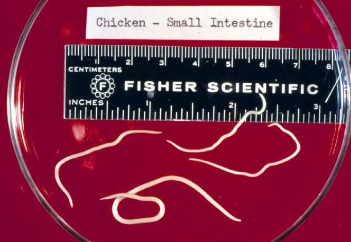
*Ascaridia galli
ORDER ASCARIDIDA
-
HOST: birds
-
MORPHOLOGY:
heavy bodied, white
eggs: smooth shelled
-
LIFE CYCLE: PPP 30-50d
L3 larvated eggs > hatch > L3 into mucosal > L4 > re-enters lumen of SI > matures
-
PATHOGENESIS + TREATMENT:
hemorrhage/diarrhea during larval mucosal migration
decreased production, low mortality
eggs can be found in feces of birds
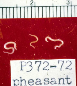
*Heterakis gallinarum (cecal worm)
ORDER ASCARIDIDA
-
HOST: chickens, turkeys, etc.; earthworms (PH)
-
MORPHOLOGY:
slender, in ceca
eggs: thick, smooth shell
-
LIFE CYCLE: PPP 24-30d
eggs in feces > L3 > oral ingestion > gastrointestinal tract
migrate in mucosa of cecum > re-enters lumen > molts 3x > mature
-
SIGNIFICANCE:
true IH of Histomonas meleagridis
*Baylisascaris procyonis
ORDER ASCARIDIDA
zoonotic threat
-
HOST: raccoons, dogs (DH); various (PH)
-
MORPHOLOGY:
large worms
eggs: thick, smooth shell
-
SIGNIFICANCE:
visceral larval migrans
long period for eggs to become infective
sanitation, raccoon latrines
*Order Rhabditida
PHYLUM NEMATODA
strongyloides spp (threadworm)
-
GENERAL:
usually free living, infect vertebrates during part of life cycle or accidentally
mainly small
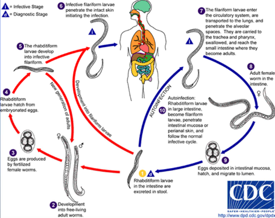
*Strongyloides spp. (threadworms)
ORDER RHABDITIDA
HOST: various species with assorted hosts
-
MORPHOLOGY:
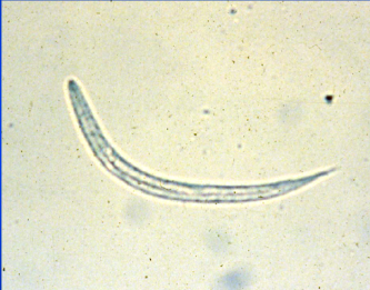
thread-like, no parasitic males
eggs: oval, thin shelled, L1 inside
S. stercoralis = L1 free in feces
NOT freeze tolerant
-
LIFE CYCLE:
parasitic with/without free living stage
free-living: complete, M+F produce eggs
Routes of infection
mucosal: ingestion of L3
tracheal/somatic: skin penetration
transmammary
S. stercoralis can cause autoinfection in humans
generally cleared from young animals at 6mo
L1 in feces > L3 > mature > oral/skin > tracheal/somatic/mucosal> embed in mucosa SI villi > lumen
-
PATHOGENESIS + DIAGNOSIS:
non-bloody enteritis, worms deeply embedded
Baermann for larva in feces
*ORDER ENOPLIDA
PHYLUM NEMATODA
Trichurus spp. (whipworm)
Capillaria spp.
Diocytophyme renale (giant kidney worm)
Trichinella spiralis
-
GENERAL:
elongate esophagus surrounded by stichocytes
host specific, many hosts, location varies by genus
deeply embedded in epithelium
-
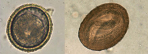
MORPHOLOGY:
slender, white
eggs: oval, distinct bipolar plugs
larvae: only in Trichinella spp. in intestinal tract/muscles
-
LIFE CYCLE:
direct and indirect, oral transmission
larvae mature in egg > DH or IH
trichinella larvae > intestine > tissue > hypobiotic
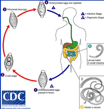
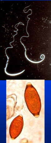
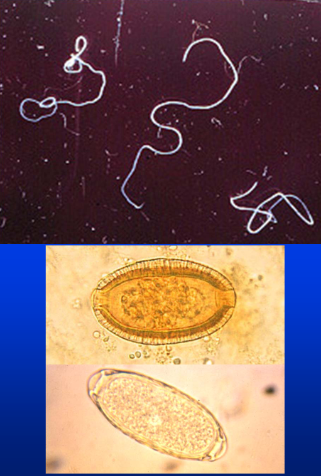
*Trichuris spp. (whipworm)
ORDER ENOPLIDA
humans can become infected
-
HOST: highly specific
each host has its own species that will not infect other hosts
-
MORPHOLOGY:
whip shapped, anterior 2/3 narrow
egg: smooth, oval shell, prominent plug, fills shell, yellow-brown
eggs are frost and enviro resistant
-
LIFE CYCLE (direct): - PPP long
larvae in egg (infective) > oral transmission > mucosal migration in SI > L1 in cecum > mature
-
PATHOGENESIS + DIAGNOSIS:
thin anterior portion deeply embedded
malabsorption, diarrhea, protein loss, blood feeders
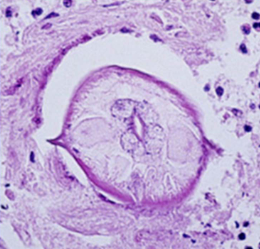
*Capillaria spp.
ORDER ENOPLIDA
-
HOST: highly specific (host and location), some have IH
can be infected by multiple species at different sites
-
MORPHOLOGY:
slender, uniform, fine, hair-like, prominent stichocytes
egg: smooth (less than Trichuris), oval shell, prominent plug, fills shell
-
LIFE CYCLE:
if IH, many use earthworms
eggs in feces/urine depending on location of adults
C. hepatica - lays eggs throughout liver and released when host dies
-
IN BIRDS:
C. contorta
wall of crop thickened, weakness, unthriftiness
C. caudinflata + obsignata
edematous intestinal wall with reddish fluid
necrosis of mucosa - heavy
reddish diarrhea, emanciation
decreased production
*Giant Kidney Worm (Diocytophyme renale)
ORDER ENOPLIDA
can infect humans
-
HOST: mink (primarily), dogs, humans (DH); invertebrate (IH)
found locally in Thames river valley
-
MORPHOLOGY:
bright red, usually seen coild in (right) kidney
females up to 100cm long and 1cm diameter
males have bell shaped copulatory bursa
eggs: bipolar plugs, pitted outer shell
-
LIFE CYCLE: PPP 3-5mo
eggs in urine > IH > larva (infective)
may go up food chain > DH oral transmission > intestine > kidney > mature
-
PATHOGENESIS + TREATMENT
affected kidney destroyed - surgery
*Trichinella spiralis
ORDER ENOPLIDA
can infect humans
-
HOST: swine, meat eating animals
-
MORPHOLOGY:
females: 3-4mm, slender, uterus with larvae
males: 1.5mm, 2 small cloacal flaps on tail
-
LIFE CYCLE:
each host is both DH and IH
larvae > oral transmission eating meat > mature > lay ~1500 larvae > distribute around body
-
PATHOGENESIS + DIAGNOSIS:
fatal dose of larvae in humans, cannot tolerate
gastroenteritis, diarrhea, muslce pain, inflammation, eosinophilia
muscle biopsy to identify heavy burden
preventative, no viable treatment
trichinoscope
*ORDER OXYURIDA
PHYLUM NEMATODA
enterobius vermicularis (human pinworm)
oxyuris equi (horse pinworm)
-
GENERAL:
highly host specific, large intestine
usually not overtly pathogenic, irritating
-
MORPHOLOGY:
most are small (except O. equi)
long tapering tail in both sexes
eggs: oval, some operculate, asymmetrical (flat + round sides)
-
LIFE CYCLE:
direct, simple
eggs in feces > deliver to perianal region > larva
fecal-oral transmission > eggs hatch in intestine > large intestine > mature
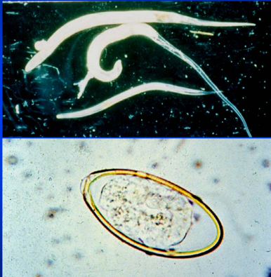
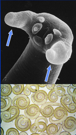
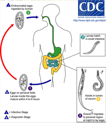
*Enterobius vermicularis (human pinworm)
ORDER OXYURIDA
-
HOST: humans (large intestine)
-
MORPHOLOGY:
long tapering tail in both sexes
eggs: oval, some operculate, asymmetrical (flat + round sides)
-
LIFE CYCLE:
direct, simple
eggs in feces > deliver to perianal region > larva
fecal-oral transmission > eggs hatch in intestine > large intestine > mature
-
PATHOGENESIS:
egg laying gives highly puritic perianal region = scratching
abdominal upset
IH of Dientamoeba fragilis
*Oxyuris equi (horse pinworm)
ORDER OXYURIDA
-
HOST: horses, ponies, donkeys
-
MORPHOLOGY:
larger, long tapering tail in both sexes
eggs: oval, some operculate, asymmetrical (flat + round sides)
-
LIFE CYCLE:
direct, simple
eggs in feces > deliver to perianal region > larva
fecal-oral transmission > eggs hatch in intestine > large intestine > mature
-
PATHOGENESIS:
egg mass deposition gives puritis = local abrasions, rat tails
inflammation and ulceration (heavy)
wide ragne of treatments
*ORDER STRYONGYLIDA
Trichostrongyles
Strongyles
Hookworms
Lungworms
-
GENERAL:
most have single host (except lungworms)
wide range of hosts, varies
-
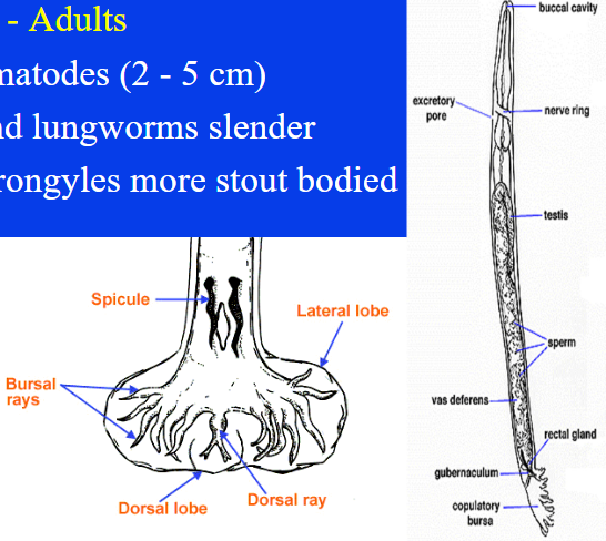
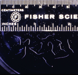
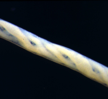
MORPHOLOGY:
smaller
slender (trichostrongyles and lungworms) vs stout bodied (hookworms and strongyles)
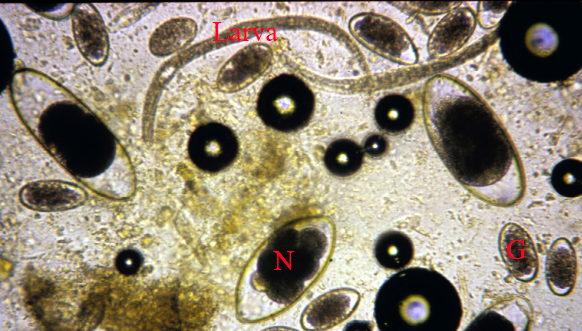
Eggs: thin-shelled morulated; GIN eggs
-
LIFE CYCLE:
direct (except lungworms)
eggs/larvae in feces > oral transmission (sometimes ksin) > extra-intestinal migrations
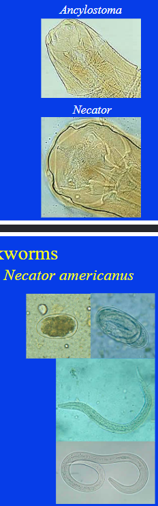
*HOOKWORMS
ORDER STRONGYLIDA
Ancylostoma duodenale and Necator americanum (human)
Ancylostoma caninum
Uncinaria stenocephala
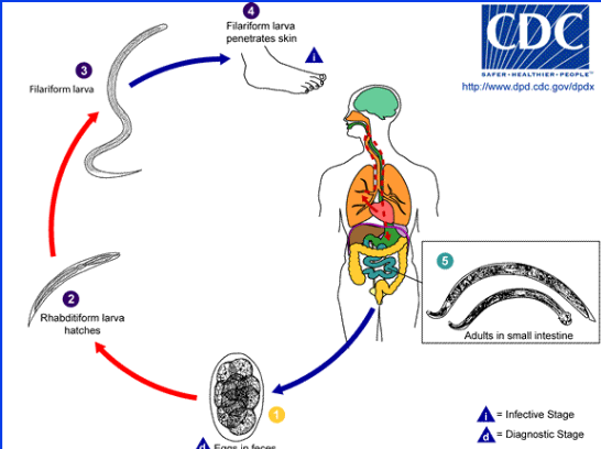
*Ancylostoma duodenale and Necator americanus (human hookworm)
ORDER STRONGYLIDA, HOOKWORM
-
HOST: humans; warm moist climate
easy to acquire, modify host behaviour
-
MORPHOLOGY:
large buccal cavity, teeth (A), plates (N), moderate size
males: colulatory bursa
eggs: morulated, L1 in egg
rhabditiform L2 and L3
filariform L3 infective
-
LIFE CYCLE:
eggs feces > rhabiditiform larvae > L3
penetrate skin > tracheal migration > intestine > mature
A.duodenale may undergo somatic and potentially transmammary
-
PATHOGENESIS + DIAGNOSIS:
blood loss at attachment sites, anemia
bulbous abdomen
ground itch (L3 penetration), respiratory symptoms (pulmonary migration)

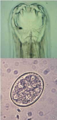
*Ancylostoma caninum (canine hookworm)
ORDER STRONGYLIDA, HOOKWORM
-
HOST: dogs
-
MORPHOLOGY:
stout, bend in body at buccal cavity - 3 pairs of teeth, red if filled with blood
egg: morulated, LESS THAN 70umI
environment will affect L3
-
LIFE CYCLE:
eggs feces > L1 > L3 > penetrate skin
tracheal migration (young), somatic (older)
possible patent if ingest L3 > mucosal migration
transmmary infection possible (hypobiotic larvae)
-
PATHOGENESIS + DIAGNOSIS:
blood loss, anemia, protein loss, enteritis, melena, emaciation
can die before patency (heavy infection) due to blood loss
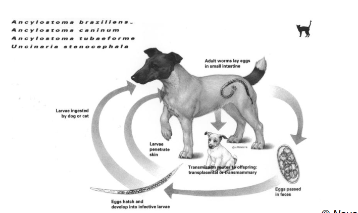
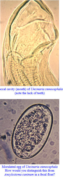
*Uncinaria stenocephala
ORDER STRONGYLIDA, HOOKWORMS
-
HOST: dogs, cats; northern
-
MORPHOLOGY:
similar to Ancylostoma but has pair of cutting plates in buccal cavity (vs 3 pairs of teeth)
eggs: morulated, oval, MORE THAN 70um
L3 are freeze tolerant and hardy
-
LIFE CYCLE:
eggs in feces > L1 > L3
L3 ingested (skin uncommon)
no extra-intestinal migration
-
PATHOGENESIS + TREATMENT:
much less pathogenic than Ancylostoma spp.
anemia, protein loss, mild enteritis
antihelmintics used for ascarids
*LUNGWORMS
ORDER STRONGYLIDA
Muelerius capillaris
Filaroides and Oslerus spp.
Aelurostrongylus spp.
Crenosoma spp.
*Muellerius capillaris
ORDER STRONGYLIDA, LUNGWORMS
-
HOST: sheep/goats (DH), snails/slugs (IH)
-
LIFE CYCLE (indirect):
eggs in feces > L1 > penetrate IH > L3
oral transmission into DH > gastrointestinal tract > lungs via bloodstream > alveoli and terminal bronchioles > mature
-
PATHOGENESIS + DIAGNOSIS:
alveolar rupture, focal interstital pneumonia, granuloma
L1 in feces = diagnostic stage
raised regions on lungs, calcification
no registered compounds to treat
*Filaroides and Oslerus spp.
ORDER STRONGYLIDA, LUNGWORMS
-
HOST: dogs, foxes, wolves
-
MORPHOLOGY:
small, slender
eggs: thin walled, fully larvated eggs laid by female, hatch quickly
larvae: small, passed in feces
-
LIFE CYCLE: PPP 10w (O), 5w (F)
L1 in feces > tracheal migration
O. osleri = trachea/bronci
F. hirthi = bronchioles/alveoli
-
PATHOGENESIS + TREATMENT:
usually asymptomatic (F), nodules and wheezing cough + trachitis/bronchitis (O)
visualize nodules via bronchoscopy
dyspnea (heavy)
diagnosis of larva by sputum/Baermann
treatments available, nodules surgically
*Aelurostrongylus spp.
ORDER STRONGYLIDA, LUNGWORM
HOST: cats (DH); snal/slug (IH)
MORPHOLOGY:
small, slender
larvae: bent tail, dorsal spine
LIFE CYCLE:
larvae in feces > IH > stomach > lungs > terminal respiratory/lung parenchyma > mature
PH common source of infection
frog, rodent, lizard, bird
PATHOGENESIS + TREATMENT:
focal pneumonia with granuloma
chronic cough and dyspnea (heavy)
larva diagnosed in sputum/Baermann
treatments available
*Crenosoma spp.
ORDER STRONGYLIDA, LUNGWORM
HOST: dogs/wolves/foxes/racoons (DH); mollusk (IH)
MORPHOLOGY:
small, slender, anterior cuticle has crenations (folds)
egg: thin-shelled, morulated,
eggs with L1 or first stage larvae released
LIFE CYCLE:
larvae/egg in feces > IH > DH ingests L3 larvae
migrate stomach > lungs > bronchi > bronciole > mature
PATHOGENESIS + TREATMENT:
focal lesions
rhinotracheitis, bronchitis (heavy)
larva diagnosed in sputum/Baermann
treatment available
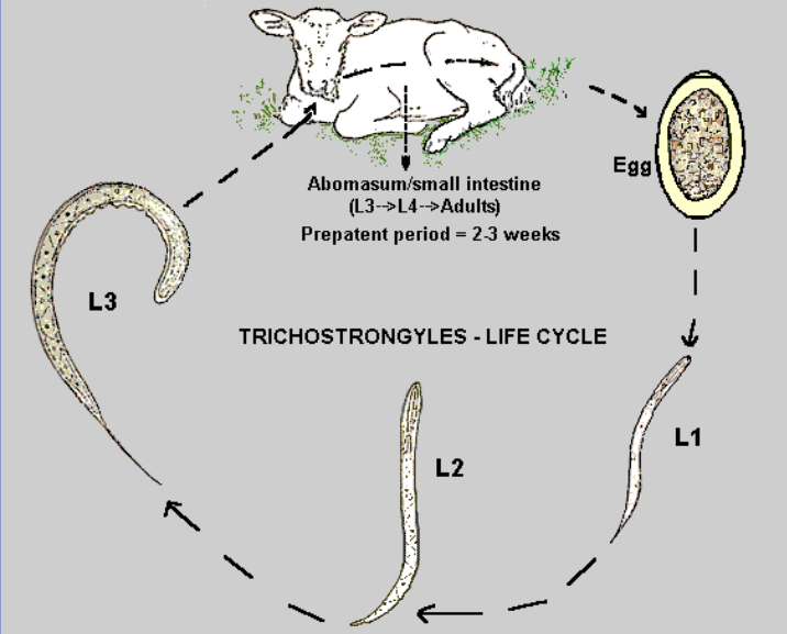
*TRICHOSTRONGYLES
ORDER STRONGYLIDA
Ostertasia/Teladorsagia spp. (medium-sized/brown stomach worm)
Haemonchus spp.
Nematodirus spp.
Others: cooperia and Trichostrongylus spp., (ruminant) Hyostrongylus rubidus (swine)
-
GIN EGGS

*Ostertagia/Teladorsagia spp. (medium-sized or brown stomach worms)
ORDER STRONGYLIDA, TRICHOSTRONGYLES
-
HOST: cattle (O. ostertagi), sheep/goats (T. circumcincta)
-
MORPHOLOGY:
slender, brown
egg: thin shelled, morulated, GIN egg
larvae and egg can overwinter and survive on pasture
LIFE CYCLE:
larvae in eggs > pasture hatch > L3
ingested > gastric pits > lumen > mucosal migration
hypobiosis possible
depends on environment
-
PATHOGENESIS:
largely associated with developing L3 in gastric pits and sudden emergence from hypobiosis
PH rises to neutral, gut leakage, emaciation
altered mucosa with nodules
type 1: summer, no hypobiosis
type 2: winter, hypobiosis
pre type 2: fall into winter

*Haemonchus spp
ORDER STRONGYLIDA, TRICHOSTRONGYLES
-
HOST: cattle, sheep (H. contortus), goats
-
MORPHOLOGY:
slender, spiral - R&W with blood
egg: thin-shelled, morulated, GIN egg
larvae and eggs no overwintering on pasture
-
LIFE CYCLE:
larvae in eggs > pasture hatch > L3
ingested > brief mucosal migration
hypobiosis is major factor
depends on environment
-
PATHOGENESIS:
largely associated with developing L4 and adults blood feeding on mucosa of abomasum
anemia, hypoproteinemia, focal areas of hemorrhage
pale carcass, watery blood, edema (acute)
continuing blood loss, bottle jaw, muscle weaness, dark and hard feces (chronic)
*Nematodirus spp.
ORDER STRONGYLIDA, TRICHOSTRONGYLES
-
HOST: cattle, sheep, goats
-
MORPHOLOGY:
very slender, multiple worms found coiled together
egg: thin-shelled, morulated, GIN egg, visible in adult female, hatch after freezing stimulus (some)
can overwinter on pasture
-
LIFE CYCLE (direct):
larvae in eggs > L3 > hatch on pasture
larvae > develop deep between villi of SI > adults > lumen
no hypobiosis
-
PATHOGENESIS
villus atrophy, diarrhea, anorexia
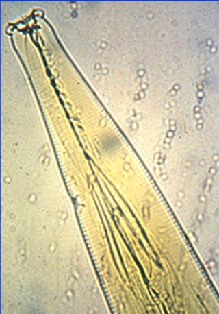
*STRONGYLES
ORDER STRONGYLIDA
Strongylus vulgaris
Strongylus equinis and edentatus
Cyathostomes
Oesophagostomum spp.
Syngamus trachea (gapeworm)
-
GENERAL:
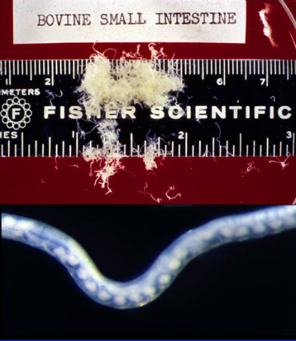
migratory (strongylus spp.) vs non-migratory (cyathostome)
all parasites of equids with adults in cecum and colon
-
MORPHOLOGY:
Strongylus spp buccal cavity = deeper than whide
Cyathostome buccal cavity = wider than deep
-
LIFE CYCLE:
larvae in eggs > pasture hatch > L3
ingested > dramatic extra-intestinal migration (S) OR mucosal migration (C) > mature and feed in cecul and colon
some hypobiosis, some can overwinter on pasture
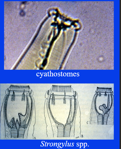
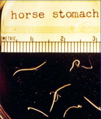
*Strongylus vulgaris
ORDER STRONGYLIDA, STRONGYLES
-
HOST: equids
-
MORPHOLOGY:
dark red, large stoma, lobed teeth in deep buccal capsule base (mickey mouse teeth), medium
egg: thin-shelled, morulated, GIN eggs
-
LIFE CYCLE (migratory):
larvae in eggs > pasture hatch > L3
ingested > extra-intestinal migration > adults in cecum and colon
-
PATHOGENESIS:
largely associaed with L4 as they move extraintestinally in crainal mesenteric artery (long development 2mo there)
larvae induce arteritis and thrombosis
insufficient number usually to cause lesions
acute disease from arteritis (heavy)
intermittent colic- obstruct bloodflow (chronic)
*Strongylus equinis and edentatus
ORDER STRONGYLIDA, STRONGYLES
-
HOST: equids
-
LIFE CYCLE (migratory):
larvae in eggs > pasture hatch > L3
ingested > extra-intestinal migration > adults in cecum and colon
-
GENERAL:
bowl-shaped buccal cavity, no teeth, large
undergo long life cycle in equid host
not highly pathogenic during larval migration
hemorrhage, colon inflammation (heavy)
adults feed on blood
larvae re-enter intestinal tract
*Cyathostomes
ORDER STRONGYLIDA, STRONGYLES
-
HOST: equids
many species, can often co-occur in same host
-
MORPHOLOGY:
cylindrical buccal cavity (flat bottomed), small sized, large stoma, may/may not have teeth
egg: thin shelled, morulated, GIN egg, smooth shell
LIFE CYCLE (non-migratory):
larvae in eggs > pasture hatch > L3
ingested > mucosal migration > adults in cecum and colon
hypobiosis possible
PATHOGENESIS:
primarily associated with larval mucosal migration
nodules around encysted larvae = impede mobility, poor weight gain, inflammation, edema, ulcer when emerging
anorexia, weight loss, diarrhea, colic, acute larval cyathostomiosis (when hypobiosis ends)
non-specific clinical signs
unthriftiness, upset digestion (large adult numbers) = intermittent and recurring
*Oesophagostomum spp.
ORDER STRONGYLIDA, STRONGYLES
-
HOST: ruminants, swine
-
GENERAL:
typical life cycle, prominent nodule formed during larval migration
no overwintering, hypobiosis in nodules
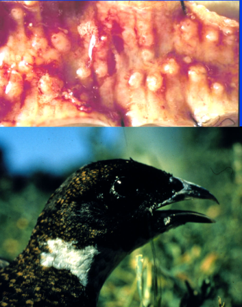
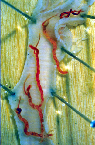
*Syngamus trachea (gapeworm)
ORDER STRONGYLIDA, StRONGYLES
-
HOST: gallinaceous birds
-
MORPHOLOGY:
males (2-6mm), females (5-40mm) - permanent copulation
Y-shaped structure attached to trachea
egg: elongate, ellipsoidal, smooth, operculum at each end, morulated
-
LIFE CYCLE:
larvae in eggs (some hatch) or soil, or some PH are infective
penetrate intestines > lungs > alveoli > trachea > attach to mucosa
suck on blood and mate in pairs
-
PATHOGENESIS:
worms in trachea > gape and gasp
increase in mucous, mild anemia
reduced production
mortality from asphyxiation (heavy)
raised, ulcerated lesions at attachment site
*ORDER SPIRURIDA
PHYLUM NEMATODA
Spirurids
Habronema spp and Draschia megastoma
Filarids:
Dracunculus medicinus (guinea worm)
Wucheria bancrofti and Brugia malayi (lymphatic filariases)
onchocera volvulus (river blindness)
dirofilaria immitis (canine heartworm)
-
GENERAL:
mainly stomach worms, some in eyes/connective tissue
indirect life cycles, simple life cycle in DH (no complex migrations)
use arthopodan intermediate host
PH commonly used for food chain
lengthy PPP
FILARIDS:
highly host-specific, require blood-sucking insect IH OR arthropod
infect conenctive tissue/body cavity
long-slender adults
microfilaria
*Habronema Spp. and Draschia megastoma (stomach worms of horses)
ORDER SPIRURIDA, SPIRURIDS
-
HOST: equids (DH); house/stable fly (IH)
-
LIFE CYCLE (indirect);
eggs in feces > L1 > IH > L3 in fly larvae
L3 exit from proboscis or whole fly eaten
L3 move directly to gastric pits > mature
-
PATHOGENESIS + TREATMENT:
Habronema
chronic gastritis with mucous
adults and larvae treatable
Draschia
tumour-like nodules, lesions largely asymptomatic
only larvae treatable
summer sores > lesions and inflammation (attracts more flies)
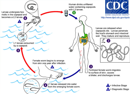
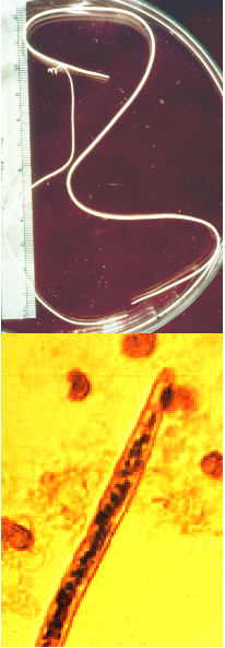
*Dracunculus medicinus (Guinea worm)
ORDER SPIRURIDA, FILARID
-
HOST: humans (DH), freshwater copepod (IH)
-
MORPHOLOGY:
slender, females ~1m long
within subcutaneous tissue
-
LIFE CYCLE:
unfiltered water > larvae penetrate stomach and intestinal wall
fertilized female > skin surface > emerge
larvae released in water as female emerging > IH > L3
-
PATHOGENESIS + TREATMENT:
non-pathogenic and asymptomatic during subcutaneous migration
painful burning blister and ulcer
no antihelmintic, simple sieve prevention
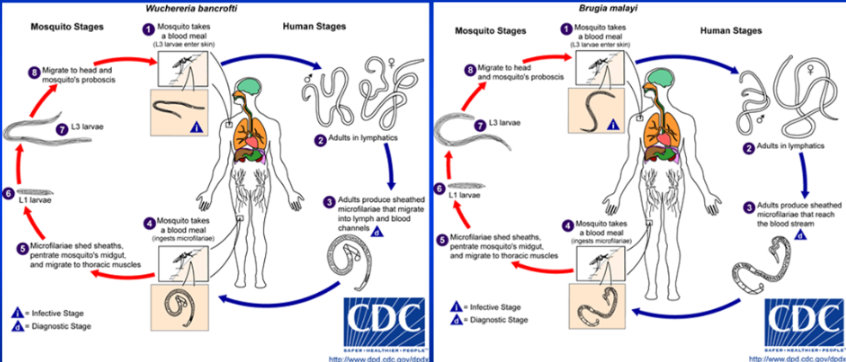
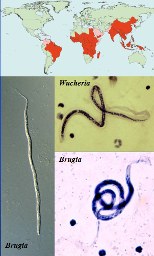
*Wucheria bancrofti and Brugia malayi (lymphatic filariases)
ORDER SPIRURIDA, FILARIDS
-
HOST: humans (DH), mosquitoes - culicine (IH)
-
MORPHOLOGY:
slender, live 10-15y
restricted to tropics (temperature)
microfilariae are sheathed, live 3-5y
-
LIFE CYCLE:
mosquito > L3 enter skin > mature in lymphatics > sheathed microfilariae > lymph and blood channels
mosquito > microfilariae penetrate gut > larvae > L3 > migrate to proboscis
-
PATHOGENESIS + DIAGNOSIS
lymphodema, lymph accumulation at body sites
diagnosed by finding microfilariae in blood
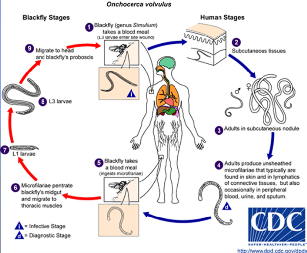
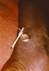
*Onchocera volvulus (river blindness)
ORDER SPIRURIDA, FILARIDS
-
HOST: humans (DH), blackflies (IH)
-
MORPHOLOGY:
slender, within subcutaneous nodules
microfilariae unsheathed, in subcutaneous connective tissue
-
LIFE CYCLE:
blackfly > L3 into bite wound > mature in subcutaneous nodules > unsheathed microfilariae
blackfly > microfilariae penetrate gut > larva > L3 > migrate to proboscis
-
PATHOGENESIS + DIAGNOSIS:
subcutaneous nodules form around adults
retinal lesions = blindness
from migrating microfilariae
diagnosed through skin snips to isolate microfilarea from subcutaneous location
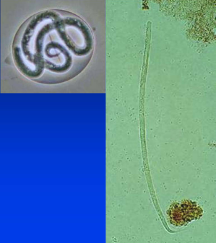
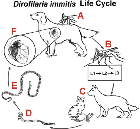
*Dirofilaria immitis (canine heartworm)
ORDER SPIRURIDA, FILARIDS
-
HOST: dogs (primarily) (DH); culicine mosquitoes (IH)
-
MORPHOLOGY:
slender, ~35cm long, right caudal lobar artery
microfilaria tapered anterior end
infections can also be in pulmonary arteries and right ventricle
temperature dependent development (>14C)
Canada = June-October
-
LIFE CYCLE (indirect)
mosquito > microfilaria in blood > circulate
mosquito > L3 > migrate to proboscis
DH > larvae > pulmonary circulation > arteries > mature
-
PATHOGENESIS + TREATMENT:
inflamation and arteritis, thromboemboli (lung capillaries)
increased vascular resistance = hypertension
heart dilation, hyperthropy, congestive heart failure
damage may take time to develop
antigen testing and knotts test available
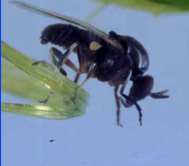
ARTHOPODA LIFE CYCLES
Simple
Complex
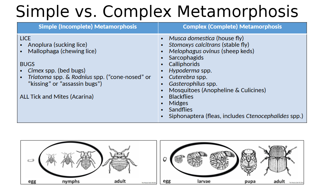
CLASS INSECTA (general)
Jointed appendages - paired bilaterally, symmetrical
Chitinous exoskeleton (most important identifier)
Body segmented: exoskeleton does not flex
3 body divisions: head, thorax, abdomen
3 pairs of legs
Sometimes wings – usually 2 pairs
Separate sexes

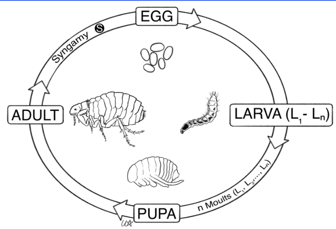
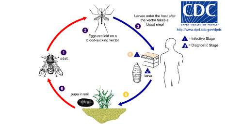
ORDER DIPTERA life cycle
complex metamorphosis
egg > larva (1) > larva (n) > pupa > adult
Culcids, simulids, ceratopogonids Psychodids, tabanids - require aquatic/semi-aquatic habitats for egg laying and development of immatures
Muscids - breed in immature animal manure/decaying organic matter
Hippoboscids - most of life on host
Sarcophagid, Calliphorid - egg laid on host/decomposing carcass, larvae develop in decomposing flesh in living/dead animal, pupae in soil
Bot flies - egs laid on hair of host/near burrows, larvae develop in host, pupate in soil

SUBORDER NEMATOCERA (general)
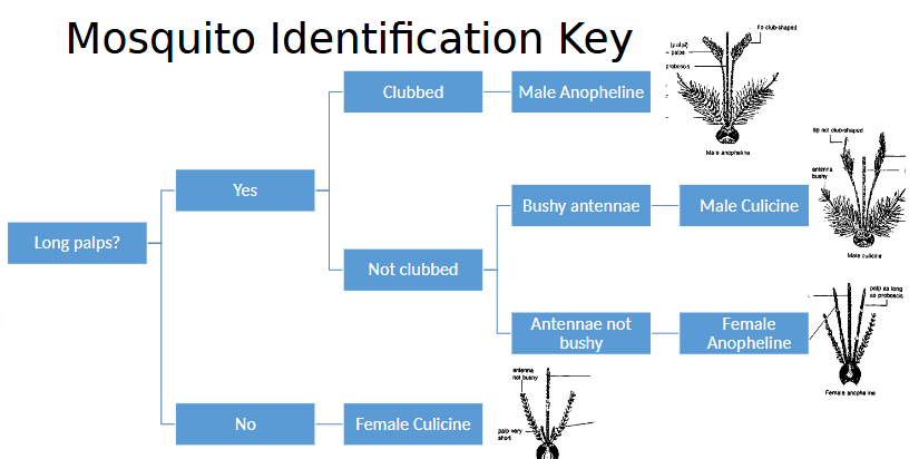
Family Culicidae: Culcinae, Anophelinae (mosquitoes)
Family Simulidae: Stimulium (blackfly/buffalo gnat)
Family Ceratopogonidae: Culcoides (midges/no-see-ums)
Family Psychodidae: Lutzomiya (sandflies)
characteristics
Long, segmented antennae
Piercing-sucking mouthparts
Females only blood feed to generate eggs
Males nectar feeders
Worldwide distribution
Significance
Annoying, causes worry
Stress generated by attacking insects leads to a physiological response > will change behaviour to avoid getting attacked
May cause anemia
Some transmit pathogens
Are neat blood feeders (siphoned), transmitted usually through saliva during next blood meal
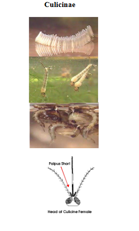
CULCINAE
Diptera > Nematocera > Family Culcidae
“Northern” mosquitoes
Transmit viruses – West Nile, Yellow Fever, non-human malarias
Biological vectors of filarial worms
Common “pest” mosquitoes
Frequently numerous even in northern temperate areas
Strong fliers, can frequently bite in both day and night
Sight feed and track by carbon monoxide
LIFE CYCLE:
Females feed with abdomen held horizontally
Eggs laid in groups forming rafts
Females lay eggs while on surface tension of water
Larvae hang vertically in water

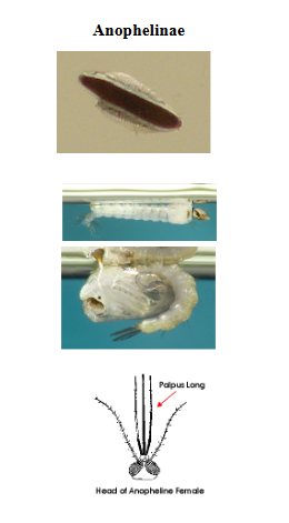
ANOPHELINAE
Diptera > Nematocera > Family Culcidae
Anophelinae (‘anophelines’): warm temperate and tropical areas
Transmit viruses – human malaria vectors (some)
Biological vector of some arboviruses (ex: malaria)
Relatively weaker fliers, frequently bite in evening/night
Crepuscular – twilight feeders
Feed when air is still and more humid – follow carbon monoxide
LIFE CYCLE:
Females feed with abdomen in raised position
Eggs laid singly
Larvae hang horizontally in water with a siphon for breathing
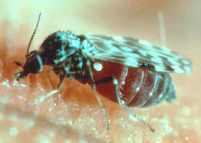
STIMULIUM (blackfly/buffalo gnat)
Diptera > Nematocera > Family Simulidae
Gets name from thorax hump that resembles buffalo
Usually associated with running water
Larvae found attached to rocks
Biological vectors of river blindness (Onchocera volvulus – nematode-caused)
Parasite causes damage to retina
Biological vector of Leucocytozoon simondi
Painful and annoying biters
CULCOIDES (midges/no-see-ums)
Diptera > Nematocera > Family Ceratopogonidae
Larva sually associated with still water
Feeds when air is still and humid
Dappled wings
Quite small, painful bites
Some transmit arboviruses and other agents of disease (ex: malaria-like parasites – birds)
Biological vector of Haemoproteus
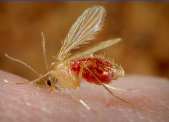
LUTZOMIYA (sandflies)
Diptera > Nematocera > Family Psychodidae
Dainty, hairy flies
Biological vectors of parasitic disease Leishmania (caused by flagellates)

SUBORDER BRACHYCERA (general)
Characteristics
Stylate antennae
Slicing mouthparts, feed by telmophagy
Both sexes blood feed
Significance
Painful bites – can get repetitive bites from the same fly
Loud fliers, persistent, can cause worry
Are good at mechanically transmitting parasites
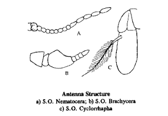
SUBORDER CYCLORRHAPHA (general)
Family Muscidae: Musca domestica (house fly), Stomoxys calcitrans (stable fly)
Family Hippoboscidae: Melophagus Ovinus (sheep ked)
Family Sarcophagidae (flesh fly)
Family Calliphoridae (screworm/blow fly/bottle fly)
Family Hypodermatidae: Hypoderma (cattle grub/warble fly)
Family Cuterebridae: Cuterebra (rodent/rabbit bot)
Family Gasterophilidae: Gasterophilus (stomach bot)
Characteristics
Aristate (feathered) antennae
Various mouthparts depending on what they feed on
Include various robust flies
House, stable, face, flesh, warble, bot flies
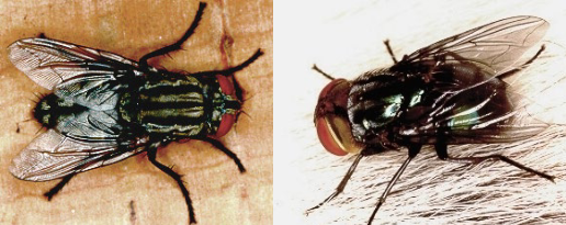
MUSCA DOMESTICA (house fly)
Diptera > Cyclorrhapha > Family Muscidae
Characteristics
Robust, hairy body
Aristate (feathered) antennae
Sponging-lapping mouthparts - non-biting
Strong flier: usually uses sight to navigate, can feed during day
Life cycle (complex, extremely prolific)
Eggs are laid in undisturbed cattle feces or other rotting organic matter
Maggots eat organic debris
Pupation occurs in soil
Significance
Annoying, nuisance
Efficient mechanical vector for pathogens – macroscopic structures get stuck on hairs around mouthparts while feeding
Feeding habits promote transmission: regurgitation from past feeding items
Biological vector – Habronema and Draschia of horses
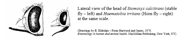
STOMOXYS CALCITRANS (stable fly)
Diptera > Cyclorrhapha > Family Muscidae
Characteristics
Body similar to that of Musca spp.
Slightly smaller and have a proboscis for blood feeding
Aristate (feathered) antennae
Bayonet-shaped mouthparts – less mechanical transmission than sponging-lapping, biting
Life cycle (complex)
Eggs laid in decaying vegetation
Maggots develop in damp straw, etc.
Pupation in soil, overwinter as pupae
Adults attack mammals during midday – strong fliers, rely on sight for feeding
Adults pierce the skin for blood, are persistent and annoying
Significance
Painful bite, persistent feeders that are easily disturbed
Can cause annoyance and worry
Could lead to reduced milk production and weight gains in production animals
Intermediate host for Habronema sp. (nematode of horses)

FAMILY MUSCIDAE CONTROL
Sanitation Measures
Timely removal of manure to reduce number of breeding sites
General cleanliness to remove sites with rotting vegetation
Ensure proper drainage to remove areas of standing water/muck
Insecticides
Ear tags with topical organophosphates or botanicals against face and horn flies
Spray/pour-on formulations
Back rubbers
Chitin inhibitors in feed of cattle to make manure unsuitable for larval development
Efficient for fly control but pasture productivity may be affected negatively
Are rarely used for flies anymore
MELOPHAGUS OVINUS (sheep ked)
Diptera > Cyclorrhapha > Family Hippoboscidae
Characteristic
Wingless fly
Flattened dorso-ventrally
Leathery, hairy
Permanent ectoparasite of sheep in their wool
Life cycle (complex)
Mature larvae laid by females spin cocoons immediately
No free-living larvae except for when it is laid before immediately going into pupa form
This method was adapted for survival in wool surroundings
Significance
Irritation, blood loss
Wool damage through rubbing and staining
Control
Topical insecticides
Usually a winter issue
higher proximity of animals and weakened immune systems/stress
humidity and cold temperatures can increase parasite survivability
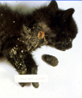
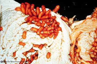
Myiasis
invasion of healthy or damaged flesh by the larvae of dipteran flies
invasion is usually accidental, role is to get rid of rotting composition
facultative: usually accidental, not necessary to continue life cycle
generally start with a wound of some kind that attracts flies
accidental egg disposition on living animal, mistakenly recognizes would as a dead animal
Family calliphoridae, Family Sarcophagidae
obligate: do not need a wound, invasion of tissue needed to survive, on live animals only
Bot flies > Hypoderma, Cuterera, Gasterophilus
FAMILY SARCOPHAGIDAE & FAMILY CALLIPHORIDAE
Diptera > Cyclorrhapha > Facultative Myiasis (2)
larger than a housefly
sponging lapping mouthparts
C: metallic colouring (adult), screw worm (larvae) - large and robust
S: checkboard patterning (adult), maggots (larvae) - large and robust
larva have pointed anterior end with mouth hooks, posterior end broad and flat with spiracular plates
life cycle
eggs usually laid in or around wounds, soiled skin/wool, decaying carcasses
species-specific level of decomposition preferred
larvae develop at or near site of oviposition
mature larvae leave host to pupate
predisposing causes for fly attack
open wounds from any cause, especially putrefying flesh
decomposing wool on soiled/wet sheep (wool strike)
may result from heavy rain, urine, uterine discharge, diarrhea, sweat, etc.
wool is a type of protein, wool can start rotting in certain conditions
locally, intestinal parasitosis resulting in diarrhea are a significant cause of wool decay and subsequent fly attack
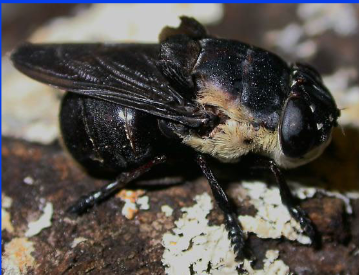
BOT FLIES (general)
Hypoderma, Cuterebra, Gasterophilus, Dermatobia hominis
characteristics (obligate myiasis)
all energy required for life cycle are obtained in larval form
large larvae must store energy for metamorphosis and egg-laying
large, bee-like adults (protective mimicry) – do NOT feed
do not sting, have no working mouthparts
usually well-prescribed sites of development, adapted specific hosts and locations
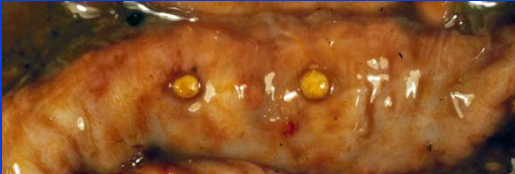
HYPODERMA (cattle grub/warble fly)
Diptera > Cyclorrhapha > Family Hypodermatidae
Hypoderma bovis: Northern Cattle grub
Larvae overwinter in spinal canal or epidural fat
Hypoderma lineatum: Southern/common cattle grub
Larvae overwinter in esophageal connective tissue
Life cycle (complex, once cycle per year in ON)
Eggs laid on legs of cattle by non-feeding adult flies
Larvae penetrate skin > migrate to overwintering sites by winter
Have thin exoskeletons, no need for large air source
In late Feb to Apr > migrate to back and produce warble (subcutaneous cyst)
Larvae mature, drop out of cyst and pupate
Pathogenesis
Gadding – disperse flies, results from oviposition, reduced production (takes energy)
holes in hides reduce value, lesions may require trimming
in horses, loss of use if warble is in saddle area
CUTEREBRA (rodent/rabbit bot)
Diptera > Cyclorrhapha > Family Cuterebridae
characteristics
adults – robust and bee-like flies with vestigial mouthparts
larvae – large with prominent spines, found within subcutaneous cysts of host
are strong fliers, tend to affect ground rodents and squirrels
life cycle (complex)
eggs found near burrow openings
larvae enter host via nose/mouth (most likely through animal grooming habits)
when they detect CO2 = means that animals are near
migrates to skin and cuts breathing hole
needs air once it reaches a particular size
breathing hole is location of emergence = area depends on size of animal
pupation on ground (over-wintering)
adults mate and lay eggs the following year
pathogenesis
subcutaneous fibrotic cyst – never gets septic (larvae produce antibiotic substances) - usually around neck in cats and dogs
wet coat in neck region, small hole
larvae can, but rarely, enter CNS
DERMATOBIA HOMINIS (human bot fly)
Diptera > Cyclorrhapha > Family Cuterebridae
Life cycle
female bot flies capture blood sucking arthopods and lay eggs on their bodies
use glue-like substance for adherence
bot fly larvae develop within eggs but remain on paratenic host until it takes a blood meal
larvae hatch in response to heat of vertebrate’s skin and penetrate host tissue
feed for a few weeks and cut breathing hole in skin
mature larvae drop into ground and pupate
GASTEROPHILUS (stomach bot)
Diptera > Cyclorrhapha > Family Gasterophilidae
Gasterophilus intestinalis: most common in horses
Gasterophilus nasalis: in horses, more uncommon locally
Life cycle (complex)
Most eggs laid on forelegs
Intestinalis: feed on food inside stomach but do not invade stomach lining
Oral migration > 2nd stage larvae is swallowed
nasalis: hatch from eggs near mouth and crawl into mouth
intestinalis: hatch from eggs only when horse links/muzzles area containing eggs > larvae enter mouth
Larvae overwinter in stomach and pass out with feces
Pathogenesis
Usually asymptomatic, gastric lesions can be significant
Rarely see perforations (can occur giving abscesses and peritonitis)
Control: Many registered compounds are available (ex: insecticides, endectocides)
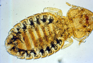
ORDER MALLOPHAGA (biting/chewing lice)
Characteristics
Highly host-specific
Feed on skin, hair, secretions, feathers
rersults in rubbing and scratching > hair loss and hide damage
Wingless, flattened dorsoventrally
Head is as broad or broader than the thorax (looks like letter M)
Large head holds muscles to help with biting and chewing
Life cycle (simple)
Eggs (nits) cemented to hairs/feathers
Life cycle around 2-4w
Entire life cycle on host, generally short survival of of the host
Are usually active or highly active
Fast moving, can move around if disturbed > difficult to see on living animal