Human Anatomy - Chapter 1
1/43
Earn XP
Description and Tags
A First Look At Anatomy
Name | Mastery | Learn | Test | Matching | Spaced |
|---|
No study sessions yet.
44 Terms
1. Distinguish between ‘anatomy’ and ‘physiology’.
Anatomy ("to cut apart")
→ The study of the structure of the body and how its parts are organized. (What things look like and where they are).Dissection
→ Cutting open a body carefully to study its parts.
Physiology
→ The study of how body parts work and what they do. (How things function).
2. Distinguish between ‘microscopic anatomy’ and ‘gross (macroscopic) anatomy’
Microscopic Anatomy
Looks at body parts that are too small to see without a microscope.
Includes:
Cytology → study of cells
Histology → study of tissues
👉 Example: Looking at blood cells under a microscope.
Gross (Macroscopic) Anatomy
Looks at larger body parts that you can see with the naked eye.
Examples: Heart, spleen, muscles, nerves
👉 Example: A doctor looking at your muscles or organs during surgery.
✨ Quick Memory Trick:
Micro = tiny (cells + tissues)
Gross = big (organs you can see)
3. Students will describe several approaches to gross anatomy and distinguish between.
** Know the name of the branch and the study of
1. Comparative - Examine the similarities and differences across species.
2. Developmental anatomy - It studies how the body changes as it grows and develops. From the very beginning (conception) → through embryo, fetus, childhood, and into adulthood.
3. Embryology - Examines the developmental changes occurring prior to birth
4. Regional anatomy - Studies everything in one body area.Example: Looking at all the bones, muscles, nerves, and blood vessels in the head/skull.
5. Surface anatomy- Studies outside markings of the body and how they connect to inside structures. Example: Checking a pulse on the wrist or drawing blood from a vein.
6. Systemic anatomy - Studies the gross anatomy of each organ system in the body. Studies the body by organ systems. Example: Studying the digestive system (stomach, intestines, liver) or the urinary system.
Surface (topographical) anatomy - surface marking of the body to understand the relationships of deep or internalres that can be visualzied with x-rays, CT scans, MRI, and other technologies.
9. Clinical anatomy - The application of anatomy to the practice of medicine, dentistry, and other health-related sciences; for example, to aid in the diagonsis and treatment of disease.
10. Pathological anatomy diagnosis(path= = disease) - Structural changes (from gross to microscopic) associated with disease.
11. Pathologic Anatomy
Anatomic changes resulting from disease
Studies how diseases change body structures.
Example: Looking at lung tissue damaged by cancer.
12. Radiographic Anatomy
Uses medical imaging (X-ray, MRI, CT scan, ultrasound) to study body parts.
Example: Using an X-ray to see if a bone is broken.
13. Surgical Anatomy
Uses anatomic landmarks (visible or felt parts of the body) to guide surgery.
Example: Using the hip bones to find the L4 vertebra before back surgery.
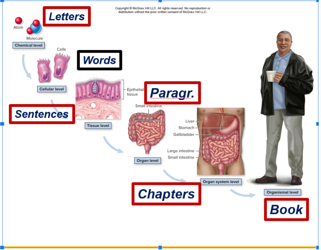
4. Identify the various levels of structural organization of the body from atom to organ system and say how each contributes to the formation of the entire organism.
Question exam
Identify name of organ system
Organ systems that belong to that organ system
And all its functions
Individual Atoms (H+H+O = H2O (becomes a molecule)(letters) → Molecules (letters) → Cells (words) —> Tissues (sentences ) —> Organs (paragraphs) —> Systems
(Chapters) —-> Organism (Book).
Tissue - Has the group of the cells
Organ level - when more than one level of tissues come together
Organ system level - urinary systems filters liver - each come together like bladder, liver, urethra
Chemical level (letters of alphabet)
Atoms and molecules - Na+, H,C,H+O2 & H2O, NaCI
Cellular level (Words)
Nerve, muscle, bone cell, blood cell, liver cell
Tissue level (sentences)
Epithelial, connective, muscular & nervous
Organ level (paragraphs)
Small intestine, stomach, spleen, liver, brain
Organ System level (Chapters)
Respiratory, digestive, urinary systems
Organismal level (‘ a book’)
The human
Identify the following organ systems (Figure 1.4) and describe their major functions
There are 11 commonly organized organ systems in the human body
Each contains organs that work together to perform specific functions
All organ systems make up the organsim
Integumentary - Largest organ of the body (covers entire surface, ~7–8% of body weight).
Made of different tissues:
Epidermis → stratified squamous epithelium (outer protective layer).
Dermis → dense connective tissue (strength, elasticity, blood supply).
Subcutaneous (hypodermis) → loose connective + fat (anchors skin, insula
Functions of the Integumentary System
Protection
Barrier against injury, chemicals, microbes, heat/cold, UV radiation.
Selectively permeable (lets some things in, blocks others).
Contains melanin → protects against sun damage.
Prevents Water Loss & Gain
Skin is water-resistant, prevents swelling and dehydration.
Controls trans-epidermal water loss (TEWL).
Sweat glands regulate water/salt balance.
Temperature Regulation
Hot: Blood vessels widen (vasodilation) → more sweat → heat released.
Cold: Blood vessels narrow (vasoconstriction) → less blood near surface → conserve heat.
Metabolic Regulation
Skin makes Vitamin D₃ (cholecalciferol) when exposed to UV light.
Converted in the kidney to calcitriol, which regulates calcium & phosphate for bone health.
Immune Defense
Skin contains immune cells (epidermal dendritic cells) that fight pathogens and detect cancer cells.
Sensory Reception
Nerve endings detect touch, temperature, pressure, pain, vibration, texture.
Example: Tactile (Merkel) cells sense fine touch.
Secretion & Excretion
Sweat glands → release water, salts, and urea (waste).
Sebaceous (oil) glands → release sebum, which lubricates and waterproofs skin & hair.
Story Method (Imagine Your Skin as a Superhero Suit 🦸)
Protection → The suit is your armor, blocking punches, germs, and UV rays.
Water Balance → The suit is waterproof, so you don’t puff up in the rain or dry out in the sun.
Temperature Regulation → The suit has a thermostat: cooling fans (sweat) when hot, insulation (vasoconstriction) when cold.
Metabolic Regulation → The suit has solar panels that turn sunlight into Vitamin D power.
Immune Defense → Tiny security guards (immune cells) patrol the suit to fight invaders.
Sensory Reception → The suit has built-in sensors to feel heat, pain, touch, vibration.
Secretion → The suit has waste vents (sweat glands) and oil spray (sebum) to keep it smooth.
1. What are the two major layers of the integument and the general components of each?
Epidermis → Outer layer, made of stratified squamous epithelium.
Function: Protection, barrier, waterproofing.
Dermis → Deeper layer, made of dense irregular connective tissue + areolar connective tissue.
Contains: Blood vessels, nerves, sweat glands, sebaceous glands, hair follicles, sensory receptors.
Function: Strength, flexibility, support, sensation, and nutrient supply to the epidermis.
2. In what ways does the skin protect us, and why is it said to be selectively permeable?
Protection:
Shields against physical injury (bumps, scrapes).
Blocks harmful chemicals, toxins, and microbes.
Protects from UV radiation (via melanin).
Prevents deeper tissues from damage.
Selectively permeable:
Some things can pass through (like drugs from a patch or certain chemicals).
Most harmful substances, water, and microbes are blocked.
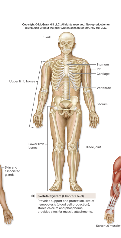
Skeletal (Ch. 6-9)
Provides support and protection, the site of hemopoiesis (blood cell production), stores calcium and phosphorus, and provides sites for muscle attachments.
Organs that belong to this organ system are the skull, upper limb bones, sternum, Rib, Cartilage, vertebrae, sacrum, upper limb bones, lower limb bones, knee joint.
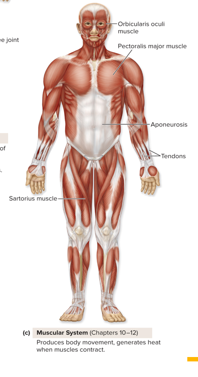
Muscular (Ch. 10-12)
Produces body movement and generates heat when muscles contract.
Organs: Orbicularis oculi muscle, pectoralis major muscle, aponeurosis, tendons, sartorius muscle.
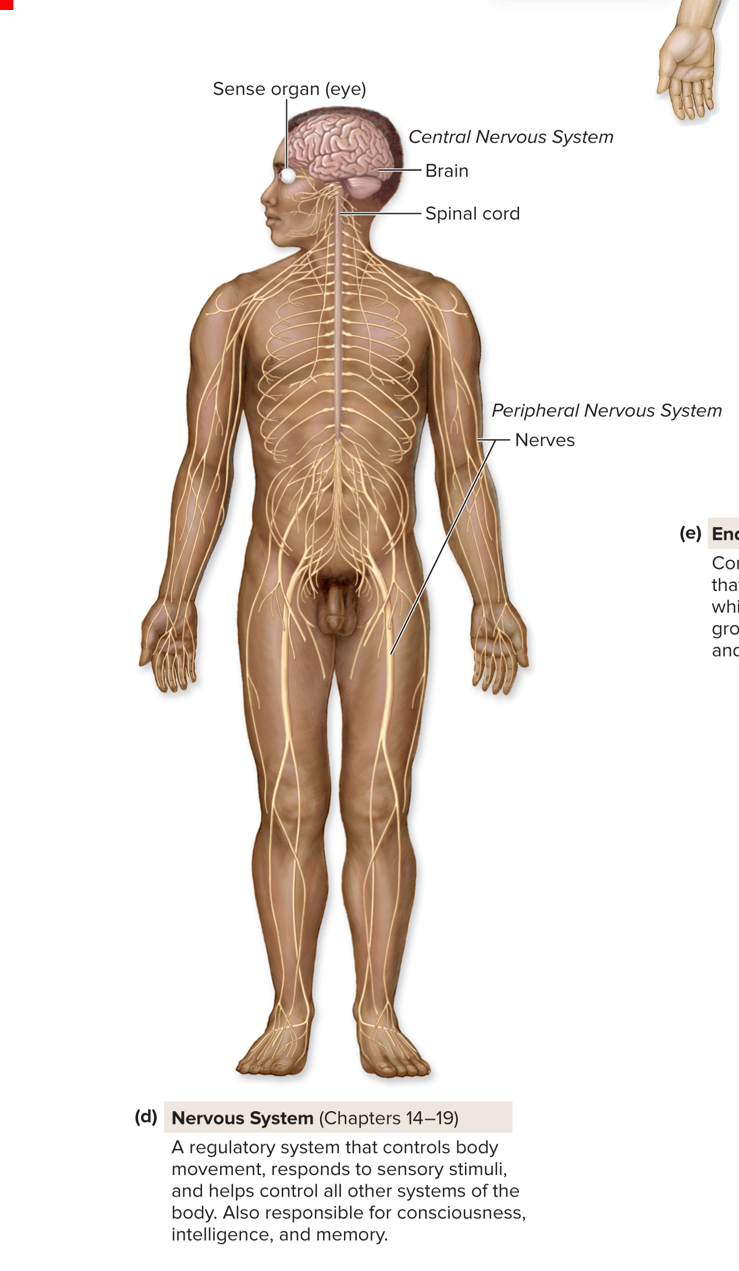
Nervous system (Chapter 14-19)
A regulatory system that controls body movement, responds to sensory stimuli, and helps control all other systems of the body. Also responsible for consciousness, intelligence, and memory.
Organs: sense organs (eye), central nervous system (brain and spinal cord), Peripheral nervous system (Nerves).
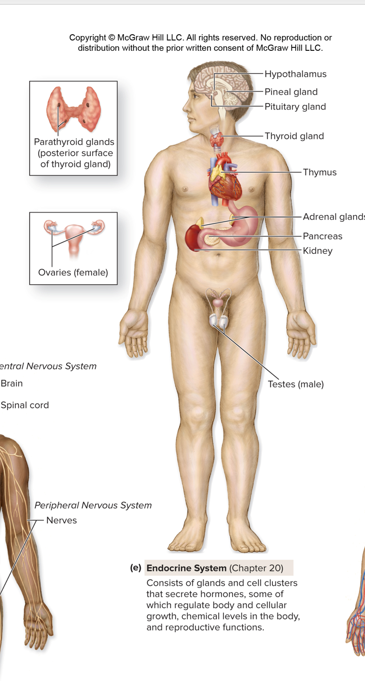
Endocrine system (Chapter 20)
Consists of gland and cell clusters that secrete hormones, some of which regulate body and cellular growth, chemical levels in the body and cellular growth, chemcial levels in the body, and reproductive functions.
Organs:
Hypothalamus – Links the nervous system to the endocrine system; controls pituitary gland.
Pineal gland – Regulates sleep-wake cycles (produces melatonin).
Pituitary gland – “Master gland”; controls growth, reproduction, and other glands.
Thyroid gland – Controls metabolism, energy, and growth.
Parathyroid glands – Regulate calcium levels in blood and bones.
Thymus – Helps immune system by maturing T-cells (in children).
Adrenal glands – Control stress response (adrenaline, cortisol), blood pressure, and metabolism.
Pancreas – Regulates blood sugar by releasing insulin and glucagon.
Kidneys – Filter waste; also release hormones (like renin, EPO) for blood pressure and red blood cells.
Ovaries (female) – Produce eggs and female sex hormones (estrogen, progesterone).
Testes (male) – Produce sperm and male sex hormone (testosterone).
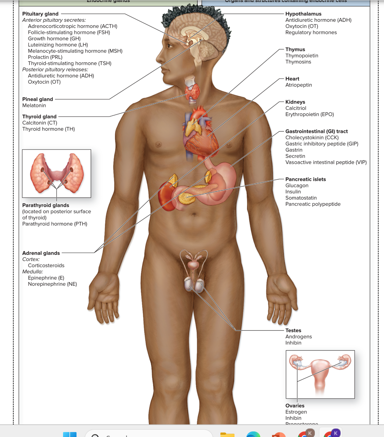
Hypothalamus → ADH, Oxytocin, regulatory hormones.
Pituitary gland
Anterior → ACTH, FSH, GH, LH, MSH, PRL, TSH.
Posterior → ADH, Oxytocin.
Pineal gland → Melatonin.
Thyroid gland → Calcitonin (CT), Thyroid hormones (TH: T3 & T4).
Parathyroid glands → Parathyroid hormone (PTH).
Thymus → Thymopoietin, Thymosins.
Heart → Atrial natriuretic peptide (Atriopetin/ANP).
Kidneys → Calcitriol (vitamin D hormone), Erythropoietin (EPO).
Adrenal glands
Cortex → Corticosteroids (e.g., cortisol, aldosterone).
Medulla → Epinephrine (E), Norepinephrine (NE).
GI tract → CCK, GIP, Gastrin, Secretin, VIP.
Pancreatic islets → Glucagon, Insulin, Somatostatin, Pancreatic polypeptide.
Testes → Androgens (mainly Testosterone), Inhibin.
Ovaries (not labeled here but included) → Estrogens, Progesterone, Inhibin.
Comparison of the Nervous system and the Endocrine system
Nervous system
Communication Method - A nerve impulse causes neurotransmitter release from a neuron into a synaptic cleft.
Effector - Neurons, muscle cells, and gland cells with a receptor for the neurotransmitter.
Response time - Rapid reaction time: Typically milliseconds or seconds.
Range of Effect: Typically has localized, specific effects in the body.
Duration of response: Short-term: Millisecond; terminates with the removal of stimulus.
Endocrine System
Communication Method - Endocrine glands secrete hormones into blood; hormones are transported withing the blood to effectros throughout body.
Effector: Any cell in the body with a receptor for the hormone
Response time: Relatively slow reaction time: seconds to minutes to hours.
Range of effect:Typically has widespread effects throughout the body.
Duration of response: Long-lasting: minutes to days to weeks; may continue after stimulus is removed.
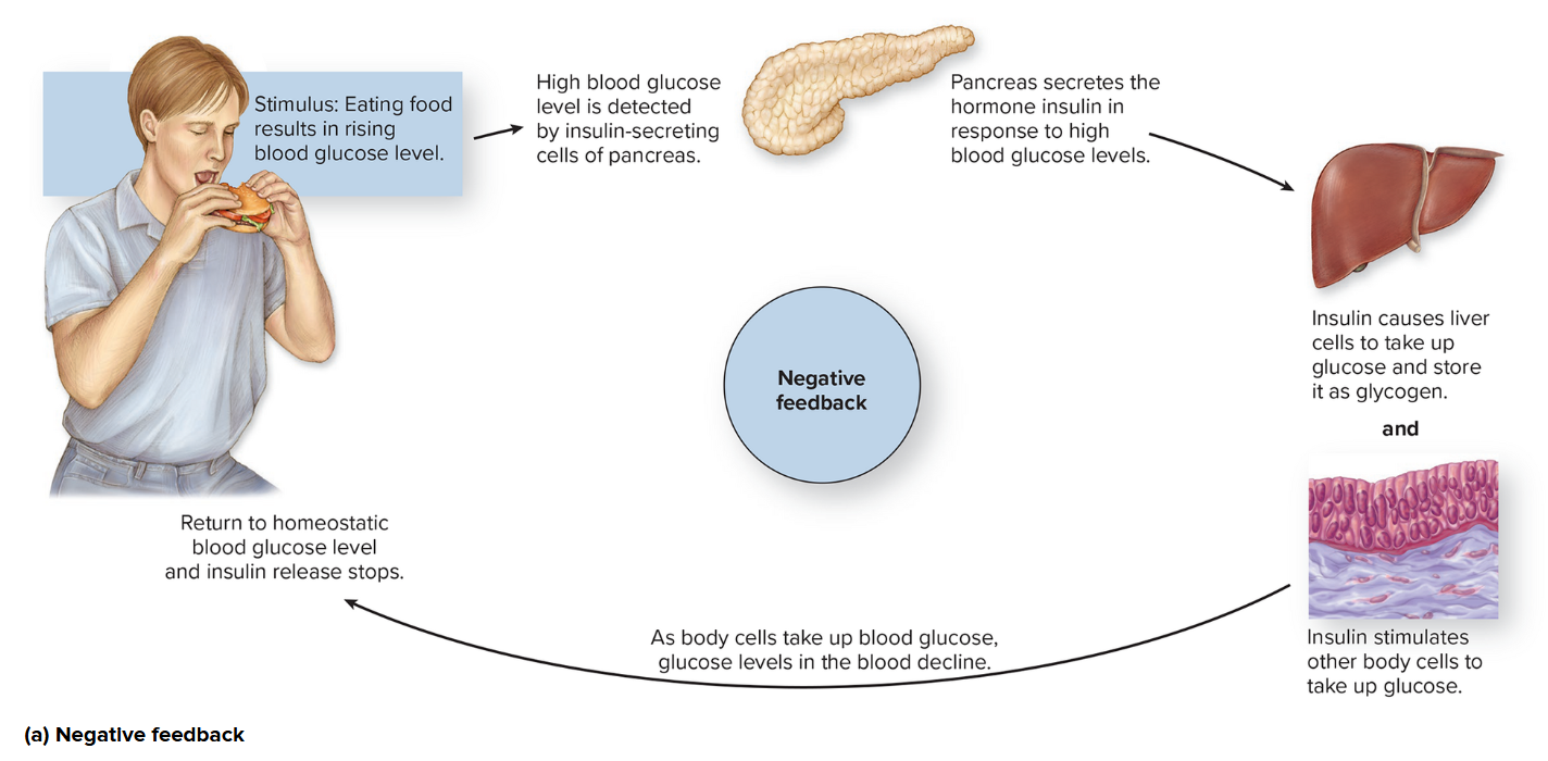
Negative feedback
Stimulus: Eating Food results in a rising blood glucose level.
High blood glucose level is detected by insulin-secreting cells of the pancreas.
Pancreas secretes the hormone insulin in response to high blood glucose levels.
Insulin causes liver cells to take up glucose and store it as glycogen. and insulin stimulated other body cells to take up glucose.
As body cells take up blood glucose, glucose levels in the blood decline.
Return to homeostatic blood glucose level, and insulin release stops.
Negative feedback occurs when the end product acts tot turn off or slow down the pathway.
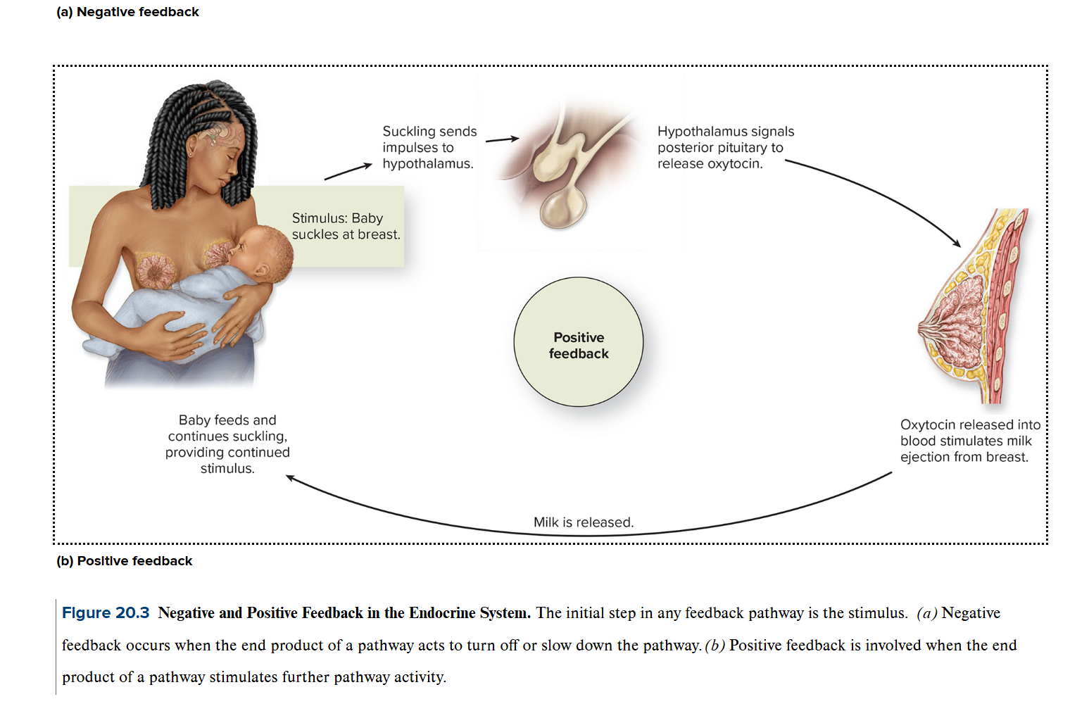
Postive Feedack
Positive feedback is involved when the end product of a pathway stimulates further pathway activity.
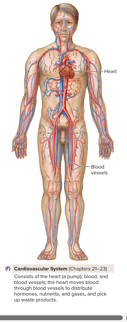
Cardiovascular Lymphatic (Ch 21-23)
Consists of the heart ( a pump), blood, and blood vessels; the heart moves blood through blood vessels to distribute hormones, nutrients, and gases, and pick up waste products.
Main parts:
Heart → the pump.
Blood → carries oxygen, nutrients, hormones, and waste.
Blood vessels → arteries (carry blood away), veins (carry blood back), and capillaries (exchange).
Functions:
Moves blood through the body.
Delivers oxygen, nutrients, and hormones to cells.
Removes carbon dioxide and waste.
Helps regulate body temperature and maintain homeostasis.
Organs: Heart & Blood vessels;
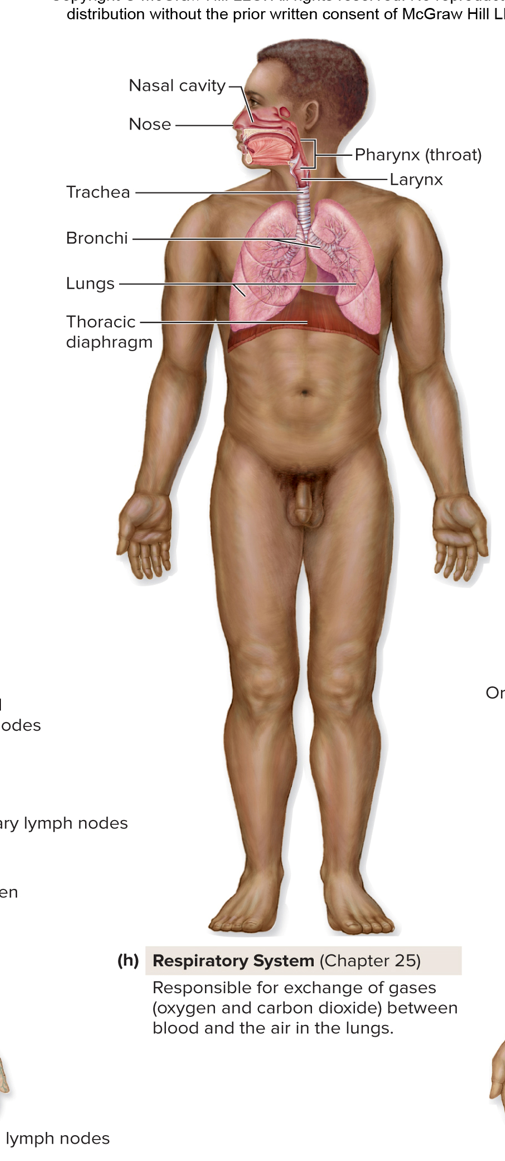
Respiratory System (Chapter 25)
Respiratory System – Main Functions
Breathing (Ventilation)
Inhalation (air in) and exhalation (air out).
Gas Exchange
External respiration: Exchange of gases between air in lungs and blood (O₂ in, CO₂ out).
Internal respiration: Exchange of gases between blood and body cells.
Gas Conditioning
Incoming air is warmed, humidified, and cleaned before reaching lungs.
Sound Production
Air moving through larynx + upper respiratory tract → produces speech & other sounds.
Olfaction (Smell)
Airborne molecules stimulate olfactory receptors in nasal cavity → sense of smell.
Defense
Nose hairs, mucus, cilia, enzymes (like lysozyme), and immune cells trap dust, microbes, and allergens → protect against infection.
Organs: Nasal cavity, Nose, Pharync(throat), larynx, Trachea, Bronchi, Lungs, Thoracic Diaphragm.
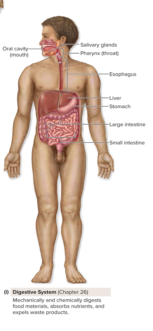
Digestive system (Chapter 26)
Digestive System Overview
Two groups of organs:
GI tract (digestive tract / alimentary canal): Continuous tube:
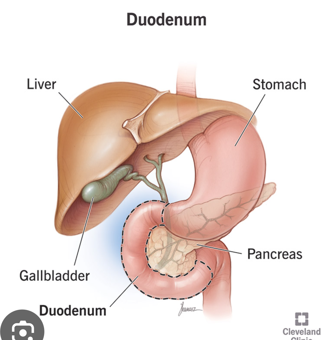
Mouth → Pharynx → Esophagus → Stomach → Small intestine → Large intestine → Anal canal → AnusAccessory digestive organs: Teeth, tongue, salivary glands, liver, gallbladder, pancreas.
IngestionTaking in food & drink (mouth).
Motility
Movement of food along GI tract:
Peristalsis → wave-like contractions push food forward.
Mixing/Segmentation → churning back-and-forth (esp. in small intestine).
Secretion
Release of digestive juices (enzymes, bile, acid, mucus) to aid digestion & protect GI tract.
Digestion
Mechanical digestion → physically breaking down food (chewing, churning).
Chemical digestion → breaking food into molecules with enzymes.
Absorption
Nutrients, water, and electrolytes absorbed into blood or lymph from small & large intestine.
Elimination
Undigested material + waste compacted into feces and removed by defecation.
Organs: Oral Cavity(mouth), Salivary glands, Pharynx (throat), esophagus, Liver, stomach, large intestine, small intestine.
Urinary System (Chapter 27)
Filters the blood and removes waste products from the blood, concentrates waste products in the form of urine, and expels urine from the body.
Organs of the Urinary System
Kidneys
Bean-shaped, filter blood, remove waste, form urine.
*** Kidneys maintain blood pressure
Ureters
Tubes that carry urine from kidneys → bladder (via peristalsis).
Urinary Bladder
Muscular sac, stores urine until ready to urinate.
Urethra
Tube that carries urine from bladder → outside of body.
Functions of the Urinary System
Excretion of wastes → Removes urea, toxins, and extra water.
Storage of urine → Bladder holds urine until convenient to release.
Elimination of urine → Urethra expels urine (urination).
Regulation of blood volume & pressure → Kidneys adjust fluid levels (affects blood pressure).
Regulation of red blood cell production → Kidneys release erythropoietin (EPO) when oxygen is low → stimulates bone marrow to make RBCs.
Regulation of ion levels → Controls sodium (Na⁺), potassium (K⁺), calcium (Ca²⁺), phosphate (PO₄³⁻).
Regulation of acid-base balance → Balances hydrogen ions (H⁺) and bicarbonate (HCO₃⁻) to maintain pH.
Kidenys
Posterior abdominal wall; right kidney placement is inferior (lower) to left kidney placement.
Paired, bean-shaped organs; composed of puter cortex and inner medulla.
Function: Filter blood and process filtrate to tubular fluid, then urine. Processing in the nephron: The filtrate then enters the renal tubule, where it is known as tubular fluid. As it travels through the nephron's segments, the fluid is continuously modified through two key processes:
Reabsorption: Necessary substances, including almost all the glucose and amino acids, and a significant amount of water and electrolytes, are transported from the tubular fluid back into the bloodstream.
Secretion: Waste products and excess ions that were not filtered initially are actively secreted from the blood into the tubular fluid for disposal.
Final product: By the time the tubular fluid reaches the end of the collecting duct, it has been concentrated and its composition has been finalized. At this point, it is called urine and is ready to be excreted from the body.
Ureters
Extend from kidneys to trigone of the bladder, along the posterior abdominopelvic wall.
Transport urine from kideny to urniary bladder via peristalsis (series of involuntary wave-like muscle contractions which move food along the digestive tract. )
Male reproductive system (chapter 28_
Produces male sex cells (Sperm) and male hormones (testosterone); transfers sperm to the female.
Female reproductive system (chapter 28)
Produces female sex cells (oocytes) and female hormones (estoregn and progesterone), receives seprm from male, site of fertilization of oocyte, site of growth and development of embryo and fetus, and produces and secretes breast milk for nourishment of newborn.
3. Describe the ‘anatomical position’.
Exam Question - Which one of the following is the correct standing anatomical position expect ?
Purpose:
Provides a universal way to describe locations and relationships in the body.
Features:
Standing upright.
Feet parallel, flat on the floor.
Head level, eyes looking forward.
Arms relaxed at sides.
Palms facing forward.
Thumbs pointing away from the body.
Advantages:
Makes it easier to visualize the body.
Helps understand the body’s organization into regions.
Allows clear description of the relationship between structures (e.g., “the heart is medial to the lungs”).
4. Describe the following planes and sections of the body? Exam Q - sections and plane
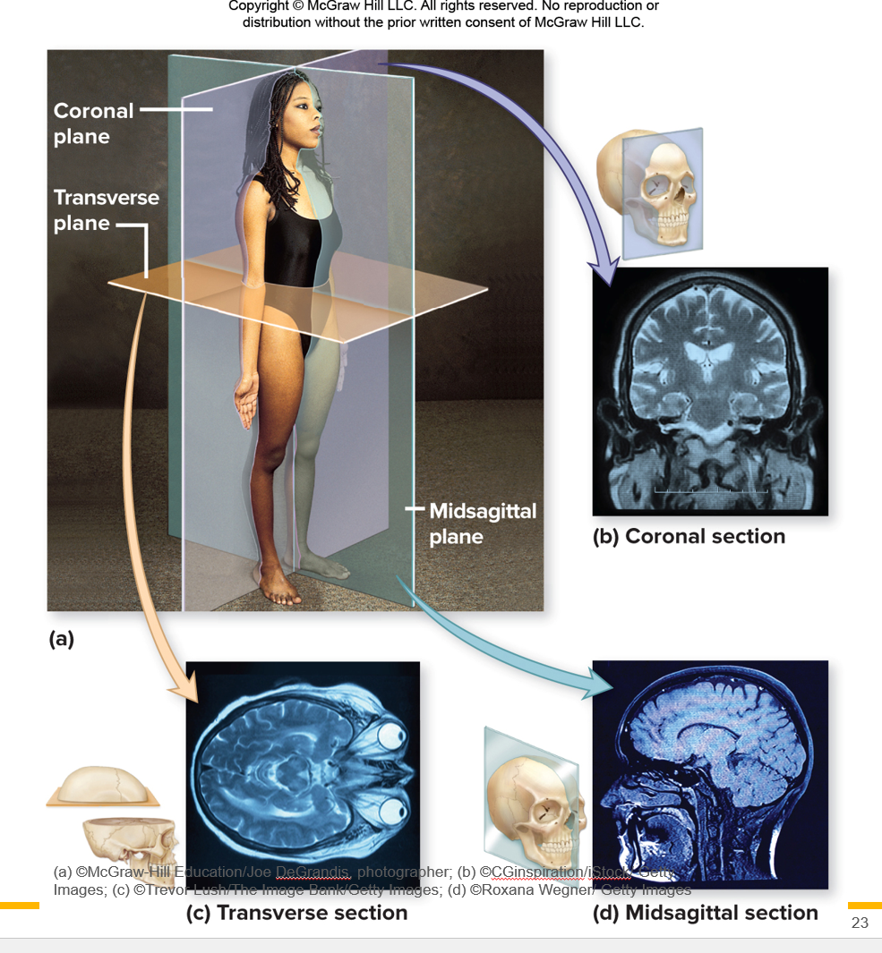
*** Planes of the Body = imaginary flat cuts through the body to help describe where things are. (Called the midline).
Coronal Plane (Frontal Plane)
Divides body into front (anterior) and back (posterior).
Example: A cut through both shoulders → front half vs back half.
Transverse Plane (Horizontal / Cross-Sectional Plane)
Divides body into upper (superior) and lower (inferior) parts.
Example: A cut at the waist → top half vs bottom half.
Midsagittal Plane (Median Plane)
Divides body into equal left and right halves.
Example: A line straight down the middle of your nose.
Other sagittal planes: Divide into unequal left and right parts.
Oblique Plane
Divides body at an angle.
Example: A diagonal cut, not straight up-down or side-to-side.
✨ Easy way to picture them:
Coronal = Crown cut 👑 (front vs back).
Transverse = Table cut 🍰 (top vs bottom).
Midsagittal = Mirror cut 🪞 (left vs right).
Oblique = Odd angle cut ✂ (diagonal).
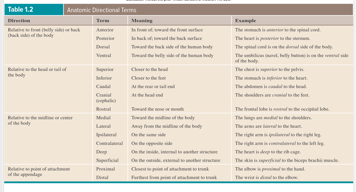
5. Compare and contrast the following pairs of anatomical directional terms and be prepared to describe the relationship of parts of the body one to the other using these terms. (Table 1.2 and Figure 1.7)
1. Anterior (ventral) vs. Posterior (dorsal)
Anterior (ventral) = front side
Posterior (dorsal) = back side
👉 Example: The sternum (breastbone) is anterior to the heart; the heart is posterior to the sternum.
2. Dorsal vs. Ventral
These terms are often the same as posterior vs anterior (especially in humans).
👉 Example: The spine is on the dorsal side; the belly button is on the ventral side.
3. Superior vs. Inferior
Superior = toward the head (up).
Inferior = toward the feet (down).
👉 Example: The head is superior to the chest; the stomach is inferior to the lungs.
4. Caudal vs. Cranial
Cranial = toward the head.
Caudal = toward the tail (or lower body in humans).
👉 Example: The neck is cranial to the chest; the pelvis is caudal to the stomach.
5. Medial vs. Lateral
Medial = closer to the midline (center).
Lateral = farther from the midline.
👉 Example: The nose is medial to the eyes; the arms are lateral to the chest.
6. Deep vs. Superficial
Superficial = toward the surface.
Deep = farther inside the body.
👉 Example: The skin is superficial to muscles; bones are deep to muscles.
7. Proximal vs. Distal
Proximal = closer to the trunk (body’s main part).
Distal = farther from the trunk.
👉 Example: The shoulder is proximal to the elbow; the fingers are distal to the elbow.
✨ Quick Memory Phrases:
Anterior = front, Posterior = back
Superior = up, Inferior = down
Medial = middle, Lateral = side
Superficial = surface, Deep = inside
Proximal = near trunk, Distal = away from trunk
How it works in humans (upright):
External → Toward the outside of a structure.
Ex: The outer ear is external to the inner ear.
Internal → Toward the inside of a structure.
Ex: The brain is internal to the skull.
Cranial = Upward (toward the head).
Caudal = Downward (toward the tailbone/feet).
👉 So:
The lungs are cranial to the stomach (lungs are higher).
The intestines are caudal to the lungs (intestines are lower
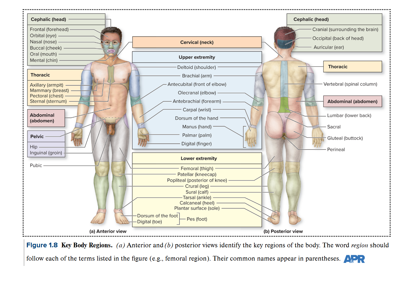
6. Be prepared to label a diagram of the body regions using the correct anatomical terms.
(Figure 1.8, p.13 and Table 1.3)
*** Oral region instead of mouth and so on…
7. Locate the major body cavities (posterior aspect and ventral cavity) and identify the major divisions and organs found in each cavity.
Exam Question - Name of the major body cavities and what organ they enclose and function
The empty spaces - They are not exactly empty spaces- they are cavities
Empty spaces in the body that house internal organs
Body cavities protect, cushion, separate, and allow organs to move and work properly.
Functions of Body Cavities
Protect organs
Cavities surround delicate organs (like brain, heart, lungs) with bones, muscles, and membranes for protection.
Example: The skull protects the brain in the cranial cavity.
Allow organ movement & expansion
Organs inside cavities can change shape/size without damage.
Example: Lungs expand when breathing, stomach expands after eating.
Separate organs for organization
Cavities divide the body into regions so organs don’t interfere with each other’s function.
Example: The diaphragm separates the thoracic cavity (lungs, heart) from the abdominal cavity (digestive organs).
Provide cushioning & reduce friction
Many cavities are lined with serous membranes that produce fluid.
This fluid reduces friction between moving organs (like the beating heart and lungs).
Maintain stable environment (homeostasis)
Cavities help keep temperature, pressure, and organ function stable by enclosing them in controlled spaces.
The thing that separates the cavities are bones, muscles, and ligaments
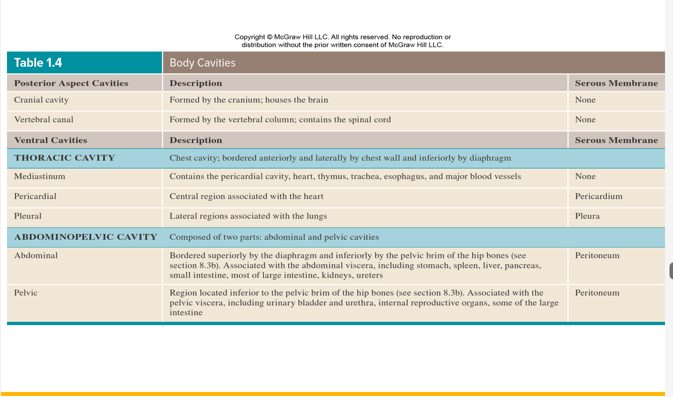
1. Posterior Aspect (Dorsal Cavity)
Located along the back of the body.
Divisions: the brain
Cranial cavity → contains the brain.
Vertebral (spinal) canal → contains the spinal cord.
Membranes: Protected by meninges (brain and spinal cord coverings).
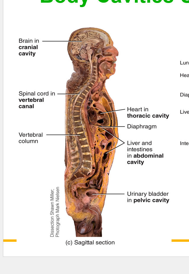
Ventral Cavities (Thoracic cavity & Abdominopelvic cavity)
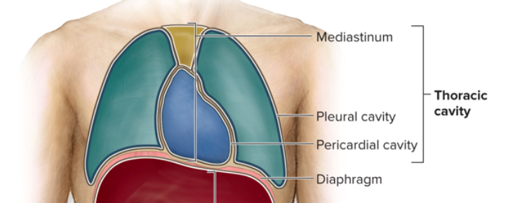
Thoracic cavity (chest)
Mediastinum → heart, thymus, trachea, esophagus, major blood vessels (no serous membrane).
Pericardial cavity → heart (serous membrane = pericardium).
Pleural cavities → lungs (no serous membrane = pleura).
(separated by the diaphragm)
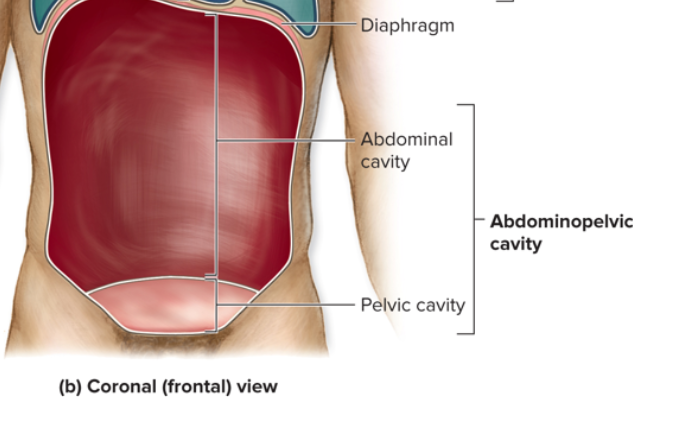
Abdominopelvic cavity
Abdominal cavity
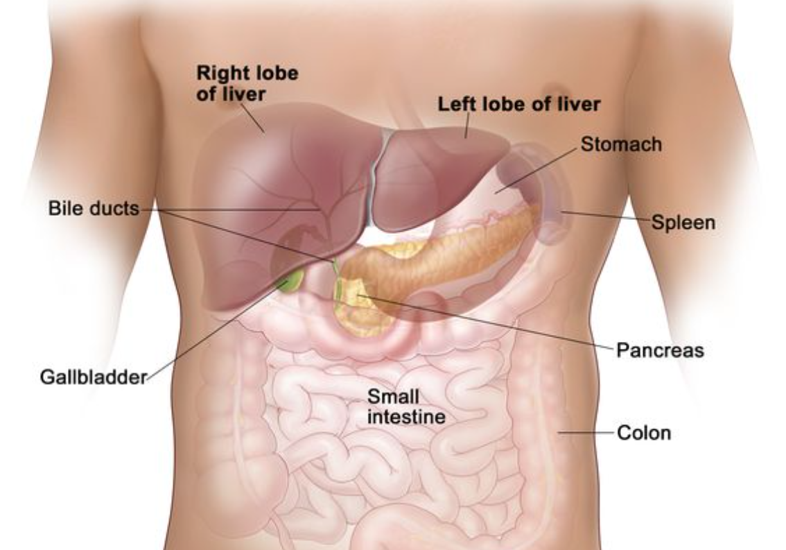
Organs: stomach, spleen, liver, pancreas, small intestine, most of large intestine, kidneys, ureters.
Serous membrane = peritoneum.
Pelvic cavity
Organs: bladder, urethra, reproductive organs, part of large intestine.
Serous membrane = peritoneum.
Ventral Cavity
The ventral cavity is the large front cavity of the body.
It includes:
Thoracic cavity (chest) → lungs, heart, thymus, esophagus, trachea.
Abdominopelvic cavity (below diaphragm) → abdominal + pelvic organs (stomach, intestines, liver, bladder, reproductive organs, etc.).
What divides it?
The diaphragm (the large dome-shaped muscle) divides the ventral cavity into:
Thoracic cavity (above diaphragm)
Abdominopelvic cavity (below diaphragm)
✅ So, the ventral cavity encloses:
Thoracic cavity organs (heart, lungs, etc.)
Abdominal cavity organs (stomach, liver, intestines, etc.)
Pelvic cavity organs (bladder, reproductive, rectum).
Body Cavities
1. Pleura (lungs)
A thin membrane that surrounds the lungs.
Function: Reduces friction when lungs expand and contract during breathing.
2. Pericardium (heart)
A thin membrane that surrounds the heart.
Function: Protects the heart and reduces friction as it beats.
3. Peritoneum (abdominal cavity)
A thin membrane that lines the abdominal cavity and covers abdominal organs (stomach, intestines, liver, etc.).
Function: Cushions, supports, and reduces friction between digestive organs.
✨ Easy memory trick:
Pleura → Lungs 🫁
Pericardium → Heart ❤
Peritoneum → Abdominal organs 🍔
8. Describe the function of ‘serous membranes’ and locate these in the thoracic and abdominopelvic cavities locating the following
1. visceral pleura
2. parietal pleura
visceral pericardium
parietal pericardium
visceral peritoneum
parietal peritoneum
Function of Serous Membranes
A serous membrane is a thin, slippery lining that reduces friction between moving organs (like lungs, heart, intestines) and body walls.
They secrete serous fluid → acts like oil to let organs glide smoothly when they move (breathing, heartbeat, digestion).
Layers of Serous Membranes
Visceral layer → directly covers the organ.
Parietal layer → lines the cavity wall.
Serous cavity → small space between them, filled with fluid.
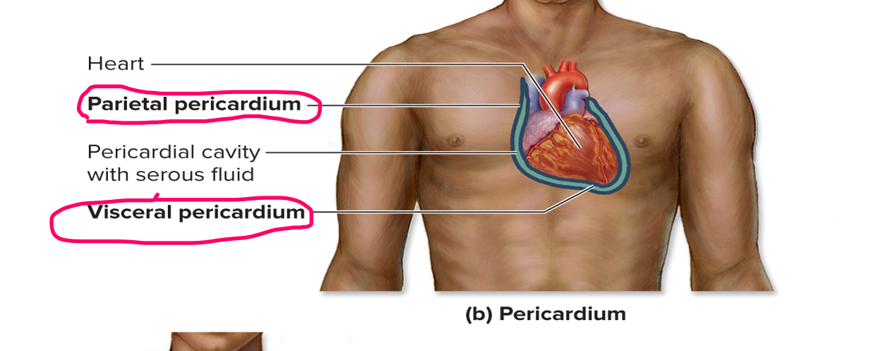
Heart (Pericardium)
Visceral pericardium → directly on the heart’s surface.
Parietal pericardium → lines the outer sac around the heart.
Pericardial cavity → space between them, filled with fluid to prevent friction during heartbeats.
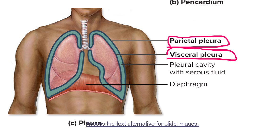
Lungs (Pleura)
Visceral pleura → directly covers the lungs.
Parietal pleura → lines the chest wall & diaphragm.
Pleural cavity → space between them, with fluid so lungs can expand and contract smoothly during breathing.
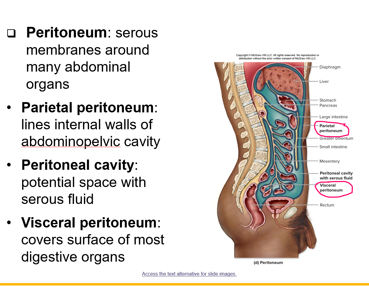
Abdominal (Peritoneum)
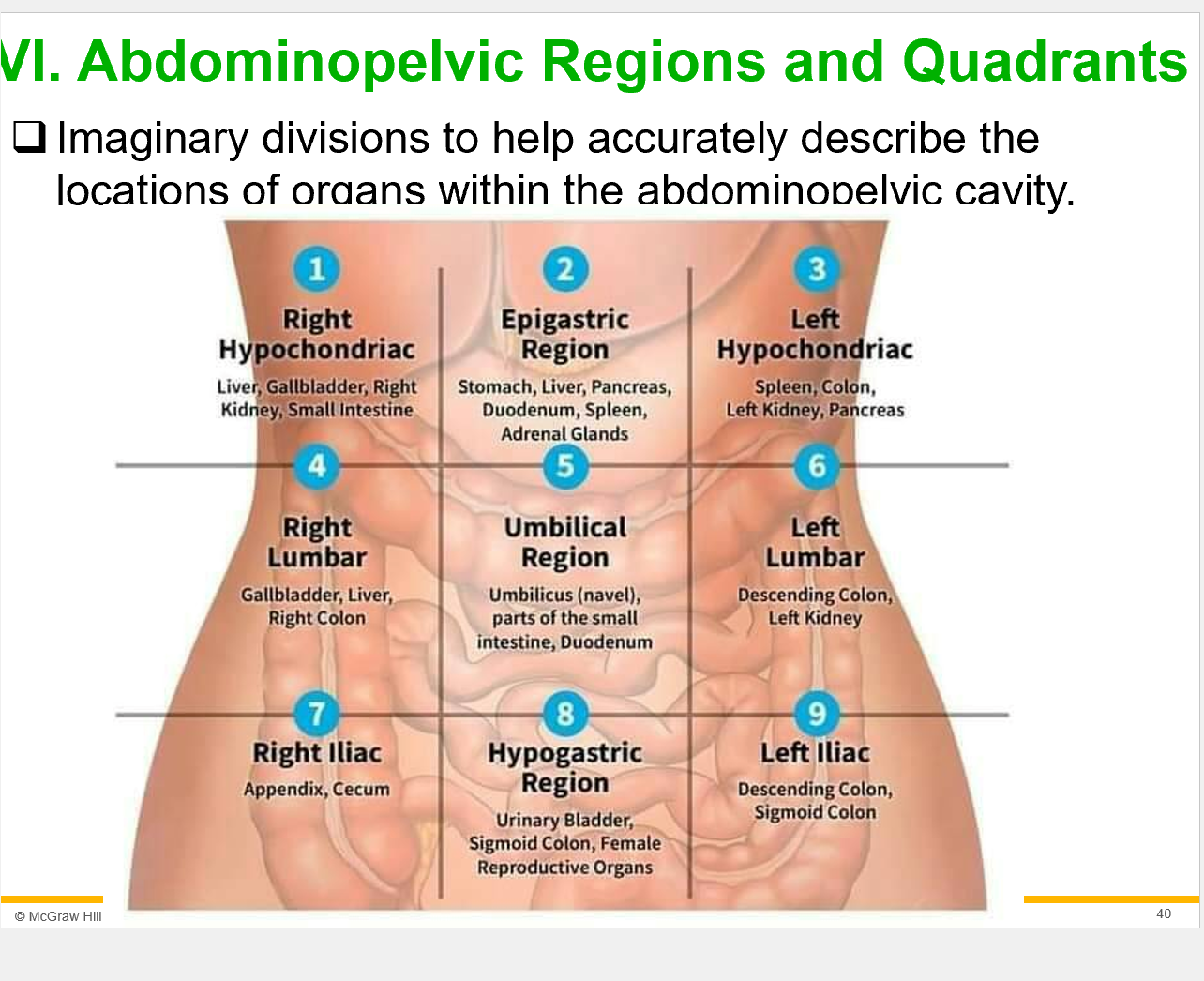
9. Name the 9 body regions and identify the major organs found in each region.
Exam question - name the region and the list of organs
Right Hypochondriac
Liver, Gallbladder, right kidney, small intestine.
Epigastric Region
Stomach, Liver, Pancreas, Duodenum, Spleen, Adrenal Glands.
Left Hypochondriac
Spleen, colon, Left kideny, Pancreas
Right Lumbar
Gallbladder, liver, right colon
Umbilical Region
Umbilicus (navel), parts of the small intestine, dudenum
Left Lumbar
Descending colon, left kideny
Right lliac
Appendix, cecum
Hypogastric Region
Urinary Bladder, sigmoid colon, Female reproductive oragns.
Left lliac
Descending colon, sigmoid colon