ANHB2214 The Digestive System
1/62
There's no tags or description
Looks like no tags are added yet.
Name | Mastery | Learn | Test | Matching | Spaced | Call with Kai |
|---|
No analytics yet
Send a link to your students to track their progress
63 Terms
From the esophagus to the rectum, the wall of the alimentary canal has 4 layers. What are they?
1. Mucosa
• surface epithelium
• lamina propria
• muscularis mucosa
2. Submucosa
3. Muscularis externa
• inner circular layer of smooth muscle
• outer longitudinal layer of smooth muscle
4. Serosa (or Adventitia)
Layers of the GI tract and functions
Surface epithelium
Promote the absorption of the products of digestion.
Facilitate the transport of food.
Produce mucus.
Aid in digestion.
Lamina Propria
Protection. The lamina propria can be rich in lymphoid tissue.
• Plasma cells in the lamina propria produce IgA antibodies, which are transported across the epithelium into the gut lumen. • Capillaries nourish the overlying epithelium and glands. • Houses the mucosal glands.
Muscularis mucosa
• Moves the mucosa locally to improve its contact with food.
• A muscle layer in a mucosa is very rare, and in humans always confirms that the mucosa belongs to the GI tract.
Submucosa
• Provides a pathway for arteries, veins and nerves.
• This layer of loose connective tissue allows the mucosa and muscularis externa to move independently.
• Lymphocytes and lymphatic nodules abound in some places.
Muscularis externa
• Mixes and propels the food in the digestive tract.
• Contraction of this muscle is co-ordinated by the myenteric and submucosal nerve plexuses.
Adventitia
• The segments of the gut that are directly attached to surrounding tissues are covered with an adventitia. Elsewhere the adventitia is replaced with a serosa to allow the intestines and stomach to slide around freely during digestive contractions.
Glands of digestive system
the small mucosal glands = simple tubular glands w/o ducts
confined to lamina propria and may fill layer
larger glands that invade submucosa = compound
prod. mucus —> surface through ducts
very large sxtrinsic glands (liver, pancreas, major salivary glands) burrow through organ wall + remain attacahed by only a duct
mucosal glands func = secretion and cytogenesis epitheliual lining turns over rapidly (harsh conditions)
stem cells deep in mucosal glands get pushed up as they differentiate
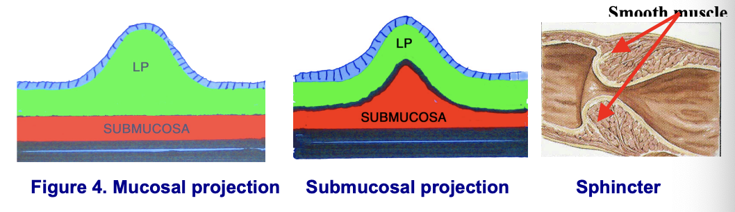
Projections into the lumen
surface epithelium can bulge into lumen due to LP thickening or submucosal projection or muscularis externa circular layer
sphincter sbetween organs of GI racr - thickening of circular layer of muscularis externa
tonic contraction of SM keep sphincter closed (may be change in epithelium at pshincter)
GALT (gut associated lymphatic tissue)
lymph tissue protect from antigens + infectious agents
lymphocytes here diff to rest of body
GALT includes tonsils, Peyer’s patches, appendix, lymph nodes + local deposit in GI tract
DNES (Diffuse NeuroEndocrine System)
GI tract mucosa had assortment of DNES cells —> prod. chem messengers
segments of tract distinguished by particular DNES chem messengers they prod.
most messengers have paracrine activities
let DS coordinate activity locally and across organs
chem. messengers ar polypeptides and most also func in CNS as neurotransmitter
Eg. secretin stimulated secretion by pancreas and cholecystokinin (CKK)
CKK also in brain and acts on appetie centr ein ht. —> satiation
Enteric NS
two extensive plexuses of autonomic ganglia innervate organs of DS
myenteric pllexus interconnected ganglia network between inner circular and outer longitudinal layers of muscularis externa
submucosal plexus controls SM and DNES cells of mucosa
enteric ganglia have sensory neurons - GI initiate and control its own activities intrinsically eve if CNS connection severed
The esophagus is basically a muscular tube to convey food to the stomach. The lumen is partially collapsed but you can image the muscle layers relaxing somewhat and it expanding out when food passes along it.
It has the same complement of layers as the rest of the gut:
Mucosa
Submucosa
Muscularis externa
but with two obvious specializations:
The epithelium is a non-keratinized, stratified squamous epithelium. This more resistant epithelium protects the mucosa from abrasion by swallowed material.
Mucous-secreting glands in the mucosa and submucosa lubricate the passage of food.
The epithelium of the esophaggus is non-keratinized stratified squamous. This more relatively thick and resistant epithelium protects the mucosa from abrasion by swallowed material.
The muscularis mucosa is smooth muscle all of the way along the esophagus. Towards the upper end, the layers of muscle become disorganized and you will have to be careful when trying to distinguish the muscularis mucosa from the muscularis externa. This image was taken from the histological section of the esophagus provided so view this section and verify that it is from the middle of the organ (Yes?....or No?).
Make sure you can identify the stratified squamous (non-keratinised) epithelium supported by the lamina propria, then the small muscle bundles of the muscularis mucosa. The submucosa exhibits a lot of artifactual spaces due to tissue processing but it has many blood vessels (e.g. a small vein?) and loose connective tissue (collagen).
Compared to the myenteric plexus (between the layers of the muscularis externa), the neurons of the submucosal (Meissner's) plexus are a lot more difficult to locate. But, if you examine the submucosa very thoroughly you may see a few ganglion cells and nerve fibres sectioned longitudinally (or obliquely) and also transversely. In this image, they are located in the submucosa close to the skeletal muscle fibres of the muscularis externa.
If you examine the histological section of the esophagus note that the submucosa contains a lot of dense irregular connective tissue and some large blood vessels. Lymphatic vessels will also be present but probably collapsed and so not able to be detected.
Submucosal glands are usually quite common and their secretions are delivered to the luminal surface by a duct.
The muscularis externa consists of an inner circular layer and an outer longitudinal layer. Check this when you view the histological section remembering the section has been cut transversely through the esophagus. Also check what type of muscle is in the muscularis externa in the section because the upper one third of the esophagus has striated muscle continuing down from the pharynx. The lower (or distal) third of the muscularis externa is entirely smooth muscle. So that leaves the middle one third being a mixture of both types of muscle.
The muscularis externa is unusual in being composed of striated muscle fibres at the top of the esophagus. Proceeding down the esophagus, the amount of striated muscle dwindles and is replaced by increasing bundles of smooth muscle. But as you know, the muscularis mucosa stays as smooth muscle all of the way through the esophagus.
Look between the layers of the muscularis externa for components of the myenteric plexus. You may see the odd ganglion cell and nerve fibres sectioned.
It is difficult to get well-preserved, human fundic stomach because the tissue autolyses rapidly. The section for you to view (and imaged here) is from a monkey and illustrates the essential features but on a much reduced scale. Distinguish even at this very low magnification the main layers of the wall - mucosa, submucosa, muscularis externa and serosa. Now learn specific details about these layers as they appear in the stomach in the items that follow.
Food enters the stomach from the esophagus and passes into the duodenum when the pyloric sphincter is relaxed. The muscularis externa of the anterior surface of the stomach has been taken away in this image of the distended stomach to reveal gross folds of the mucosa and supporting submucosa called rugae. A gross view of the stomach indicates 4 separate regions. The cardia, fundus, body and pyloris. The fundus and the body have a similar histological structure so only three histological regions are described in the items that follow.
You can examine this section later as it shows the extent of mucus-secreting cells in the gastic glands. If you examine the inner surface of the stomach you can see large folds (rugae). They are composed of the stomach submucosa and the mucosa and will disappear when the stomach is distended.
Before leaving the stomach recall that at both the cardiac (upper) and pyloric (lower) ends of the stomach the mucosal glands are purely mucous. Thus, if a bit of gastric juice leaks into the duodenum or esophagus it will be mainly mucus instead of rich in HCl and peptic enzymes. It is worth while observing this mucosal specialization. The histological section of stomach available to view is mainly from the fundus of the stomach but one end is near the cardiac sphincter. You can find this end by looking at the glands. Each gastric pit is well developed here but the glands are very short. They are devoid of parietal cells and are short on chief cells. Closer to the cardiac sphincter the chief cells would disappear.
The gastric pits are very deep. The pyloric glands that empty into these gastric pits are fairly straight for much of their length compared to the gastric glands of the main fundic region but then coil at their bases - even branch.
This section shows the pyloric-duodenal junction in the human. You will study the duodenum in the next items but you can see important features here and maybe once you study the items of the dudenum then come back to this image and also examine the histological section.
The duodenum has stubby villi in the mucosa near the pyloric sphincter. Is this a transverse or longitudinal section? Is the pyloric sphincter formed by circularly or longitudinally arranged smooth muscle? Observe the Brunner's glands. They fill up the lamina propria as well as the submucosa of the duodenum. This breaks up the muscularis mucosa into streaks of smooth muscle coursing through the glands. This is characteristic in the human. Brunner's glands are long, branched, and tubulo-alveolar in form. Ducts are sparse and those present are lined with mucous-secreting cells. Spend a moment looking at the glands and thinking about their shape.
Examine the mucosa of the fundic portion.
The surface epithelium is fundamentally different from that of the intestine. In the stomach the surface epithelium consists of "mucous cap cells".
The gastric epithelium does not form a flat surface. Instead it is criss-crossed by tiny creases several millimeters long and about a millimeter deep. Each is are called a "gastric pit". They increase the surface area of the stomach in a way different from the protruding villi of the intestine. From the bottom of each gastric pit, a simple, tubular branched gland (without a duct) extends down into the lamina propria.
Two cell types can be distinguished in the upper parts of the glands. One is the brightly eosinophilic parietal cell which secretes HCl (and intrinsic factor). The other is the mucous neck cell.
The cells at the base of the glands close to the muscularis mucosa are mainly of another type. These "chief cells" secrete the digestive enzymes for the stomach.
Other cell types are found in the stomach (and intestinal) wall, but generally require special preparation for their demonstration. These include stem cells and scattered hormone-secreting cells. As in the intestine, the surface cells are born from cell divisions deep in the mucosal glands.
In the stomach the surface epithelium consists of "mucous cap cells," a unique cell type filled with tiny secretory granules of mucus but they appear empty in this H&E section as the mucinogen is lost during processing. The mucus that they produce is biochemically distinctive in that it stays slippery at the low pH of the stomach.
The mucous neck cell (at the neck of the gastric gland) has a nice rounded nucleus, is much shorter than the surface mucous cells, and also manufactures a special form of mucus that does not precipitate at low pH. Their secretion is less alkaline than the surface epithelial cells.
The cells at the base of the glands close to the muscularis mucosa are mainly of another type. These "chief cells" secrete the digestive enzymes for the stomach. The enzymes are packaged into secretory granules but in our particular section the granules have leached out and the apical cytoplasm looks foamy and colorless. The basal ends of the chief cells is markedly basophilic attesting to the large quantities of rough endoplasmic reticulum, and therefore of, protein synthesis.
Note the distinctive eosinophilic staining of a neighbouring parietal cell.
Two cell types can be distinguished in the upper parts of the glands. Observe the brightly eosinophilic cytoplasm and central rounded nucleus of the parietal cell which secretes HCl (and intrinsic factor). The other cell type you know is the mucous neck cell.
From the bottom of each gastric pit, a simple, tubular branched gland (without ducts) extend down into the lamina propria. They run pretty much straight down except for the very distal ends, which twist around a bit.
The lamina propria is limited to supporting the numerous gastric pits and gastric glands and will contain typically plasma cells, macrophages, lymphocytes and maybe eosinophils.
Make sure you can readily distinguish the chief cells from the parietal cells and pale stained mucous neck cells.
The muscularis mucosa consists of two thin layers of smooth muscle - and an inner circular layer and an outer longitudinal layer but in some areas there may be a third almost circular layer . Some smooth muscle cells may extend into the lamina propria and may be involved with the secretion process of the gastric glands (?).
Mucous cells
The submucosa of the stomach is similar to that in other regions of the gastrointestinal tract in that it contains blood vessels (artery and vein) and lymphatic vessels embedded in supporting collagenous connective tissue.
Look at this image of the muscularis externa carefully. Identify smooth muscle cut longitudinally and cut transversely. Of course you would need to view the histological section to work out the orientation of the cut section to see which layer is a longitudinal and circular layer. These layers of muscle will vary in different regions of the stomach wall (e.g. there may be an additional inner "oblique" layer in parts) to enhance effective mechanical digestion and mixing of the chyme.
The serosa is a layer of connective tissue and a "serous membrane" of a simple squamous epithelium (mesothelium). Make sure you know the distinction between a structure having a serosa and one having an adventitia. Blood vessels (e.g. in this image - a small vein) enter or exit the serosa to supply or drain the wall of the gut.
Although at first it might seem most sensible to start histological studies of the gut with the esophagus and simply progress along the digestive tract to the rectum (as is the case with this online atlas), it is, in fact, the small intestine (particularly the duodenum) which demonstrates the fundamental features of this system most fully. If one understands this organ, it is easy to understand other parts of the alimentary canal as variant specializations. So there is more content loaded of histology of the dudoenum than the other parts of the gut.
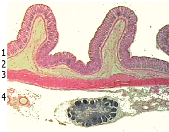
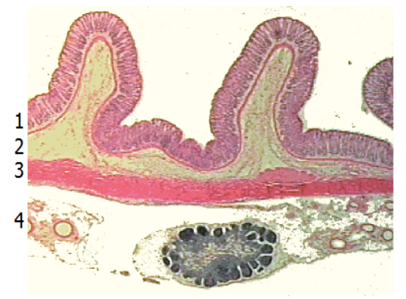
The section for you to view is a well preserved example of human lower duodenum, sectioned longitudinally. The wall is thick, but much of its thickness is accounted for by a succession of transverse folds called the plicae circulares.
Under low magnification it becomes evident that plicae are folds in the submucosa covered with mucosa (epithelium, lamina propria and muscularis mucosa). The plicae are covered on their surface by a multitude of villi. Each villus is a smaller protrusion of mucosa.
Each villus is a smaller protrusion of mucosa made up of only epithelium and lamina propria. The lamina propria is sharply demarcated from the underlying submucosa and the characteristics of these two connective tissues are totally different.
The epithelium of the villus is tall columnar. Its predominant cells are enterocytes (also called absorptive cells in view of their main function). These are noted for their "brush border", which is visible with high magnification. Look for the distinctively shaped goblet cell also sprinkled amongst the enterocyytes. Their upper parts are swollen and their cytoplasm appears more textured than that of the enterocytes. Blebs of mucus may be oozing from their apexes. The nucleus of the goblet cell is distinctively compact, darkly stained and triangular in shape in contrast with the paler, oval enterocyte nuclei.
Stubby and relatively inconspicuous mucosal glands invade between the villi down close to the muscularis mucosa. Paneth cells at the bottom of the glands contain red secretory granules (see next item) in their apical cytoplasm. These are visible under high magnification, but in some areas the granules have been leached out, leaving a foamy-looking apical cytoplasm instead.
The cells at the bottom of the glands (Paneth cells) have a basophilic cytoplasm and contain red secretory granules in their apical cytoplasm. These are visible under high magnification.
In the histological section with the link below you can view a segment of lower duodenum from a monkey which was stained with a trichrome procedure. The tissue has been cut between plicae and shows only villi. How do you know that this is a cross section instead of longitudinal?
The histological section and this image reveals the main function of the mucosal glands - the production of cells. The stem cells for the epithelium of the gut are sequestered in the crypts. Here they are far more sheltered than they would be on the surface. All cell division in the epithelium takes place in the middle to lower part of the glands. The new cells push the old ones up out of the glands and up over the villi. Here they are shed. Cells last only several days on the villi so their progenitors must divide actively in the glands. The same stem cell gives rise to enterocytes, goblet cells and Paneth cells.
You can see mitotic figures in the crypts even at fairly low magnification. When the chromosomes have condensed on the metaphase plate they show up as dark lumpy masses. These look entirely different from the pale staining, large nuclei of the surrounding interphase cells. The nuclei tend to be located at the basal end of the cells during interphase. When the cells go into mitosis, the nuclei migrate to the apical part of the cell, making it even easier to pick them out.
Briefly examine the population of cells in the lamina propria of the villi. Lymphocytes and plasma cells dominate, but some fibroblasts and smooth muscle cells can be present. Both capillary and lymphatic channels richly supply this region, so that many of the elongate nuclei are the endothelial cells that line those structures. Lymphocytes commonly invade the overlying epithelium, some work their way across the epithelium to be discharged into the gut lumen. In places with proper orientation, a well-defined basement membrane can be seen underlying the epithelium
Each villus has a central lymphatic capillary (lacteal) bound by simple squamous epithelium surrounded by the cells within the lamina propria.
A thin sheet of smooth muscle of the muscularis mucosa marks the base of the lamina propria. Imagine what this section would look like if the tissue had been sectioned transversely, in the plane of a plica, instead of longitudinally.
In some sections of the gastrointestinal tract you can see that this muscularis mucosa has both a longitudinally and a circularly arranged sublayer.
Turn to the submucosal region and follow this layer from the core of the plicae to the tissue between plicae where a rich plexus of large blood vessels and the occasional lymphatic channel is visible under low magnification. These vessels reveal the main function of the submucosa. Outside of this layer lie the two principal muscle layers of the intestinal wall (see later item "muscularis externa").
While harder to identify, very small nerve ganglia are present in the submucosa. Some of you will enjoy the challenge of looking for them. Ganglia are clusters of relatively large nerve cell bodies and supporting satellite cells and nerve fibres.
The muscularis externa consists of the inner circular layer and the outer longitudinal layer of smooth muscle.
This smooth muscle is regulated by a well developed plexus of autonomic ganglia. Even under relatively low magnification, random sections through the myenteric nerve plexus can be seen as pale islands of cells, sandwiched between the two muscle layers. View them at higher magnification (next item).
The smooth muscle of the muscularis externa is regulated by a well developed plexus of autonomic ganglia. What types of cells do they consist of?
Each plexus has been named after the persons who described them, Auerbach and Meissner. Increasingly histologists are trying to get away from personal names for structures. Thus, you are forward looking if you call Meissner's plexus the submucosal plexus and Auerbach's plexus the myenteric plexus.
The jejunum has more numerous plicae circulares, the villi are taller and lymphoid nodules are scarce.
Villi are taller in the jejunum than in the ileum and duodenum. Follow along this villus and you can imagine how tall it would project into the lumen if the smooth muscle fibres relaxed.
Identify the enterocytes (and their brush border), goblet cell and the occasional lymphocyte on "sentry duty" within the epithelium. This section is excellent to view plasma cells and smooth muscle in the lamina propria (but go to higher magnification) and also lymph vessels, blood vessels and the submucosal plexus in the submucosa.
This section of ileum shows the aggregation of lymphatic nodules known as Peyer’s patches. Its function in relation to the small intestine presumably parallels that of the tonsil to the pharynx and the appendix to the colon. It is uncertain whether or not these lymphatic aggregations along the GI tract have some special role in immunity beyond those of isolated nodules found throughout the mucosae of the body.
With the naked eye (i.e. when viewing the small intestine macroscopically) permanent folds of both the mucosa and submucosa (mostly developed in the jejunum and unlike the rugae of the stomach) are called plicae circulares (also referred to as valves of Kerckring). As a review identify a villus and crypt of Lieberkuhn.
From your knowledge of the lymphatic system (studied earlier) recall that in a lymphatic nodule within Peyer’s patches, the corona has more lymphocytes around one side than the other. The more developed side is towards the lumen, as it also is for the appendix.
The mucosal epithelium is simple columnar in type and consists of the goblet cell and the absorptive cell (enterocyte) each with a brush border or striated border (microvilli). Oh and don't forget the lymphocyte !
The goblet cell is abundant in the intestinal epithelium. Recall that their nucleus is basal and often compressed by surrounding enterocytes and the apical cytoplasm contains mucoid granules (carbohydrate-rich glycoproteins) which are released at the cell surface by exocytosis into the lumen through a space between the striated (brush) border.
The appendix was introduced in the topic on the "Lymphoid tissue".
The mucosal and submucosal layers have been entirely converted into a mass of lymphoid tissue. Note the similarity in structure between the appendix and the tonsil. Both contain enormous depots of lymphoid tissue separated from a dirty, stagnant space or lumen by an epithelium infiltrated with lymphocytes. Tonsils alert the immune system to resident bacteria in the throat. The appendix stimulates immunity to the range of bacteria that colonize the other end of the GI tract.
As you saw with "Peyer's patches" the corona of the lymph nodule has more lymphocytes around one side than the other. The more developed side is towards the lumen.
The large intestine, the colon, conforms to the essential features of the gastrointestinal tract. But, villi are gone, as are plicae and the brush border on the epithelium, but the glands, still called crypts of Lieberkühn, remain.
A principal specialization of the colon relates to its external longitudinal smooth muscle layer. This is reduced to a thin layer everywhere except for three heavy bands known as the teniae coli. These are easily found even without magnification as bulges in this cross-section of the monkey colon. What is their function? Between them and the inner circular layer of muscle are well developed ganglia of the myenteric plexus .
The teniae coli are bands of smooth muscle. Note each cell has a central nucleus (when the plane of section passes through it) and there is connective tissue binding each cell to the adjacent cell.
In the large intestine (or colon) villi are gone, as are plicae and the brush border on the epithelium, but the glands, still called crypts of Lieberkühn, remain.
In a section cut perpendicular to the surface, the glands might look like the boundaries between closely packed villi. However, if you look at an area where the knife has cut parallel to the surface you can see that they are glands organized in a regular pattern with a small sheet of connective tissue between them. Clearly, the epithelium extends down into the lamina propria instead of protruding upwards over villi. Goblet cells dominate the epithelium of the crypts.
The connective tissue of the lamina propria is heavily infiltrated by lymphocytes and plasma cells. You can also look around for eosinophils and the stromal cells. Lymph nodules are scattered in the submucosa near the junction with the mucosa.
This is a good section for observing that a muscularis mucosa has two layers of smooth muscle - an inner circular and outer longitudinal layer.
The glands, still called crypts of Lieberkühn, remain. In a section exactly perpendicular to the surface the glands might look like the boundaries between closely packed villi. However, if you look at an area where the knife has cut parallel to the surface you can see that they are glands organized in a regular pattern with a small sheet of connective tissue between them. Clearly, the epithelium extends down into the lamina propria instead of protruding upwards over villi. Goblet cells dominate the epithelium of the crypts.
The connective tissue of the lamina propria is heavily infiltrated by lymphocytes and plasma cells. You can also look around for eosinophils and the stromal cells.
This image is from a histological section specifically stained to illustrate the distribution of the goblet cells in the colon. They are abundant. The other non-mucous secreting cells of the epithelium show up as well. Their main function is to absorb water. If you compare the number of goblet cells to the number of nuclei in the epithelium it is obvious that most of the cells are absorptive. The abundance of goblet cells increases towards the distal end of the colon.
Not a well preserved section. At low magnification find the simple columnar epithelium lining the rectum. Notice the numerous glands, many have been cut in cross section. Identify the muscularis mucosa, the submucosa with a rich plexus of veins (some distended into hemorrhoids), the inner circular muscle layer, and the longitudinally sectioned fibres of the outer longitudinal muscle layer.
Again find the simple columnar epithelium lining the rectum. Now follow the epithelium to the right. Look for the abrupt transition to a non-keratinized stratified squamous epithelium at the ano-rectal junction. Examine the transition at medium magnification and at high magnification.