Exam 3 BIOL 2251
1/106
There's no tags or description
Looks like no tags are added yet.
Name | Mastery | Learn | Test | Matching | Spaced | Call with Kai |
|---|
No analytics yet
Send a link to your students to track their progress
107 Terms
Functions of the skeletal system
Support, protection, movement (leverage), mineral storage (Ca2+, phosphate), blood cell formation (hemopoiesis), fat storage

Osteogenic cells
Stem cells that divide to produce osteoblasts; found in periosteum and endosteum

Osteoblasts
Bone-forming cells; secrete osteoid (organic matrix)

Osteocytes
Mature bone cells; maintain bone matrix and communicate via canaliculi
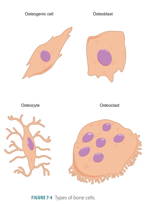
Osteoclasts
Large multinucleated cells that break down bone (resorption)
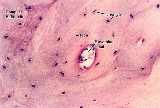
Compact bone histology
Dense bone organized into osteons (Haversian systems)
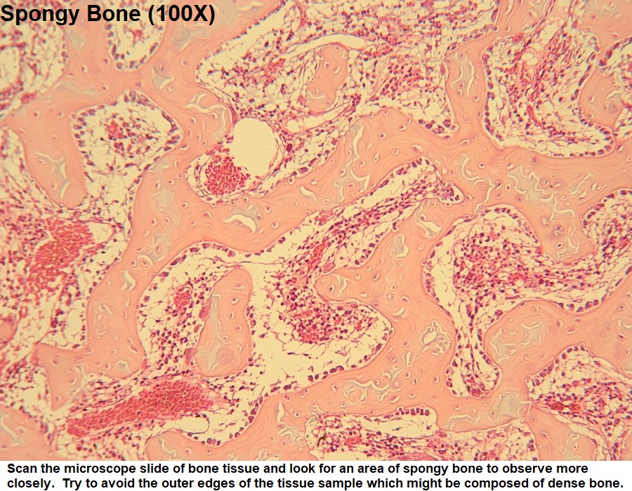
Spongy bone histology
Trabeculae network with red marrow spaces; no osteons

Osteon (Haversian system)
Structural unit of compact bone with concentric lamellae around a central canal (A)
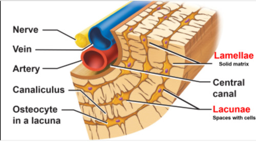
Central (Haversian) canal
Contains blood vessels and nerves
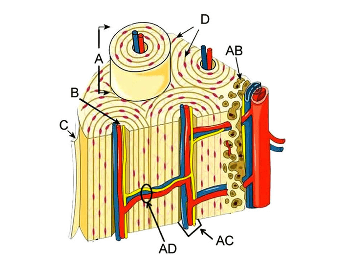
Lamellae
Concentric layers/rings of calcified bone matrix (D)

Lacunae
Small spaces housing osteocytes
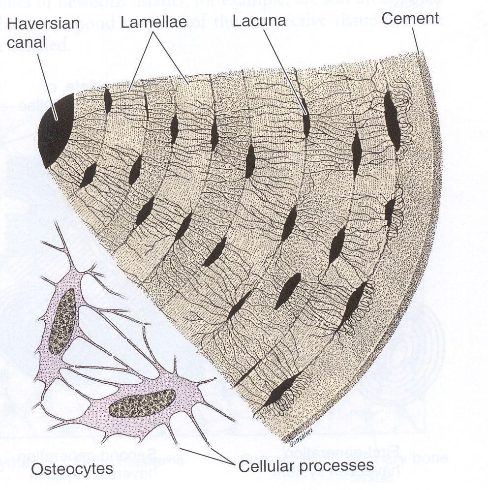
Canaliculi
Tiny channels connecting osteocytes for nutrient/waste exchange
Organic bone component
Osteoid (collagen fibers); provides flexibility and tensile strength
Inorganic bone component
Hydroxyapatite (calcium phosphate salts); provides hardness and compressive strength
Red bone marrow
Produces blood cells; found in spongy bone of flat bones and epiphyses
Yellow bone marrow
Stores fat; found in medullary cavity of long bones
Hemopoiesis
The process of creating a wide variety of blood and bone marrow cells, namely erythrocytes, platelets, granulocytes, lymphocytes, and monocytes.
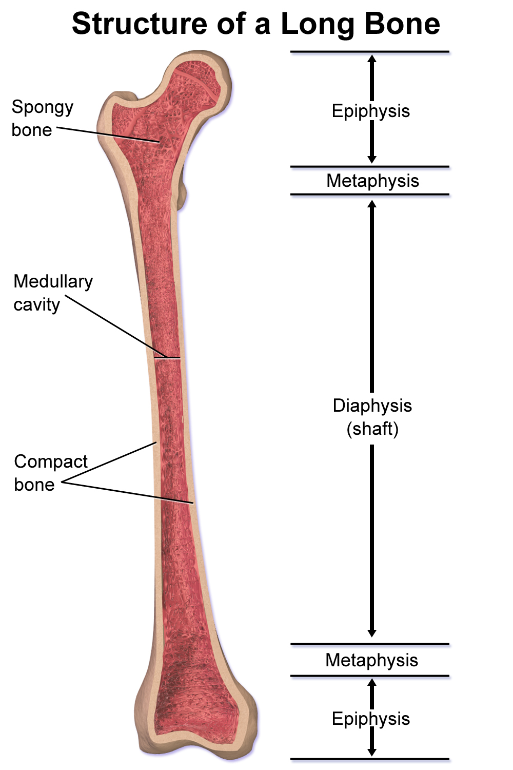
Diaphysis
Shaft of a long bone

Epiphysis
Ends of a long bone; contains spongy bone
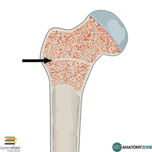
Metaphysis
Region between diaphysis and epiphysis; contains growth plate
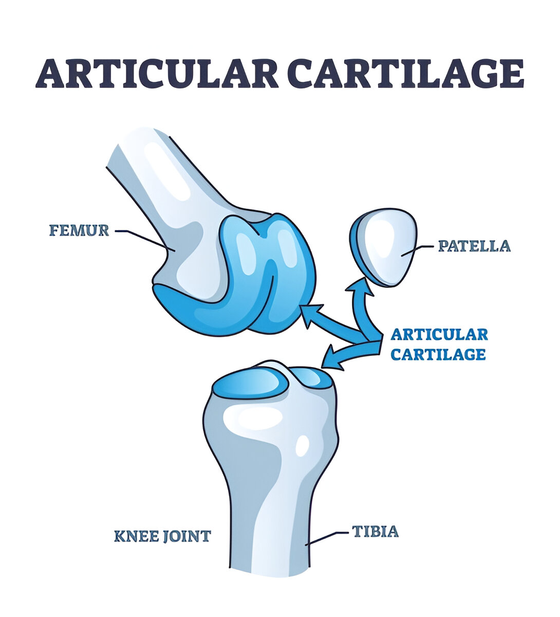
Articular cartilage
Hyaline cartilage covering bone ends; reduces friction
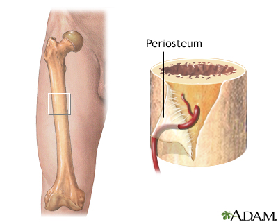
Periosteum
Outer fibrous covering of bone; contains blood vessels and nerves
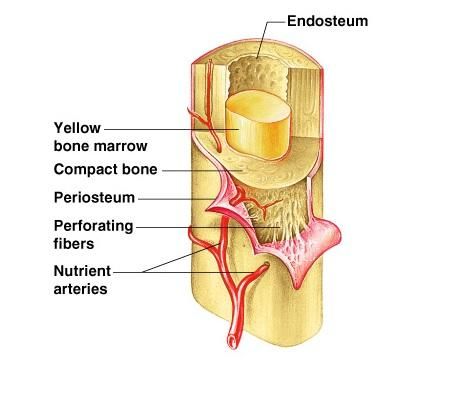
Endosteum
Inner lining of medullary cavity
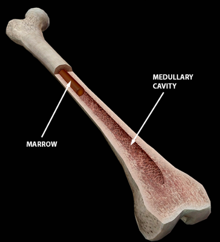
Medullary cavity
Hollow cavity in diaphysis containing marrow
Bone remodeling
Ongoing replacement of old bone with new bone
Purpose of remodeling
Maintains strength, repairs damage, regulates calcium levels
Cells in remodeling
Osteoclasts (resorption) and osteoblasts (formation)
Bone homeostasis
Maintenance of bone density via hormones, minerals, and mechanical stress
Role of calcium
Essential for bone strength and signaling
Role of vitamin D
Increases calcium absorption from intestines
Role of protein
Provides collagen for bone matrix
Parathyroid hormone (PTH) source
Parathyroid glands
PTH stimulus
Low blood calcium levels
PTH target
Bones, kidneys, intestines (indirect)
PTH effect
Increases blood calcium (bone resorption, Ca2+ reabsorption, activates vitamin D)
Calcitonin source
Thyroid gland (C cells)
Calcitonin stimulus
High blood calcium levels
Calcitonin target
Bones
Calcitonin effect
Decreases blood calcium (inhibits osteoclasts)
Calcitriol source
Kidneys (activated vitamin D)
Calcitriol effect
Increases calcium absorption in intestines
Calcitonin vs PTH
Calcitonin lowers blood Ca2+; PTH raises blood Ca2+
Low PTH effect
Bone overgrowth, low blood calcium
High PTH effect
Excess bone loss (weak bones)
PTH and calcitriol relationship
PTH stimulates calcitriol production to increase calcium absorption
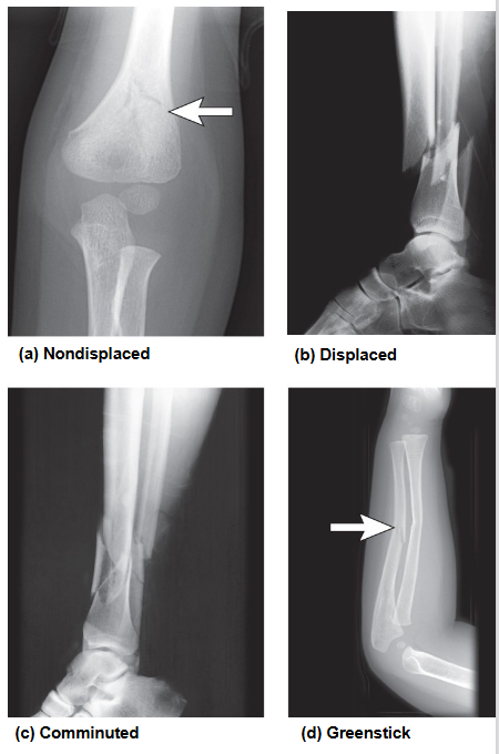
Stress fracture
Small crack from repetitive force

Comminuted fracture
Bone breaks into 3 or more pieces

Greenstick fracture
Incomplete break; one side bends, the other breaks

Transverse fracture
Break is a straight line across the bone
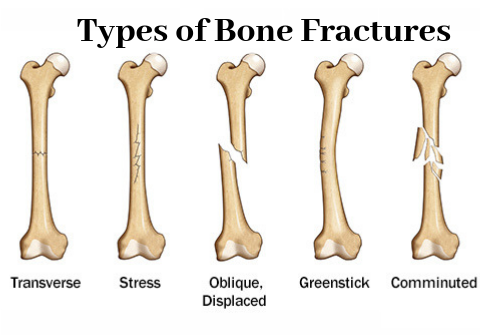
Oblique fracture
Break is at a diagonal angle
Open fracture and closed fracture
Open: bone pierces the skin
Closed: bone breaks but doesn’t pierce the skin
Pathological fracture
Break due to disease (e.g., osteoporosis)
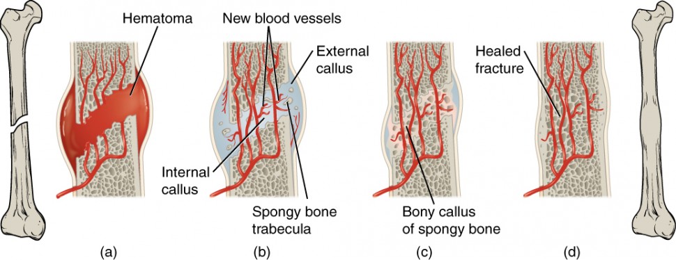
Fracture repair phases
Hematoma → granulation tissue → bony callus → remodeling
Functions of muscular system
Movement, posture, heat production, joint stabilization
Skeletal muscle
Long, cylindrical, striated, voluntary; attached to bones
Cardiac muscle
Branching, striated, involuntary; found in heart
Smooth muscle
Spindle-shaped, nonstriated, involuntary; found in organs
Muscle repair ability
Skeletal: limited; Cardiac: none; Smooth: good
Muscle control
Skeletal: voluntary; Cardiac & Smooth: involuntary
Epimysium
Connective tissue around whole muscle
Perimysium
Connective tissue around fascicles
Endomysium
Connective tissue around muscle fibers
Origin
Attachment site that remains relatively stationary
Insertion
Attachment site that moves during contraction
Muscle fiber cell
Long multinucleated cell
Myofibrils
Contractile rods within muscle fiber
Sarcoplasmic reticulum
Stores and releases calcium
T-tubules
Transmit electrical signals into fiber
Sarcomere
Functional contractile unit of muscle
Z disc
Boundary of sarcomere
A band
Dark band; length of myosin
I band
Light band; actin only
H zone
Region with only myosin
Actin (thin filament)
Anchored to Z disc
Myosin (thick filament)
Contains heads that bind actin
Sliding filament mechanism
Myosin pulls actin inward to shorten sarcomere
Troponin
Binds calcium and shifts tropomyosin
Tropomyosin
Blocks myosin-binding sites on actin
Neuromuscular junction (NMJ)
Synapse between motor neuron and muscle fiber
Acetylcholine (ACh)
Neurotransmitter that stimulates muscle contraction
Excitation-contraction coupling
Process linking electrical signal to contraction via Ca2+ release
Muscle relaxation
Calcium pumped back into SR; cross-bridges detach
Motor unit
Motor neuron + all muscle fibers it controls
Motor unit recruitment
More units = stronger contraction
Muscle twitch
Single contraction cycle
Latent phase
Time before contraction begins
Contraction phase
Muscle shortens
Relaxation phase
Muscle returns to resting state
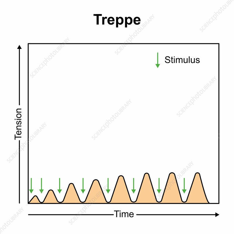
Treppe
Staircase increase in contraction strength
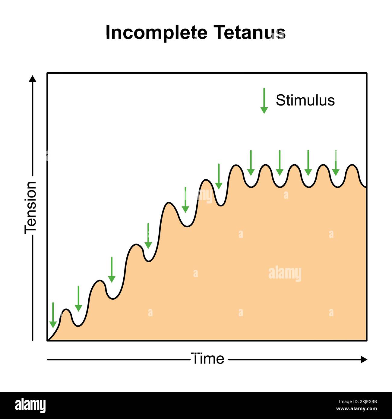
Incomplete tetanus
Sustained contraction with partial relaxation
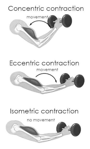
Isometric contraction
Tension without muscle length change

Isotonic contraction
Muscle changes length (concentric or eccentric)
Aerobic metabolism
Uses oxygen; produces more ATP
Anaerobic metabolism
No oxygen; produces lactic acid
Creatine phosphate
Quick ATP regeneration source
Myoglobin
Stores oxygen in muscle cells
Prime mover (agonist)
Main muscle causing movement
Antagonist
Muscle opposing movement
Synergist
Assists prime mover
Fixator
Stabilizes origin of muscle