14. RGCs
1/72
There's no tags or description
Looks like no tags are added yet.
Name | Mastery | Learn | Test | Matching | Spaced | Call with Kai |
|---|
No analytics yet
Send a link to your students to track their progress
73 Terms
What is the primary function of horizontal cells in the retina?
Horizontal cells provide lateral inhibition, shaping center‑surround antagonism to sharpen spatial contrast and “hone” the visual image.
How do ON and OFF pathways relate to fundamental visual features?
ON cells signal highlights/light increments
OFF cells signal shadows/dark decrements
Together they encode key contrast features of visual scenes.
How does center–surround antagonism enhance edge detection?
By opposing responses between center and surround, it amplifies differences at borders, enhancing the edge between highlights (ON) and shadows (OFF).
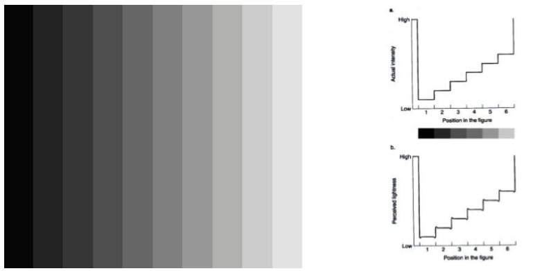
What is the mechanism underlying Mach bands?
Mach bands arise from center–surround antagonism via lateral inhibition, which exaggerates contrast at borders between regions of different luminance.
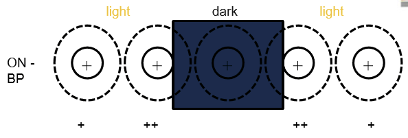
How does center-surround interaction alter perception at stripe borders in Mach bands?
Right edge of a stripe appears darker due to increased surround inhibition
Left edge appears lighter due to reduced surround inhibition
This occurs despite uniform physical intensity within each stripe.

Why are Mach bands considered an illusion, and what do they demonstrate about visual processing?
They are illusions because perceived brightness does not reflect actual luminance; they demonstrate that the visual system prioritizes edge enhancement and contrast, not absolute light levels.

What do ON‑center and OFF‑center retinal ganglion cells signal?
ON‑center cells increase firing (depolarize) with light in the center → signal brightness / light increments
OFF‑center cells increase firing (depolarize) with darkness in the center → signal darkness / light decrements
Why is this ON/OFF pairing important for visual perception?
ON and OFF pathways encode relative contrast rather than absolute luminance, allowing the visual system to distinguish brightness vs darkness efficiently across different lighting conditions.
What is contrast enhancement in the retina and what circuitry mediates it?
Contrast enhancement is the preferential signaling of local luminance differences rather than absolute intensity, mediated by lateral inhibition from horizontal cells onto bipolar cells.
What determines bipolar cell responses in contrast processing?
Bipolar cell activity depends on the relative difference between center luminance and surround luminance, not the absolute level of light.
Why can absolute luminance become irrelevant in retinal processing?
Because center–surround organization emphasizes contrast, uniform illumination produces little change in bipolar cell output, making absolute intensity poorly encoded.
How does contrast enhancement explain humans being poor “light meters”?
The visual system prioritizes edges and relative contrast, so our subjective perception of absolute brightness is inaccurate despite strong sensitivity to changes in luminance.

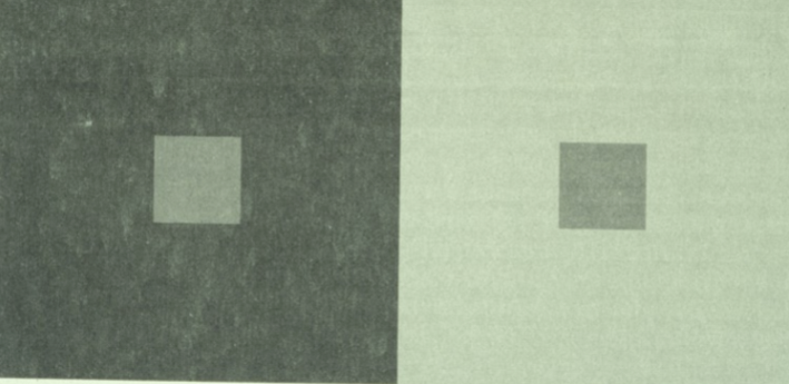
How is the apparent brightness of an object related to its surroundings?
The brightness of an object is inversely related to the luminance of its surroundings.
What is contrast enhancement and why is it a fundamental function of the visual system?
Contrast enhancement is the visual system’s emphasis on patterns of light and dark, allowing efficient object detection and recognition by prioritizing changes in luminance rather than absolute light levels.
What aspect of the visual scene is most strongly emphasized by retinal processing?
The visual system strongly emphasizes changes in the scene (edges), making humans especially good at contrast detection rather than uniform illumination.
How is “contrast” defined in visual perception?
Contrast refers to the difference in brightness (and color) of an object relative to other objects or surrounding regions in the visual field.
Why is perceived object lightness relatively invariant despite large changes in absolute luminance?
Because perception is based on relative luminance between spatial locations, not absolute luminance; this allows object lightness to remain stable across different lighting conditions.
What generates the electrical signal in bipolar cells of the retina?
Bipolar cell signals are generated in the dendrites based on photoreceptor input, with that input modulated by lateral inhibition from horizontal cells.
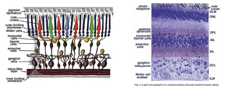
Where does the bipolar cell transmit its output signal?
The electrical signal travels down the bipolar cell to its axon terminal located in the inner plexiform layer, where it synapses onto ganglion cells.
How do bipolar cells transmit signals given that they do not fire action potentials?
Bipolar cells generate graded (electrotonic) potentials, not action potentials. Their short length (<100 µm) allows effective electrotonic spread of voltage changes from dendrites to terminals to control neurotransmitter release.
What neurotransmitter do bipolar cells release, and how is its release regulated?
Bipolar cells release glutamate.
Release occurs when the synaptic terminal is depolarized, leading to Ca²⁺-dependent exocytosis, similar to photoreceptors.
How is neurotransmitter release from bipolar cells similar to photoreceptors?
Both use graded membrane potentials and Ca-dependent glutamate release rather than action potentials. Depolarization increases Ca²⁺ influx, which increases glutamate release.
What type of synapse do bipolar cells use?
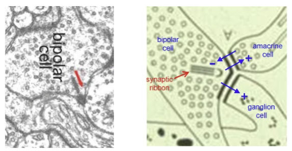
Bipolar cells use ribbon synapses, like rods and cones.

Describe the structural organization of a bipolar cell ribbon synapse.
A synaptic ribbon sits along the active zone with two postsynaptic processes, one on each side of the ribbon, allowing coordinated transmitter release to multiple targets.

Which postsynaptic cells are typically contacted at a bipolar ribbon synapse in primates?
In primates, each ribbon synapse usually contacts one amacrine cell and one ganglion cell, enabling parallel processing and modulation before signals exit the retina.
What is the functional role of retinal ganglion cells (RGCs) within the retina?
RGCs are entirely post‑synaptic within the retina, they only receive information. They integrate retinal signals before sending action potentials out of the eye via the optic nerve.
Which neurotransmitter receptors are expressed on retinal ganglion cell (RGC) dendrites, and what does this imply about their inputs?
RGC dendrites express ionotropic glutamate receptors (AMPA/kainate and NMDA), indicating they receive excitatory glutamatergic synaptic input from bipolar cells.
What is meant by the bipolar cell → RGC synapse being “sign‑conserving”?
The bipolar→RGC synapse is sign‑conserving, meaning glutamate depolarizes RGCs. Increased bipolar cell glutamate release increases RGC excitation.
Why are both AMPA/kainate and NMDA receptors important for RGC signaling?
AMPA/kainate receptors mediate fast excitatory transmission, while NMDA receptors contribute to sustained responses and coincidence detection, shaping RGC output patterns.
How do ON‑center and OFF‑center bipolar cells connect to retinal ganglion cells?
OFF‑center bipolar cells synapse onto OFF‑center RGCs, and ON‑center bipolar cells synapse onto ON‑center RGCs, preserving parallel ON and OFF visual pathways.
Are there differences in glutamate receptor expression between ON‑center and OFF‑center RGCs?
No. Both ON and OFF RGCs express ionotropic glutamate receptors, including AMPA/kainate and NMDA receptors. ON/OFF differences are not due to receptor type.
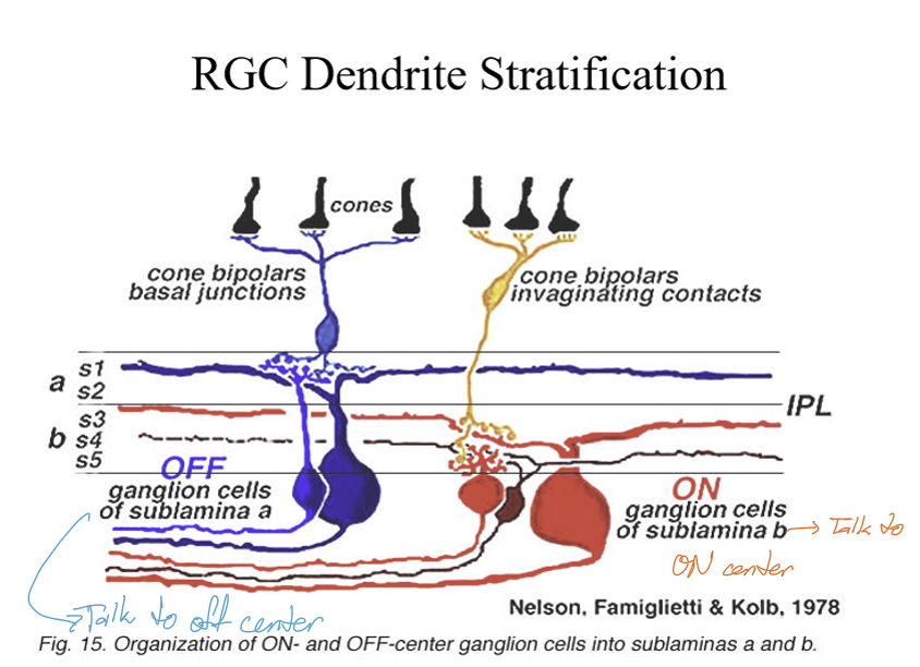
If ON‑ and OFF‑center retinal ganglion cells use the same glutamate receptors, what distinguishes their synaptic inputs?
The key distinction is laminar location within the inner plexiform layer (IPL):
ON‑pathway synapses terminate in the inner IPL
OFF‑pathway synapses terminate in the outer IPL

Why is IPL stratification important for ON and OFF retinal pathways?
IPL layering segregates ON and OFF visual processing, ensuring pathway‑specific synaptic connectivity despite similar neurotransmitters and receptors.
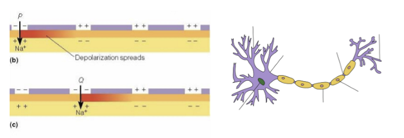
How do retinal ganglion cells (RGCs) differ fundamentally from other retinal neurons in signal transmission?
RGCs are spiking neurons. When depolarized, they fire action potentials, distinguishing them from other retinal neurons that rely on graded potentials.
How do RGC respond to depolarization?
They increase spike frequency with an increased depolarization.
How is stimulus strength encoded in retinal ganglion cells?
Stimulus intensity is encoded by spike frequency, not spike amplitude. Increased depolarization leads to a higher firing rate, not larger action potentials.
Why is the use of action potentials by RGCs functionally important?
Action potentials allow signals to be transmitted long distances without decrement along the optic nerve, unlike graded potentials, which decay with distance.
What do extracellular recording detect?
Action potentials only.
Which recording came first? Intracellular or Extracellular?
Extracellular. Intracellular recording techniques were developed after extracellular ones.
What did inital intracellular recordings measure?
RGC spiking responses. In early 1950’s, it was observed the RGC responses depended on the location of the light stimulus (center-surround antagonism).
What drives the light‑induced increase in firing of ON‑center and ON‑OFF RGCs?
Primarily ON bipolar cell input. In light, photoreceptors reduce glutamate release, causing ON bipolar cells to depolarize and release more glutamate, which excites ON‑center RGCs and the ON component of ON‑OFF RGCs.
What drives the light‑induced decrease in firing of OFF‑center and ON‑OFF retinal ganglion cells?
Primarily OFF bipolar cell input. In light, photoreceptors reduce glutamate release, causing OFF bipolar cells to hyperpolarize and release less glutamate, which reduces excitation of OFF‑center RGCs and the OFF component of ON‑OFF RGCs.
What cellular mechanisms generate surround responses in RGC receptive fields?
Surround responses arise from lateral inhibition, primarily via horizontal cell–mediated feedback onto photoreceptors, which alters glutamate release from surrounding photoreceptors and shapes bipolar cell input to RGCs.
How do amacrine cells contribute to center–surround responses in RGCs?
Amacrine cells provide inhibitory input in the inner plexiform layer (IPL), modulating RGC responses and enhancing or shaping surround inhibition.
What is the form of the electrical signal generated by retinal ganglion cells (RGCs)?
RGCs generate brief bursts of action potentials (spikes), not graded potentials. This spiking output is the final retinal signal sent to the brain via the optic nerve.
How is information encoded in ON and OFF retinal pathways at the level of RGCs?
Information is encoded by spike timing and spike frequency, not response amplitude. Stronger depolarization produces a higher firing rate.
What ionic mechanism underlies action potential generation in RGCs?
Action potentials arise from rapid opening and closing of voltage‑gated ion channels (primarily Na⁺ and K⁺), producing all‑or‑none spikes.
Why is spiking output from RGCs critical for vision?
Spikes allow rapid, long‑distance, non‑decrementing transmission of visual information from the retina to central visual targets, unlike graded potentials.
What is the overall flow of visual information in ON and OFF retinal circuits?
Light‑evoked signals are transmitted from photoreceptors → bipolar cells → retinal ganglion cells (RGCs). Bipolar cells encode ON vs OFF changes, and RGCs convert this into spiking output sent to the brain.
What determines whether an ON or OFF retinal circuit is activated by a visual stimulus?
It depends on whether the stimulus involves light onset or light offset.
ON circuits respond best to light increments
OFF circuits respond best to light decrements
How do ON and OFF circuits respond to a small spot of light flashing on and off?
Light ON: ON bipolars depolarize → ON RGC firing increases; OFF bipolars hyperpolarize → OFF RGC firing decreases
Light OFF (dark): OFF bipolars depolarize → OFF RGC firing increases; ON bipolars hyperpolarize → ON RGC firing decreases
What types of visual scenes preferentially activate ON vs OFF pathways?
ON pathways are best stimulated by light objects on dark backgrounds
OFF pathways are best stimulated by dark objects on light backgrounds
Why do the retina and visual system maintain both ON and OFF pathways?
Parallel ON and OFF circuits allow the visual system to independently and efficiently encode increases and decreases in luminance, improving contrast detection and temporal precision.
What is the overall effect of center-surround antagonism?
Lateral inhibition mediated by horizontal cells serves to sharpen edges and border in our perception of images, aka, center-surround antagonism enhances contrast.
What are midget retinal ganglion cells (RGCs), and why are they especially abundant in primates?
Midget RGCs are a major RGC class in primates, present at much higher proportions than in cats or rodents because primates evolved specializations for high visual acuity and fine spatial detail.
Where are midget RGCs most concentrated, and how does their distribution change across the retina?
Fovea: ~95% of RGCs are midget
Periphery: ~45% of RGCs are midget
This decline reflects reduced spatial resolution demands outside the fovea.
Describe the circuit connectivity of foveal midget RGCs.
Foveal midget RGCs receive input from one midget bipolar cell, which in turn receives input from one cone:
OFF pathway: flat midget bipolar
ON pathway: invaginating midget bipolar
This creates a 1 cone → 1 bipolar → 1 RGC circuit.
Why are foveal midget RGC circuits ideal for high spatial resolution?
Minimal convergence (1:1:1 wiring) preserves fine spatial detail, allowing precise localization and discrimination of visual stimuli.
How does midget RGC connectivity differ in the peripheral retina?
In the periphery, midget RGCs receive input from more than one midget bipolar cell, but still fewer bipolars than parasol RGCs in the same region, representing an intermediate level of spatial summation.
How do parasol retinal ganglion cells (RGCs) differ from midget RGCs in their synaptic input?
Parasol RGCs receive input from diffuse bipolar cells, which collect signals from multiple cones, unlike midget RGCs that receive more selective input. This leads to greater spatial summation.
Which bipolar cell types provide input to OFF vs ON parasol RGCs?
OFF parasol RGCs receive input from diffuse flat bipolars (fb or db)
ON parasol RGCs receive input from diffuse bipolar cells (flat vs invaginating is unclear)
What is the functional consequence of diffuse bipolar input onto parasol RGCs?
Input from multiple cones produces greater spatial summation, making parasol RGCs less sensitive to fine detail but better at detecting low‑contrast, rapidly changing stimuli.
What is the key functional role of bistratified RGCs?
Bistratified RGCs play a key role in color vision, integrating cone signals (classically associated with chromatic opponency), with details elaborated in later circuitry.
How do midget retinal ganglion cells (RGCs) respond to light onset or offset?
Midget RGCs show a sustained response:
ON midget RGCs fire continuously during light onset
OFF midget RGCs fire continuously during light offset
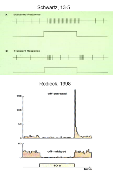
How do parasol retinal ganglion cells respond to light onset or offset?
Parasol RGCs show a transient response:
ON parasol RGCs fire briefly at light onset
OFF parasol RGCs fire briefly at light offset

What is the key functional difference between sustained (midget) and transient (parasol) responses?
Sustained responses (midget) encode steady luminance and fine detail
Transient responses (parasol) encode rapid changes, contrast, and motion

Do both midget and parasol RGCs exhibit center–surround receptive field organization?
Yes. Both midget and parasol RGCs show center–surround antagonism, enhancing contrast and edge detection, although some other RGC types lack this organization.
Why does myelination allow action potential impulse to travel down the axon faster?
Saltatory propagation: action potentials jump from node to node.
For patients with demyelination diseases, such as MS, what are the visual-related symptoms?
Decreased VA
Reduction in visual fields
Sudden-onset diplopia
pupillary defects
color vision defects
Uhthoff’s sign (VA issues after exercise/hot showers)
Pulfrich’s stero phenomenon
What is the Pulfrich Stereoeffect?
When a swinging object appears to move in an elliptical motion.
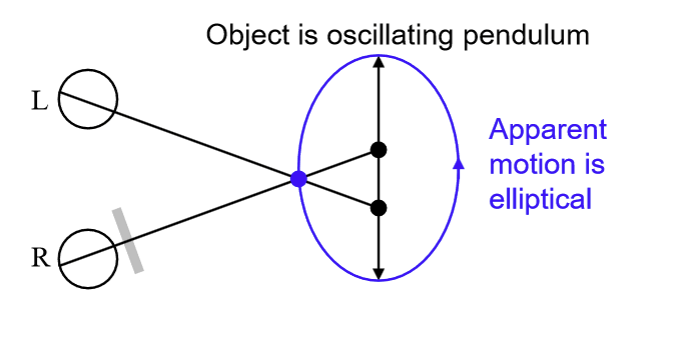
What causes the Pulfrich Stereoeffect?
A delay in visual processing between the two eyes leading to a neural timing difference that creates an illusion of depth for moving objects.
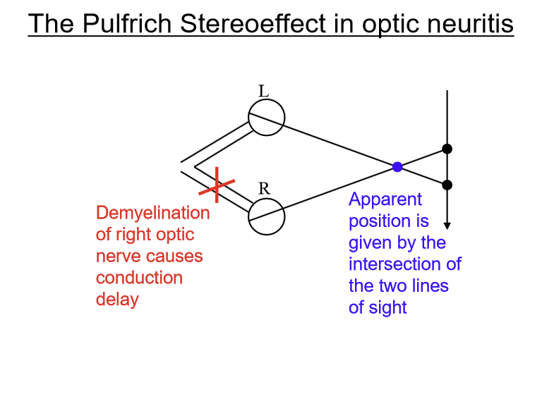
What can cause a delay in visual processing between the two eyes?
Reduced retinal illumination in one eye
Unequal velocities of signal transduction in optic neuritis with demyelination
How can symptomatic Pulfrich phenomenon be managed?
Uniocular tinted lens, or CL, can provide immediate and lasting relief from visual difficulties caused by the visual delay underlying spontaneous Pulfrich effect.