LAM II: Equine Respiratory
1/186
There's no tags or description
Looks like no tags are added yet.
Name | Mastery | Learn | Test | Matching | Spaced | Call with Kai |
|---|
No analytics yet
Send a link to your students to track their progress
187 Terms
Describe a normal equine breathing effort and pattern.
- Breathing effort: Minimal movement of nares, thorax and abdomen is normal
- Breathing pattern: Synchronic chest and abdomen movement is normal
What are signs indicative of an upper airway obstruction?
- Audible respiratory noise
- Increased inspiratory effort
- Decreased nostril airflow
What are signs indicative of lower airway disease?
- Increased expiratory effort (abdominal push)
- +/- adventitious lung sounds
What are signs of respiratory disease observed in the equine head?
- Nasal discharge (consider character, amount, unilateral/bilateral)
- Nostril flaring
- Reduce nostril airflow
- Lymphadenopathy
- Facial swelling
- Dull sinus percussion
Cause of nostril flaring?
- Increased respiratory effort
Cause of reduced nostril airflow?
- Upper airway obstruction
Broad Ddx for lymphadenopathy?
- Inflammation
- Infection
- Neoplasia
Ddx for equine facial swelling?
- Tooth root abscess
- Suture line periositits
- Neoplasia
Ddx for dull equine sinus percussion?
- Fluid (sinusitis)
- Mass
- Cyst
What areas should be ausculted during an equine respiratory examination?
- Trachea
- Lungs (at rest, with rebreathing bag, after exercise)
What are abnormal sounds which can be heard on equine lung auscultation? What do these sounds indicate?
- Crackles: Airways opening on inspiration (due to fluid or atelectasis)
- Wheezes: Vibration of airway walls (bronchoconstriction)
- Pleural friction rubs: Parietal and visceral pleura rubbing together
- Decreased bronchovesicular sounds: Pleural effusion (severe consolidation as with fibrosis)
How is a rebreathing examination performed? What is the purpose of this examination?
- Using a large garbage bag filled with air
- Leads to an increase in inspired CO2 to encourage deep breathing
What are normal changes expected in equine respiration during a rebreathing examination?
- No cough
- Increased bronchivesicular sounds
- No adventitious lung sounds
- No tracheal rattling
- Recovery within a few breaths after bag removal (4-6)
How is equine thoracic percussion performed?
- Tap a spoon in the ICS during auscultation
- Aerated tissues like the lungs should result in a resonant sound
What does a dull sound on equine thoracic percussion indicate?
- Fluid-filled structures (lung abscesses vs. consolidated lung vs. pleural effusion)
- Can be used to compare dorsal vs. ventral lung and identify the line of pleural effusion
Indications for equine thoracic US?
- Suspicion of lower airway disease (pleural effusion, pneumonia, thoracic neoplasia, pneumothorax)
- Rib fractures
What are some limitations of thoracic US?
- Does not penetrate air
- Only detects superficial lung lesions (Deeper parenchymal lesion may be missed)
- May lead to underestimation of disease severity
What are some advantages of thoracic US?
- Readily available to most practitioners
- Portable
- Allow for examination of lungs even when there is pleural effusion
What do "comet tails" indicate on thoracic US?
- Pleural roughening
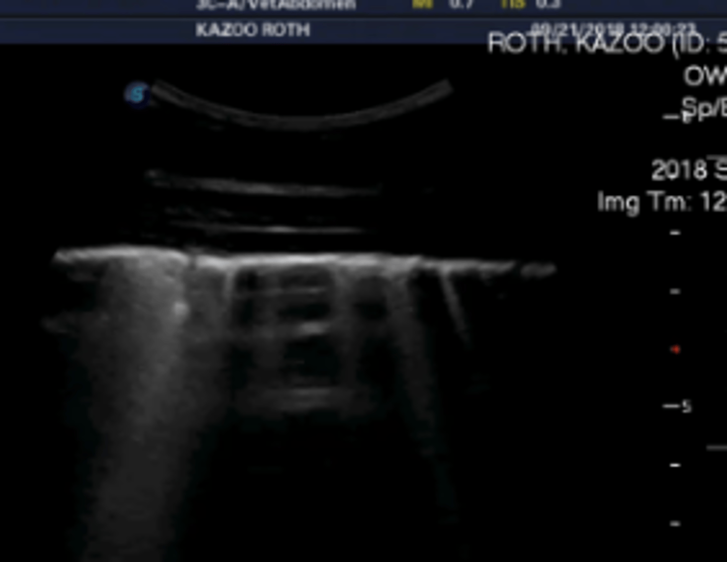
Consolidation on thoracic US can indicate what?
- Non-aerate lung (edema vs. discharge vs. mass (neoplasia or fibrosis) vs. atelectasis)
Pnuemonia/aspiration pneumonia in horses most commonly affects which lung fields?
- Caudoventral
DDx for respiratory disease in a horse with unremarkable thoracic radiographs?
- Equine asthma
- Upper airway disease
- Diaphragmatic paralysis (rare)
DDx for diffusely increased lung opacity in the caudodorsal equine lungs on thoracic radiographs?
- EIPH (exercised induced pulmonary hemorrhage)
- Focal pneumonia (FB, embolus, thromboembolus)
- Iatrogenic (BAL)
DDx for diffusely increased lung opacity in the caudoventral equine lungs on thoracic radiographs?
- Aspiration pneumonia
- Broncho-/pleuropneumonia
DDx for generalize/diffuse increased opacity of equine lungs on thoracic radiographs?
- Interstitial pneumonia
- Pulmonary fibrosis
- Pulmonary edema
DDx for one/multiple discrete tissue opacities in the equine lungs on thoracic radiographs?
- Neoplasia
- Abscess
- Bacterial or fungal granuloma
- FB (single)
- Pulmonary edema (Patchy opacities possible)
- EMPF/Equine Multinodular Pulmonary Fibrosis (multiple)
- Eosinophilic interstitial pneumonia (multiple)
- Idiopathic granulomatous interstitial pneumonia (multiple)
Indications for equine upper airway endoscopy?
- Suspicion of upper airways disease - decreased/asymmetric nostril air, upper airway noise, increased inspiratory effort, coughing when eating, dysphagia
- Localize source of nasal discharge/ epistaxis
- Perform tracheal wash
- Examine trachea and bronchus
Limitaitons of equine upper airway endoscopy?
- Unable to visualize sinus, entire ethmoids
- May exacerbate respiratory distress- providing supplement oxygen may help
Which cranial nerves are found in the medial compartment of the guttural pouch?
- 9, 10, and 12
Options for airway fluid sampling?
- Nasal swab
- Guttural pouch lavage
- Tracheal wash (TW/ TTW/ TTA)
- BAL
- Thoracentesis
Airway fluid samples can be submitted for what testing?
- Cytology
- C/S
- PCR
TTA collects a sample from where? BAL collects a sample from where?
- TTA collects a sample from entire lower respiratory tract (Gets fluids which have drained from the cranial 2/3 of the lungs to the thoracic inlet)
- BAL collects a sample from the distal terminal airways of one section of one lung (Blind); (Good for diffuse disease, i.e., asthma, but will bypass the entire cranial 2/3 of the lung)
Normal cell types in a tracheal wash?
- Predominance of macrophages, < 10% neutrophils, <1% eosinophil, occasional extracellular bacteria
What is the fluid sample of choice for diagnosis of pneumonia in horses? What findings are consistent with this?
- Tracheal wash
- Intracellular bacteria
Normal cell prevalence in BAL sample:
A. Alveolar macrophages
B. Lymphocytes
C. Neutrophils
D. Mast cells
E. Eosinophils
A. Alveolar macrophages 60-80%
B. Lymphocytes 20-35%
C. Neutrophils <5%
D. Mast cells <2%
E. Eosinophils <1%
- Note: Hemosiderophages common in racehorses
What fluid sample is preferred for the diagnosis of equine asthma? What findings are consistent?
BAL cytology
> 5% neutrophils or > 2% mast cells or > 1% eosinophils
When is a pleurocentesis performed?
- Only when there is pleural effusion (pleural fluid cannot be obtained in a normal patient)
What are normal cytology findings for pleural fluid?
- <5,000 cell/uL cells
- TP < 2.5 g/dL
- Mostly macrophages and neutrophils, low numbers of lymphocytes and mesothelial cells
What "blood" gas findings on pleural effusion can suggest sepsis?
- Elevated lactate
- Decreased glucose
What are large animal options for arterial blood collection?
- Transverse facial - ventral to zygomatic arch
- Dorsal metatarsal artery - between MT3 and MT4
- Brachial artery - medial aspect of elbow
- Femoral artery - external inguinal ring
- Carotid?
Air contamination of a blood sample will result in what findings?
- Increase in PO2 and decrease in PCO2 (Make it look better)
Plastic syringes with samples for blood gas should be analyzed how quickly after collection? What about for a glass syringe in an ice bath?
- Within 10 mins
- Within 2 hours
True or false: Venous samples for be used for blood gas analysis of the respiratory system.
- True; values are similar with the exception of PO2, so you cannot say a patient is hypoxemic while looking at a blood gas
Describe the prevalence of thoracic neoplasia in horses.
- Rare - Prevalence 0.15-0.6% of necropsied horses
Most common primary lung tumor of horses?
- Granular cell tumor
Most common thoracic neoplasia of horses?
- Lymphoma (multicentric); 54% and 74% of thoracic neoplasia in 2 studies
In what signalment of horses does thoracic neoplasia most commonly occur?
- Mature or aged horse, with exception of lymphoma which may also be observed in young animals.
Clinical signs of equine thoracic neoplasia?
- Cough
Increased expiratory effort
- Exercise intolerance
- Weight loss
- Abnormalities involving other body system such as diarrhea, cutaneous mass, ventral edema
- Paraneoplastic syndromes: hypercalcemia, hypertrophic osteopathy, hypoglycemia, unexplained fever
Methods for diagnosis of equine thoracic neoplasia?
- US: most often have pleural effusion, may see pulmonary nodules
- Radiographs: Usually nodular interstitial pattern
- Bronchoscopy
- Cytology of pleural fluid
- Cytology TTA and or/ BAL
- Aspirate or biopsy of thoracic mass - ultrasound-guided
- Biopsy of peripheral lymph node
What is HO?
- Symmetrical proliferation of connective tissue and subperiosteal bone along the diaphyses and metaphyses
Signs of HO?
- Usually lame
- Reluctance to move
- Effused joints
Etiology of HO?
- Associated with thoracic neoplasia, and other inflammatory conditions of chest such as intrathoracic abscess or granuloma
- Likely due to overproduction of vascular endothelial growth factor (VEGF)
Prognosis of equine thoracic neoplasia?
- Guarded to poor
Describe the prevalence of EIPH in racehorses.
- Over 80% of TB and STB racehorses when examined after each of 3 consecutive races
Clinical signs of EIPH?
- Epistaxis (<9% of racehorses; Not reliable)
- Poor performance (if severe bleeding)
- Frequent swallowing
What signalment of horses commonly get EIPH?
- Racehorses
- Increases in frequency with age
Describe the pathophysiology of EIPH.
- Failure of pulmonary capillaries at maximal exercise (Marked increase in stress in the alveolar wall → rupture of capillaries; High blood pressure in tiny vessels and very negative pressure in thorax leads to burst of vessels)
- Physiologic mechanisms rather than pathologic
- Additional suspected contributors are upper airway obstructions (DDSP, laryngeal hemiplegia - increase negative pressure in thorax to overcome obstruction) and lower airway obstructions (Asthma)
True or false: With EIPH, recurrence is likely. Why or why not?
- True
- Hemorrhage into airways and interstitium once causes inflammation -> develop fibrosis and decreased compliance -> Reoccurence
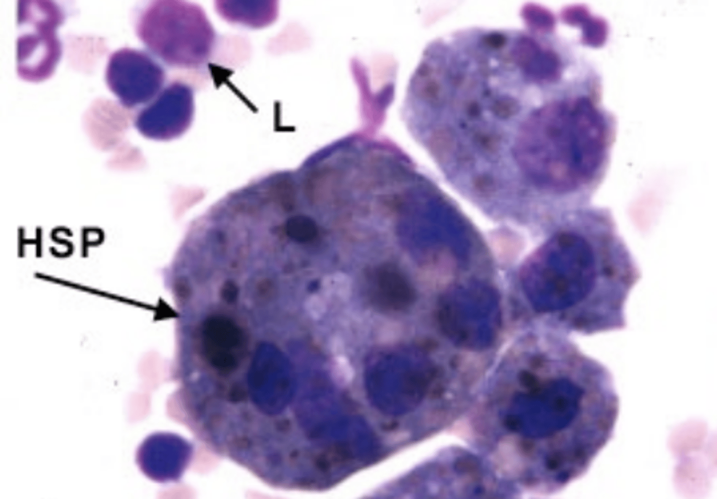
Methods for diagnosis of EIPH?
- PE
- Endoscopy (30-120 mins after exercise)
- BAL (Macrophages with hemosiderin inside = Hemosiderophages)
Treatment for EIPH?
- Furosemide 4 h pre-race to decrease severity of bleeding
- Rest - mandated by some racing jurisdiction for up to 2 weeks (optimal duration unknown)
- Anti-inflammatories (NSAIDS)
- Treat any other respiratory disease such as upper airway abnormality or equine asthma
What injury can result from smoke inhalation?
- Thermal injury - upper airway (Burns)
- Chemical injury - CO exposure
- Low PAO2 causes pulmonary vasoconstriction and hypoxemia
Following smoke inhalation, it may take ____________ to develop clinical signs of respiratory disease.
- 24 to 48 hours
In addition to respiratory signs, what other issues in other body systems can smoke inhalation lead to?
- MODS
- SIRS
- Sepsis
- Hypermetabolism
Describe phase 1 of the pathophysiology of smoke inhalation following exposure.
- 0 to 36hrs after exposure
- Acute pulmonary insufficiency due to CO and hypoxia
- Upper airway edema and necrosis (airway obstruction)
- Bronchoconstriction (due to noxious products burned)
- Altered pulmonary blood flow (hypoxic bronchosconstriction)
Describe phase 2 of the pathophysiology of smoke inhalation following exposure.
- 48-72 hours after exposure
- Pulmonary edema, lower airway obstruction, and parenchymal lesions due to release of inflammatory mediators from macrophages and neutrophils
Describe phase 3 of the pathophysiology of smoke inhalation following exposure.
- 1-2 weeks after exposure
- Bronchopneumonia: impaired immune system locally and systemic
Diagnostics for smoke inhalation?
- PE
- CBC/ Chem
- Arterial blood gas (Hypoxia?)
- Endoscopy of upper airway and tracheobronchial tree
- 1-2 weeks later - may develop pneumonia -> Do tracheal aspirate (cytology, C/S), and thoracic radiographs or US
Treatment of smoke inhalation?
- Humidified Oxygen
- Tracheotomy if upper airway obstruction
- Bronchodilation
- Furosemide (for pulmonary edema)
- Anti-inflammatories (NSAIDs vs. corticosteroids, consider immunosuppression)
- Hyperbaric oxygen chambers
- If IV fluids are needed - be judicious due to risk of pulmonary edema
- ABX if documented pneumonia
Poor prognostic indicators associated with smoke inhalation?
- Tachypnea (>50 bpm)
- Dehydration (PCV>50%)
- Opaque nasal discharge
- Persistent severe hypoxemia despite O2 supplementation (PaO2 <60 mmHg)
True or false: Horses with smoke inhalation are often in severe acute respiratory distress following exposure.
- False; Horses may be "normal" at presentation
Define bronchopneumonia.
- Infection of the bronchi extending into the parenchyma
Define pleuropneumonia.
- Extension of parenchymal infection to the pleural space
- Fluid and/or fibrin over lung/in thorax
- Presence of pleural effusion
True or false: Spontaneous pneumonia is uncommon in adult horses.
- True
Predisposing factors to adult equine pneumonia?
- Long distance transport (Stress and head elevation, poor drainage)
- Aspiration - choke, dysphagia
- General anesthesia
- Viral respiratory infections
- Strenuous exercise
- Penetrating wounds
Clinical signs associated with adult equine pneumonia are dependent on what?
- Severity/duration of illness and presence/absence of pleuritis
Possible clinical signs associated with adult equine pneumonia?
- Fever
- Anorexia
- Bilateral nasal discharge
- Cough
- Exercise intolerance
- Tachycardia, tachypnea, increased effort
- Pleurodynia: pawing, stiff, reluctant to move (Can look like colic)
- Weight loss
- Ventral edema
- Epistaxis
- Halitosis
Diagnostics for adult equine pneumonia?
- History/PE (thoracic auscultation, thoracic percussion, tracheal auscultation, +/- rebreathing exam)
- Labs: CBC, Chem, blood gas
- Imaging: US, radiographs, endoscopy
- Respiratory fluid sampling
When presented with a horse with suspected pneumonia, (pleuropneumonia or bronchopneumonia), what fluid sample is best to submit for culture?
- Tracheal wash
Methods of obtains a tracheal wash sample? Briefly describe benefits/limitations.
- Endoscopic: Convenient with good visualization, accessibility may be limiting factor
- Transtracheal: More invasive but more sterile and doesn't require specialize equipment
What cytologic findings may be consistent with pneumonia on a tracheal wash sample? What other diagnostics should be performed on the sample?
- Increased neutrophils, degenerative neutrophils, intracellular bacteria
- Perform gram stain and aerobic/anaerobic culture
When a thoracocentesis is performed, what findings are suggestive of septic pleuritis?
- High cell count and protein with degenerative neutrophils or intracellular bacteria
- Low fluid pH (<7.1) and low glucose (<40mg/dl)
A foul smell in a thoracocentesis sample is suggestive of what?
- Anerobes
What ABX therapy is appropriate for pneumonia?
- Broad spectrum antibiotics (Don't wait on culture)
- Combination antibiotic therapy: Penicillin + gentamicin
- Rifampin aids tissue penetration
Common ABX for gram positive coverage?
- Penicillin
- Ceftiofur or ampicillin
Common ABX for gram negative coverage?
- Gentamicin
- Amikacin (foals)
- Enrofloxacin
- Ceftiofur
- Trimethoprim sulfamethoxazole/sulfadiazine
- Chloramphenicol
- Tetracyclines - doxy, mino
Common ABX for anaerobe coverage?
- Penicillin (except Bacteroides fragilis)
- Metronidazole (PO)
- Chloramphenicol (PO)
- Tetra-/doxy-/minocycline
How long should ABX therapy be continued for management of equine pneumonia?
- Until resolved - clinical signs, inflammatory indicators on lab work and imaging (may take weeks to months)
What adjunct treatments can be used for management of equine pneumonia in addition to ABX?
- NSAIDs - endotoxic/SIRS & pain
- Additional analgesia - butorphanol, lidocaine
- IV fluids
- Deep bedding and ice boots if endotoxic/SIRS
- Oxygen
What is the purpose of pleural effusion drainage in management of equine pneumonia?
- Facilitate lung expansion and reduce hypoxemia
- Removes cells and protein that can create adhesions and loculation
How often should pleural effusion drainage be performed in equine pneumonia patients?
- Repeat q 1-2 days or use indwelling drains
- Note: Large volumes (20 L) often removed
When is pleural lavage indicated in equine pneumonia patients?
- If thick viscous pleural fluid is present -> Remove fibrinous effusion
Complications of equine pneumonia?
- Thrombophlebitis
- Laminitis
- DIC
- Pleural/pulmonary/mediastinal abscess
- Bronchopleural fistula (pneumothorax; Often cough during pleural lavage)
- Pericaridits
- ABX induced colitis
Prognosis of equine pneumonia?
- Overall survival of ~ 75%
- Performance (i.e., racing future) often unaffected
Negative prognostic indicators for equine pneumonia?
- Anaerobic infection
- Complications and severe disease
Common cause of equine fungal pneumonia in the PNW?
- Cryptococcus gattii
What is equine multi-nodular pulmonary fibrosis?
- Nodular form of interstitial pneumonia - fibrosis
Clinical signs of equine multi-nodular pulmonary fibrosis?
- Fever
- Cough
- Weight loss
- Exercise intolerance that can progress to respiratory distress
- Inflammatory CBC - leukocytosis, hyperfibrinogenemia
EMPF has been associated with what?
- EHV-5 (Positive PCR lung biopsies and BAL)
Treatment of equine multi-nodular pulmonary fibrosis?
- Corticosteroids
Prognosis of equine multi-nodular pulmonary fibrosis?
- Poor; Progressive condition