ANHB2214 Respiratory System
1/48
There's no tags or description
Looks like no tags are added yet.
Name | Mastery | Learn | Test | Matching | Spaced | Call with Kai |
|---|
No analytics yet
Send a link to your students to track their progress
49 Terms
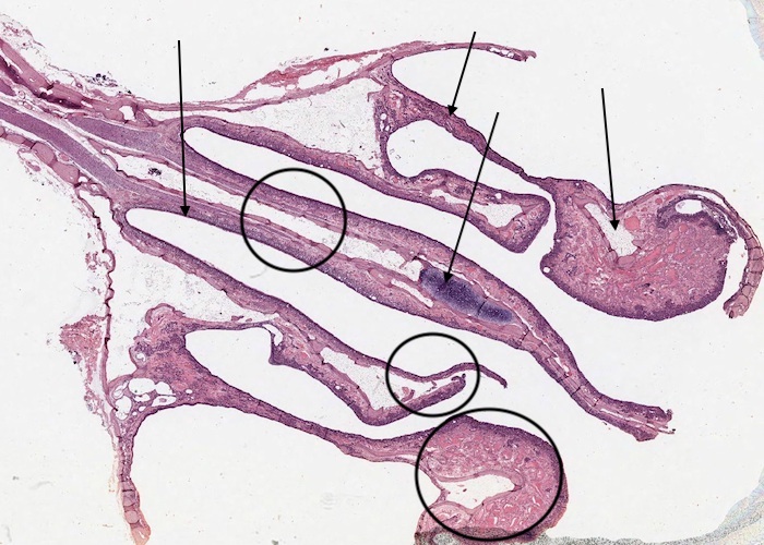
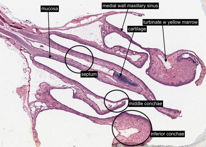
You are looking at the nasal cavities "front-on" but with the head tilted about 45 degrees to the left! Whilst this image is at very low magnification identify the major histological features then view the histological sections of the nasal cavity via the links below.
The bony nasal septum is in the midline. On either side of the nasal septum is an air passageway, and protruding into these spaces are two large masses on each side that represent the middle and inferior nasal conchae. The turbinate supports each conchae and has yellow marrow inside which makes them look hollow. In removing this block of tissue, each maxillary sinus was largely cut away, but their medial wall remained.
The mucosa of the entire nasal cavity is underlain by bone, except for the mid anterior part of the nasal septum, which is cartilage. This cartilage abuts directly with the bone of the bony septum.
Towards the anterior part of the respiratory passageway the surrounding supporting tissue shifts entirely to cartilage (you can feel its flexibility).
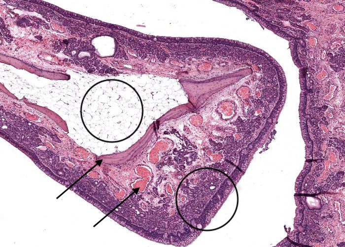
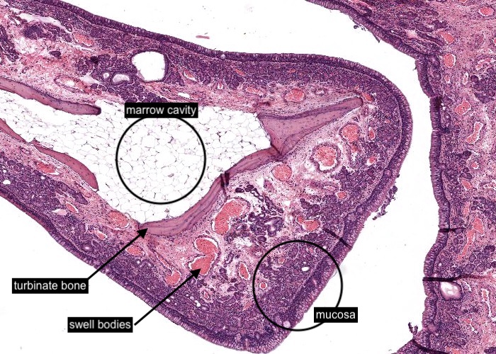
Now (at this higher magnification) you can see the nasal conchae in more detail. They protrude into the air passageways and increase the surface area of the lateral walls of the nose. A turbinate bone supports each concha. The marrow cavity inside is mostly adipose tissue.
The mucosa is lined with pseudostratified columnar epithelium with cilia and, in most places, large numbers of goblet cells. The lamina propria is richly vascularized to provide an ample supply of blood to cool or warm the inspired air as appropriate. On the conchae the blood vessels are exaggerated into large venous structures called swell bodies.
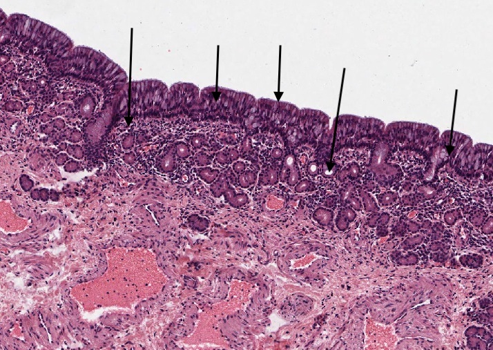
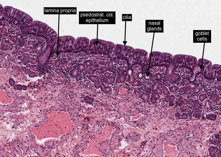
Most of the nasal cavity is the "respiratory region" lined with pseudostratified columnar epithelium with cilia and, in most places, large numbers of goblet cells (although is is hard to see both of these structures at this low magnification). This mucosa is also in the walls of the sinuses. Numerous mixed (serous / mucous) nasal glands extend into the lamina propria from the epithelium.
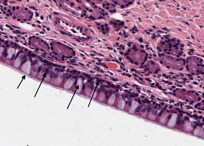
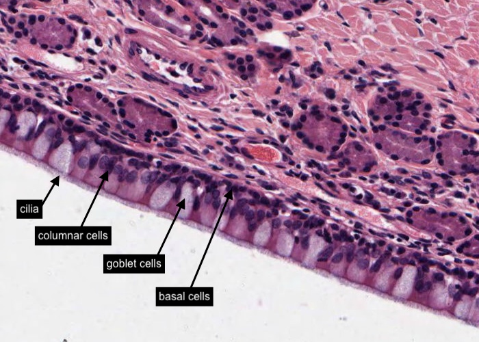
Ciliated, pseudostratified columnar epithelium is the epithelium lining the "respiratory region" of the nasal cavity and is often referred to as "respiratory epithelium". There are a number of cell types visible in this "respiratory epithelium".
Thin but tall columnar cells with cilia projecting from their apical surfaces.
Goblet cells with clear cytoplasm (as the mucus has been removed during processing).
Basal cells.
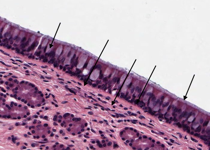
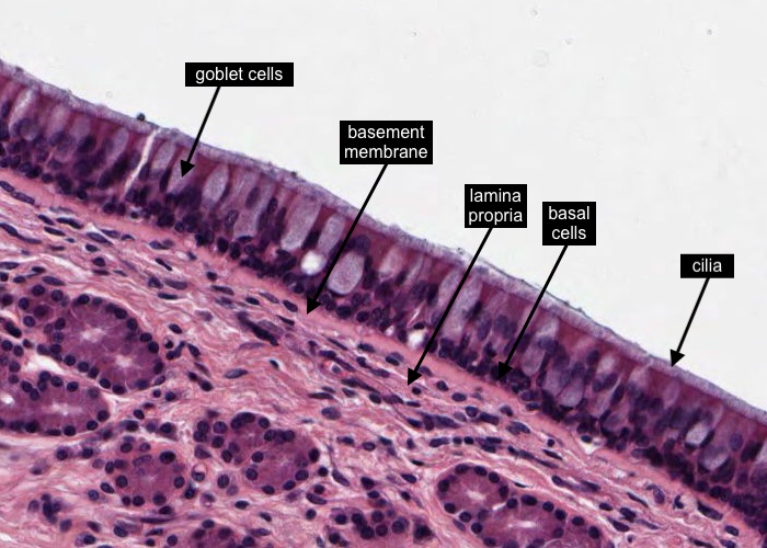
The basement membrane is quite prominent here as the amorphous layer between the basal region of an "respiratory epithelium" and the underlying lamina propria.
Once again (as a revision exercise !) identify the tall ciliated columnar cells, goblet cells and basal cells
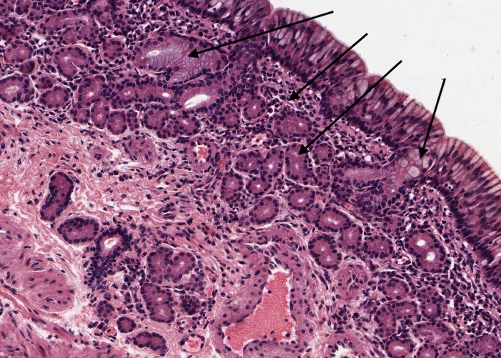
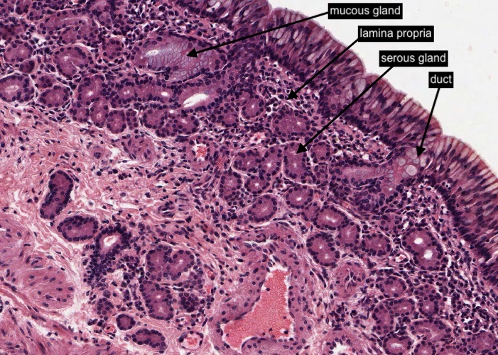
Numerous mixed (serous / mucous) glands extend into the cellular lamina propria from the epithelium. Their secretions are conveyed to the epithelial surfaces via a duct. The lumen of each serous gland is visible as a central small clear region when these glands are sectioned transversely.
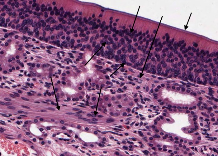
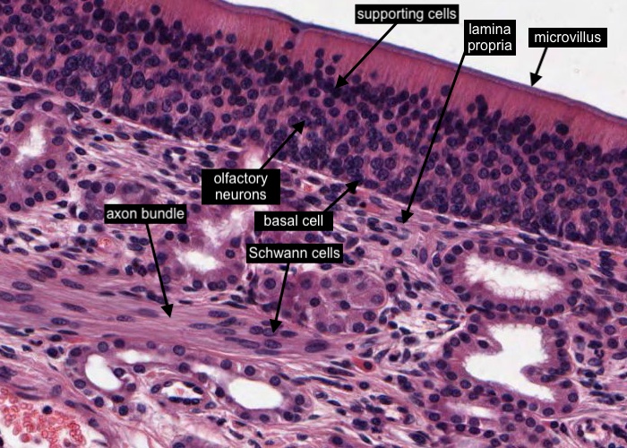
Olfactory epithelium is a pseudostratified columnar type. The lamina propria underlying the olfactory epithelium contains numerous glands.
Olfactory epithelium is made up of three cell types and I think you should be able to discern them in this image (or at least their nuclei). Most numerous are sustentacular cells = supporting cells. They extend from the basement membrane to the surface but their nuclei are relatively dense, darker stained, and located closer to the epithelial surface than the olfactory neurons. They exhibit an apical microvillus border.
Olfactory neurons lie at different levels within the epithelium and careful observation indicates they have more euchromatin than the other cell types. They are round cells with two processes. A dendrite goes to the surface where it sprouts several long non-motile stereocilia which bear the receptors for odoriferous molecules. The neurons also send an axon down through the basement membrane. It joins with others in the lamina propria to form an axon bundle which travel to the olfactory lobes of the brain (a very short distance). The nuclei in those axonal bundles, of course, belong to Schwann cells, characteristic of a peripheral nerve.
The third cell type is the basal cell. It is the stem cell, able to divide and differentiate into both sustentacular cells and olfactory neurons.
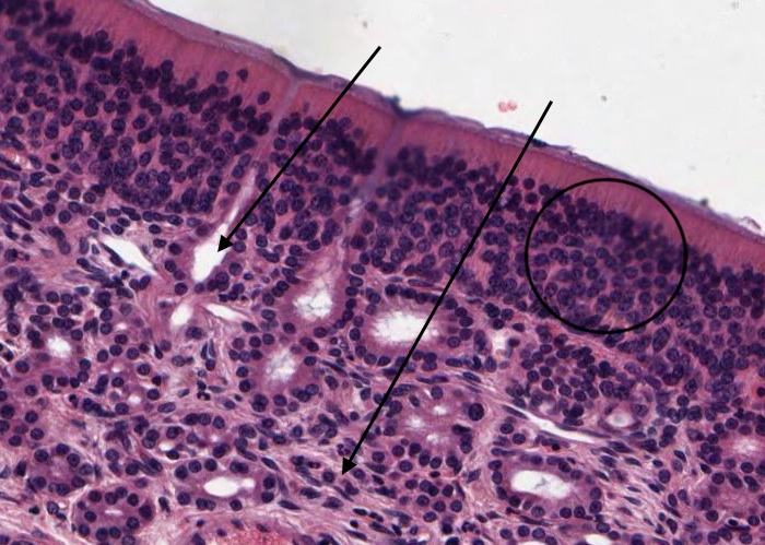
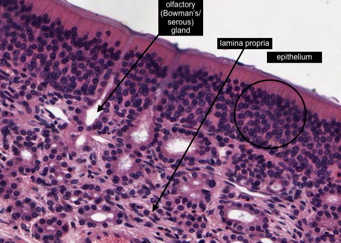
The lamina propria underlying the olfactory epithelium contains numerous glands. These are the specialized olfactory (serous) glands (Bowman's glands) that provide fluid to wash away old odoriferous molecules. One of their distinctive structural characteristics is that each short duct enlarges just under the surface epithelium.
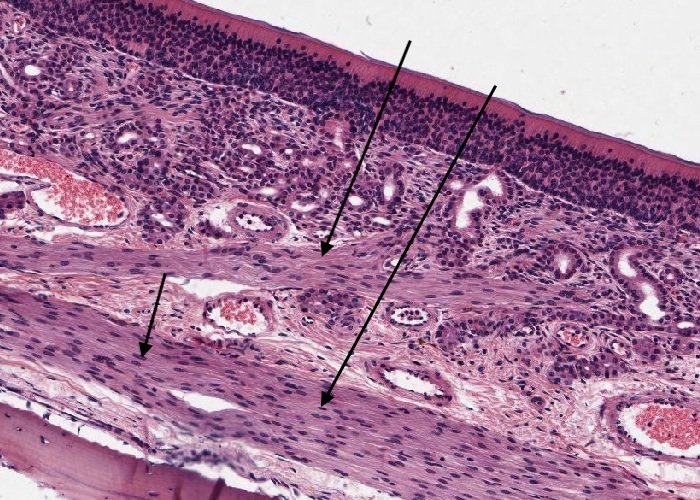
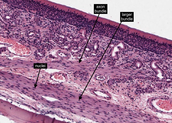
Olfactory neurons each extend an axon to form an axon bundle which unite to form a larger bundle and travel to the olfactory lobes of the brain - not a long distance as you may be able to see when you look at the histological section of the nasal cavity. The nuclei in the axonal bundles you can see in this image you now know are those of Schwann cells, characteristic of a peripheral nerve.
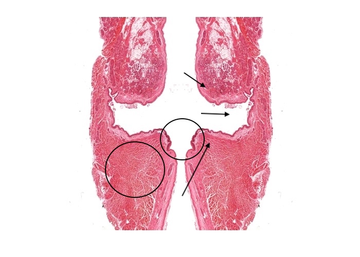
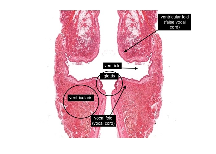
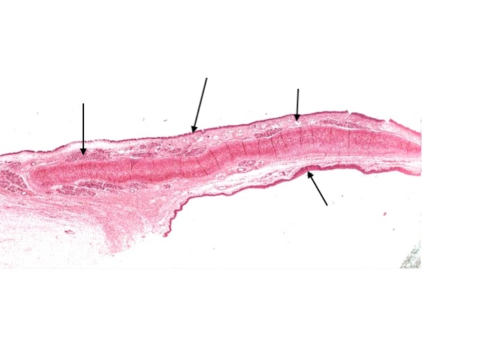
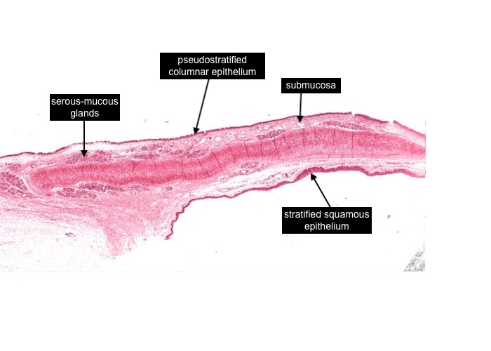
In the true anatomical position each vocal fold is oriented in an anterio-posterior direction (front-to-back; ventral-dorsal). In this frontal image (with a little bit of "fiddling" with the histological section image) each vocal fold has been cross-sectioned.
Each vocal fold (vocal cord) together with the opening between them is the "glottis".
The recess superior to each fold is the ventricle and the fold above this ventricle is the ventricular fold (or false vocal cord).
The tissue below the vocal fold (vocal cord) and lateral to the ventricle and also a little superio-lateral to the ventricular fold (false vocal cord) is the vocalis muscle which would also include in a particular region (see later item) the vocal ligament.
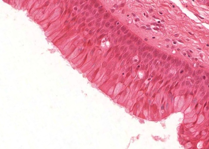
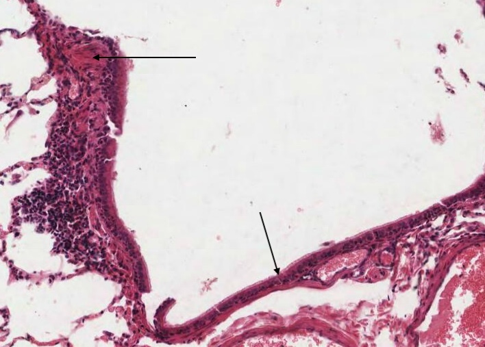
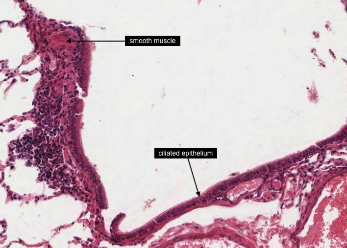
The epiglottis is a flap of tissue, which closes off the upper entrance to the larynx during swallowing. Its walls are supported by cartilage and its lumen is lined with respiratory mucosa. During breathing the epiglottis stands straight up from the anterior side of the larynx. Swallowing raises the larynx up against the epiglottis. During swallowing food passes across the upper anterior surface . As a consequece that surface is covered with stratified squamous nonkeratinized epithelium. The lower ventral surface (pharyngeal surface) is lined largely with pseudostratified columnar ciliated epithelium except along the periphery where it slaps down on the rim of the larynx during swallowing. There it can be partially converted towards a stratified squamous form. The submucosa has substantial amounts of elastic fibers, blood vessels and on the posterior surface, nicely visible mixed mucous-serous glands. Without knowing exactly how this piece of tissue was collected it is difficult to orientate anatomically but when you view the two epithelial surfaces you may understand it a bit more.
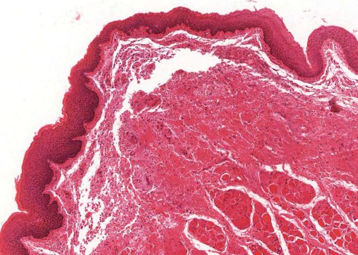
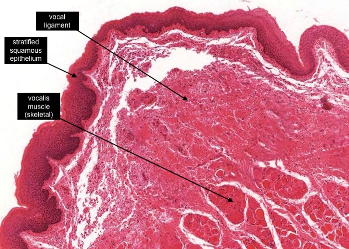
The true vocal cord contains two main functional components, the vocal ligament and an underlying vocalis muscle. Both appear in cross-section and skeletal muscle cut transversely is easily recognized, but the ligament is a bit trickier to identify. There has been some inevitable tissue shrinkage which has separated the thick round collagen fibers from each other. Directly over the vocal ligament the epithelium is a stratified squamous epithelium. The transition between the stratified squamous epithelium and the surrounding typical respiratory epithelium (not shown in this image) is relatively sharp.
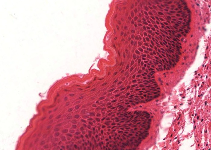
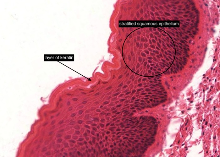
The vocal fold requires an epithelium (stratified squamous epithelium) to resist the abrasive forces of the movement of air over the vocal cord. But actually, I am sure I can see evidence of "keratinization" i.e. a layer of keratin?
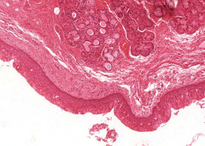
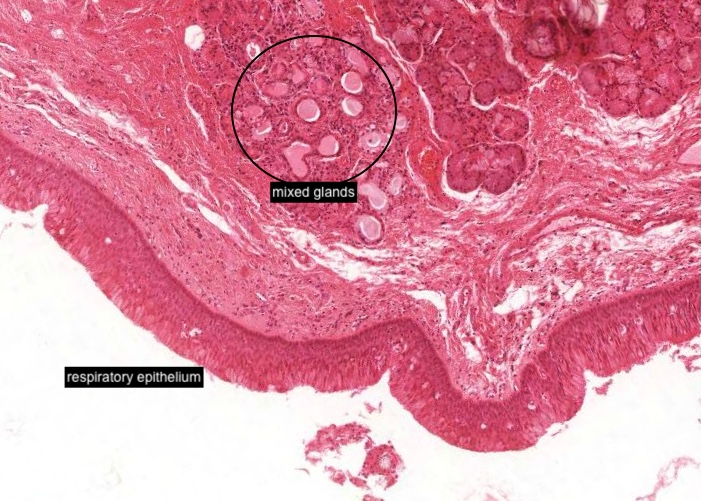
The vestibular fold (false vocal cord) is covered with typical "respiratory tract epithelium" and has a large number of mixed glands in the submucosa. Often you may find lymph nodules in this fold.

Confirm that the epithelium on the surface of the vestibular fold (false vocal cord) is "respiratory tract epithelium" even though in this image the epithelium has been cut obliquely so appears a lot thicker and even striated.
Which muscle is this in the respiratory system?
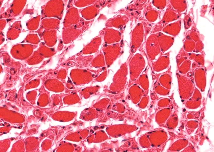
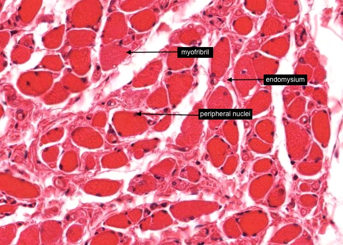
The vocalis muscle is striated/skeletal muscle. In this image of the muscle sectioned transversely each muscle fibre has at least one peripherally located nucleus and is encased by thin connective tissue - the endomysium. In some fibres you can see myofibrils appearing as red dots within the sarcoplasm.
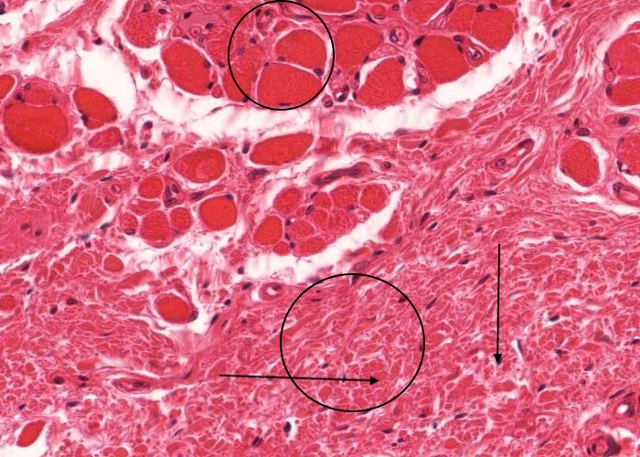
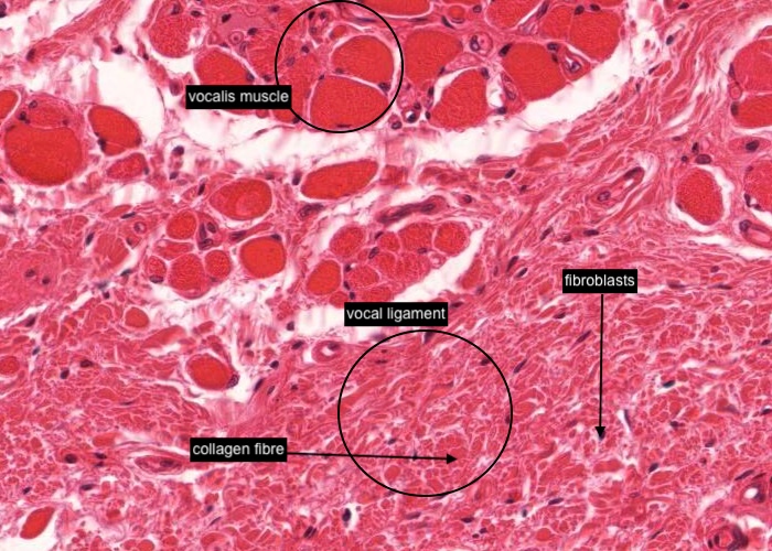
The vocal ligament is just under the epithelium at the angular part of the vocal fold (true vocal cord) and adjacent to the vocalis muscle. There has been some inevitable tissue shrinkage which has separated the thick round collagen fibres from each other. Between many of the collagen bundles the nucleus of a fibroblast is visible - some looking small and round because they are orientated in the direction of the transversely sectioned fibres; others more elongated where the fibres run obliquely in this section.
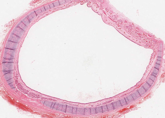
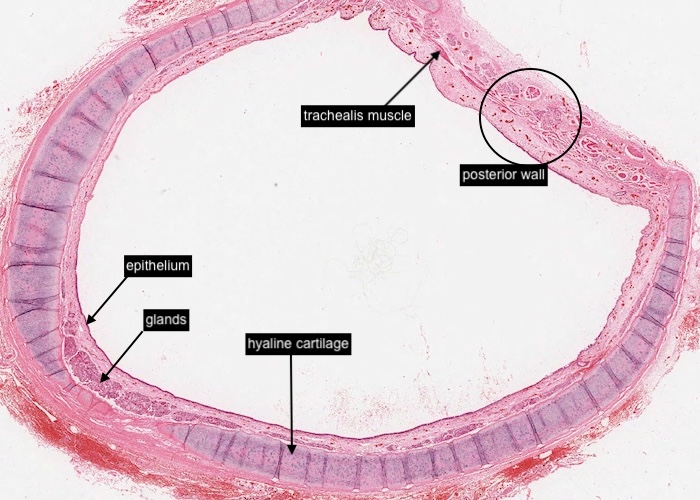
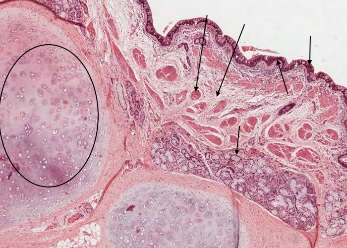
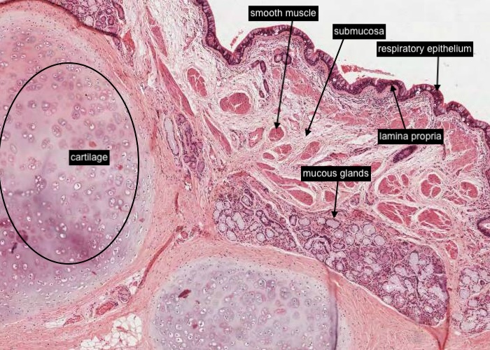
The respiratory airway begins with the trachea. Exactly what one sees of its histology depends on whether the plane of section passes mainly through a U-shaped supporting ring of hyaline cartilage, or between two neighbouring rings. Masses of glands lie deep under the epithelium but are mainly located between cartilage rings. The cartilages are U shaped instead of complete rings.
A thin band of smooth muscle, the trachealis muscle, stretches between the two ends of a cartilage ring to form the posterior wall of the trachea. The muscle fibres insert into the dense, elastic fibre bundles surrounding the tracheal cartilages and are joined to the tracheal mucosa by a layer of loose connective tissue.
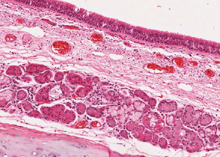
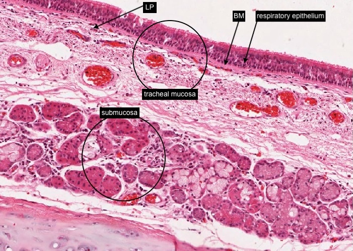
The tracheal mucosa is the inner part of the tracheal wall. As you would expect, the mucosa consists of "respiratory tract epithelium" supported by a lamina propria. Note the prominent basement membrane supporting the epithelium. The submucosa and cartilage layer just below make up two other components of the tracheal wall visible in this image.
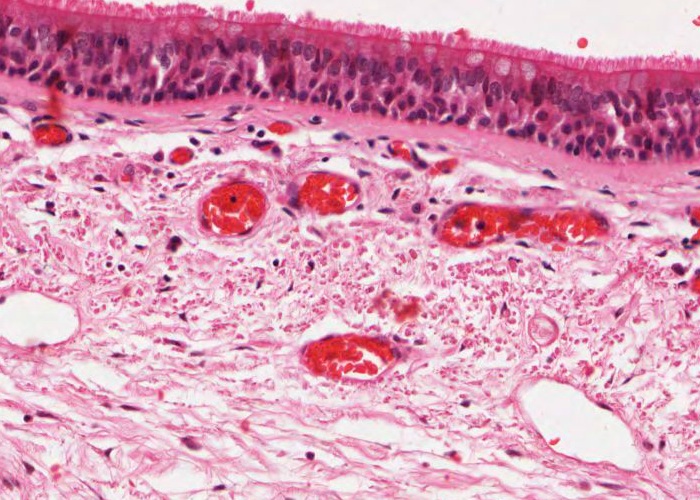
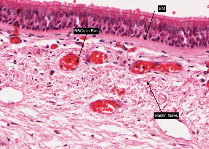
The lamina propria contains many small blood vessels (with red blood cells in their lumens) and loose connective tissue containing abundant elastic fibres seen here in this H&E stain as aggregates of tiny red dots. They are running down the length of the trachea. How do I know that?
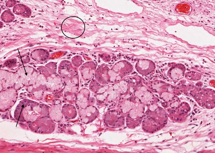
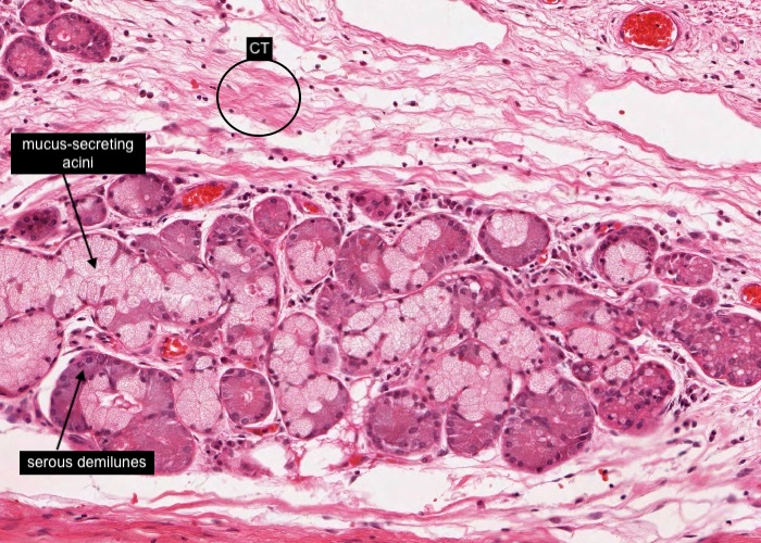
The tracheal submucosa has maybe slightly denser connective tissue than the lamina propria (?) but also contains glands comprising mucus-secreting acini and serous demilunes. As you saw earlier, these glands tend to lie in the region between the cartilage rings and in the posterior aspect of the trachea.
periochondriun? Chondrocyte? isogenous group?
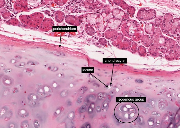
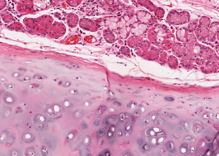
The denser connective tissue layer of the submucosa blends with the perichondrium of the U-shaped hyaline cartilage rings supporting the trachea. Remnants of a chondrocyte in its lacuna are very visible and in some places their lacunae form an isogenous group.

The trachealis muscle is a band of smooth muscle, stretching between the two ends of a cartilage ring to form the posterior wall of the trachea. The muscle fibres insert into the dense, elastic fibre bundles surrounding the tracheal cartilages and are joined to the mucous membrane by a layer of loose connective tissue.
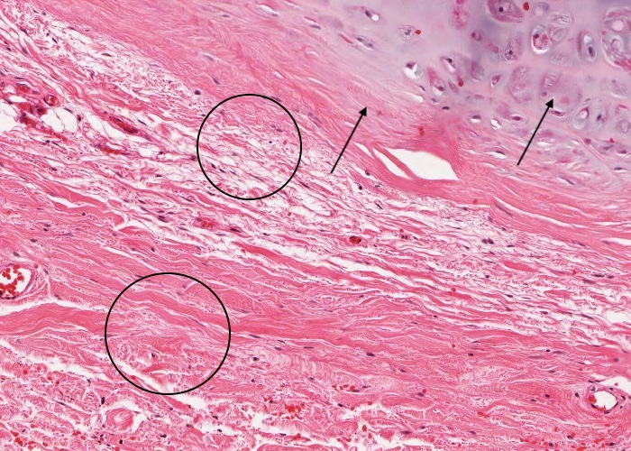
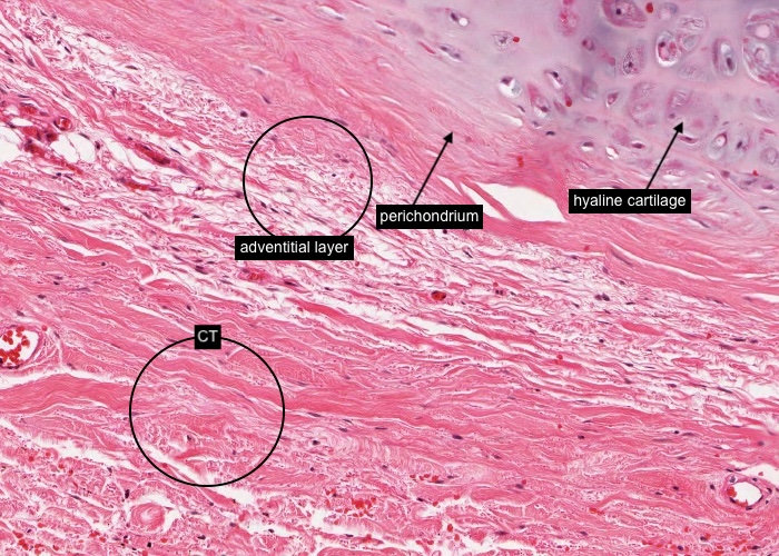
The adventitial layer is the outermost layer of the tracheal wall. It lies outside the perichondrium of the cartilage layer, and the trachealis muscle, and so blends with connective tissue supporting nearby structures. The immediate structure posterior to the trachea is the esophagus.
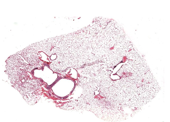
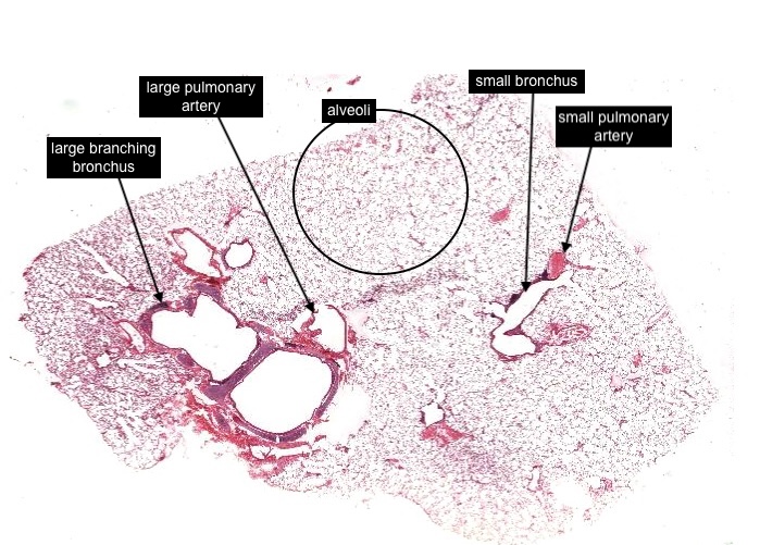
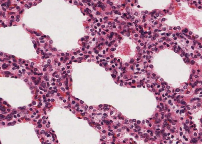
In this image of the lung you can just recognise a section though a small bronchus and a small pulmonary artery and a large branching bronchus and larger pulmonary artery but you will see more details of these structures as you view other items in the subheadings. The bulk of the tissue you see in this image, are the sectioned terminal ends of the respiratory airway - or alveoli (the sites for gas exchange).
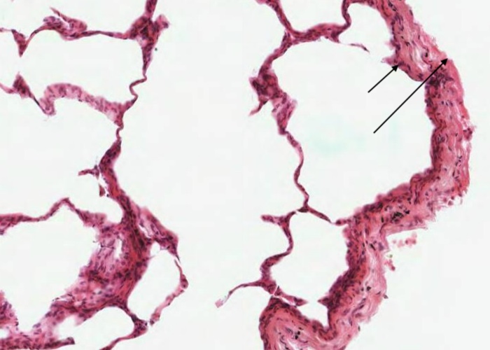
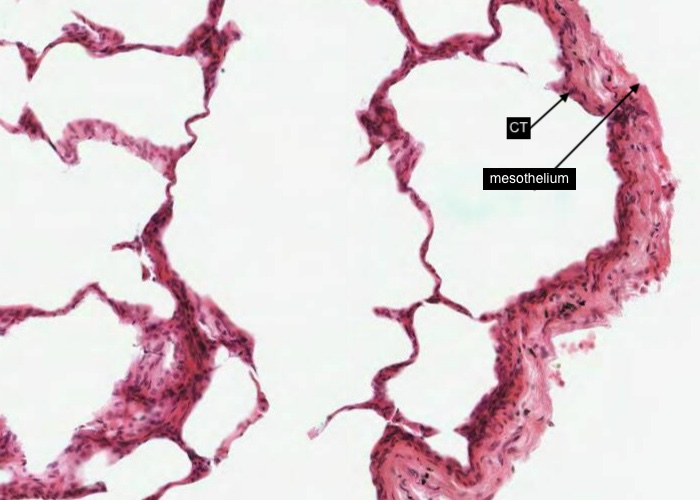
The visceral pleura is a thin layer of connective tissue lined by mesothelium = a serous membrane. Within this visceral pleura is a series of lymphatic vessels that drains the surface of the lung. The other series of lymphatic vessels drain the lung parenchyma and follow the airways and blood vessels to lymph nodes in the hilum of the lung.
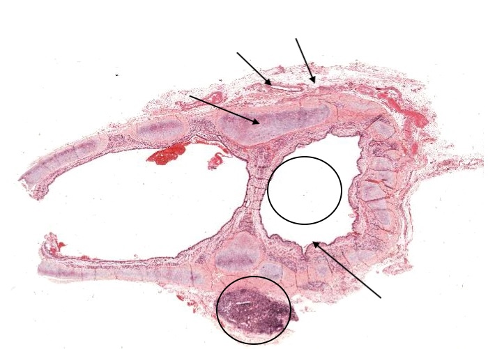
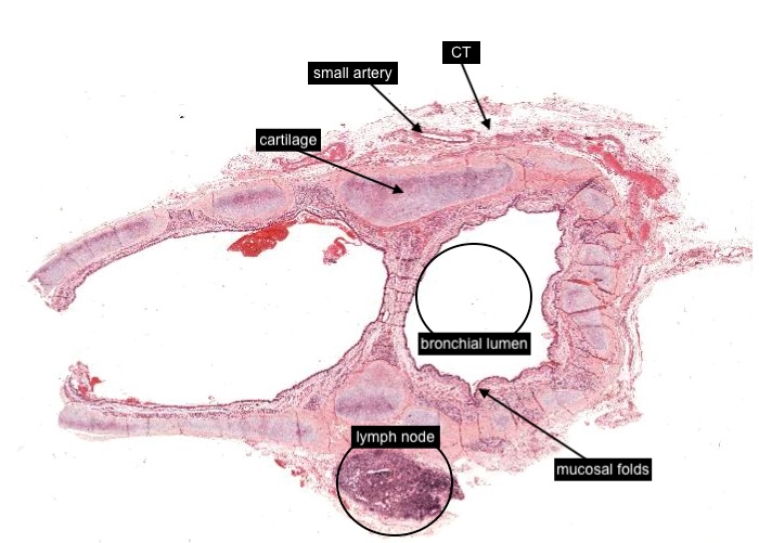
This image is of a section through the branching bronchial tree. Note the supporting plates of cartilage. The pulmonary vessels have not been included but the section has a lymph node. This section must be from the hilum of the lung where the bronchi are running in connective tissue, instead of being intrapulmonary (see later item). The small artery along the edge is a systemic artery - not a pulmonary artery. These vessels carried arterial blood to nourish the tissues of the bronchi instead of deoxygenated blood to the lung alveoli.
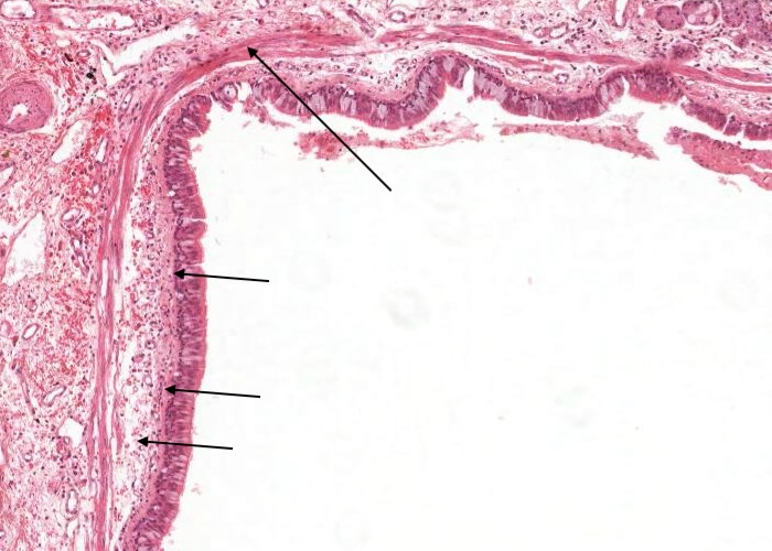
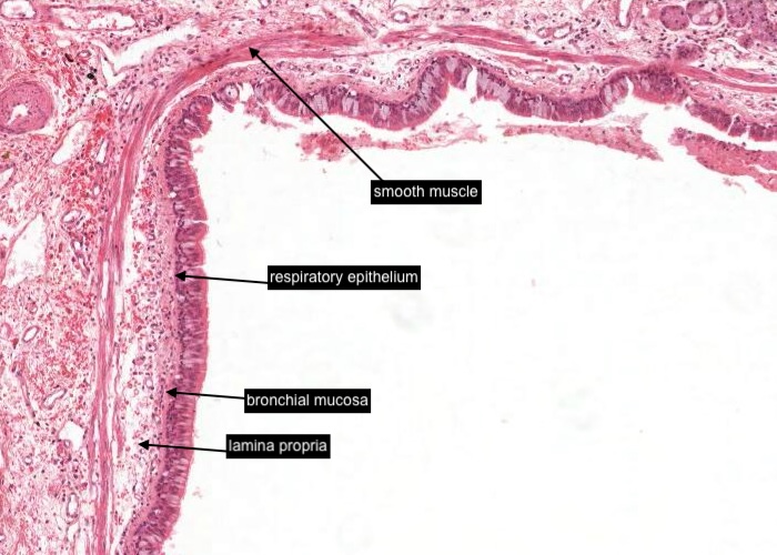
The smaller of the two sections through the bronchial lumen, cut more transversely, shows a scalloped appearance. Bronchi characteristically have pronounced mucosal folds that run longitudinally. They allow a limited expansion in volume when you inhale.
When you examine this section you will notice the mucosa has typical respiratory epithelium with goblet cells. Also, identify the plates of cartilage, smooth muscle and mucous glands in the lamina propria and submucosa of the bronchi. The smooth muscle is a particularly important component, it is distributed in the form of ribbons that spiral down the bronchus and continue along the air passageways all the way down to the alveolar sacs.
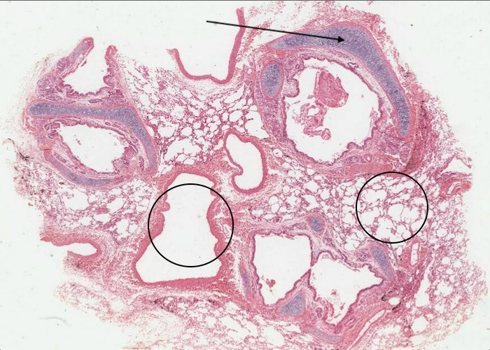
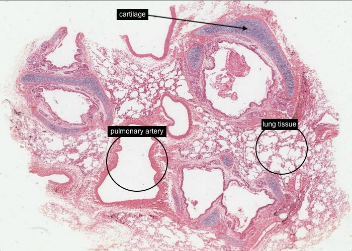
This section cuts through a branching bronchus and associated structures (maybe branches of the pulmonary artery). Again note the cartilage plates in the walls of the bronchial branches.
These bronchi are intrapulmonary because you can see lung tissue sectioned. Do not waste time examining this tissue when you view the section because the lung has been allowed to collapse, is poorly preserved, and has pathological features.
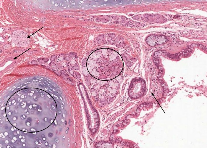
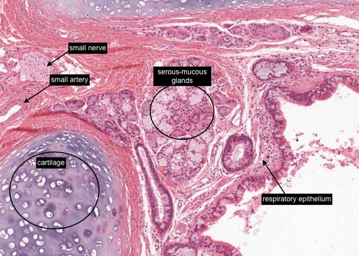
This section cuts through a branching intrapulmonary bronchus. Note its typical respiratory epithelium with goblet cells adjacent to the lumen. Also identify the cartilage, and it is an opportune time to revise the structure of hyaline cartilage so identify the remnants (nucleus) of a chondrocyte in its lacuna. The matrix is stained at various intensities to indicate the variable molecular content of the matrix components.
Now going back to focus on the bronchial mucosa/wall identify serous-mucous glands in the lamina propria and submucosa of the bronchus.
Between the two plates of cartilage you may wish to view the small nerve and artery.
Again appreciate that the smooth muscle is a particularly important component of the air passageways. As mentioned in a previous item, it is distributed as muscle ribbons that encircle the bronchial mucosa (respiratory epithelium and supporting connective tissue (lamina propria) and spirals down the bronchus and continues along the air passageways all the way down to the alveolar sacs.
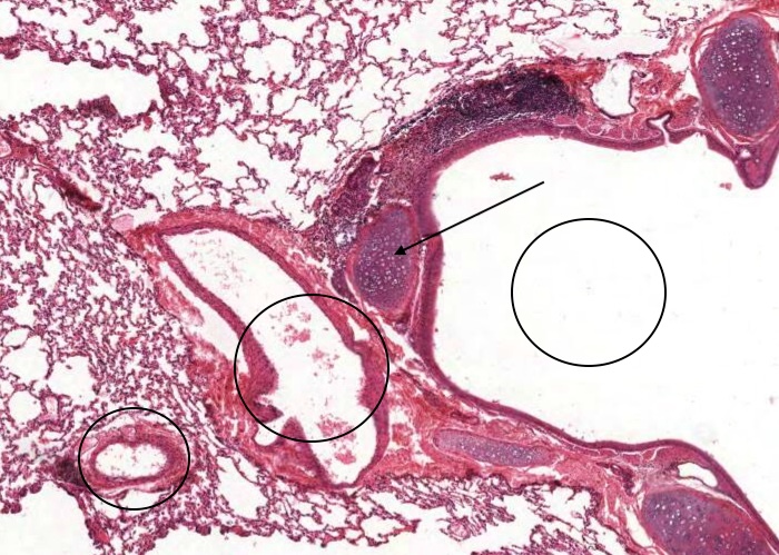
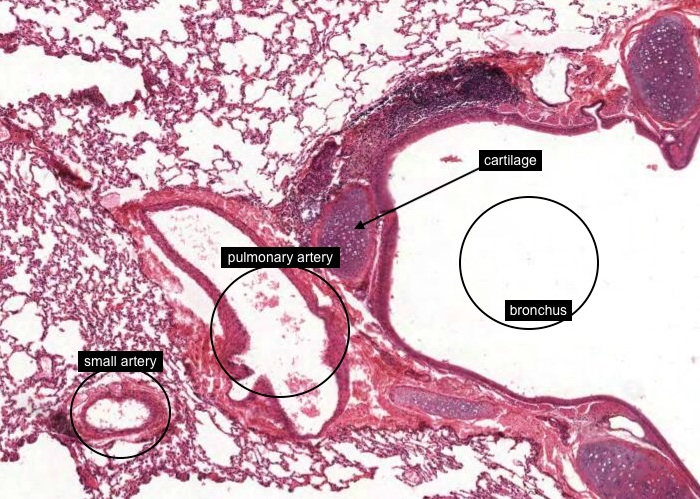
One important anatomical detail of the lung to clearly understand is its blood supply and drainage. Branches of the pulmonary artery run alongside the air passageways (bronchus in this image - note the cartilage layer) as far down as respiratory bronchioles. Pulmonary veins, in contrast, go their separate way when the bronchi become bronchioles. Be sure that you can tell pulmonary arteries and veins apart. Obviously you will look at the smooth muscle in their walls to do so. Be aware, however, that the walls of pulmonary arteries are thinner than those of systemic arteries because the pulmonary blood pressure is lower.
The small artery on the outside is probably a bronchial artery to nourish the tissues of the bronchi instead of deoxygenated blood that would be carried by the pulmonary artery. Look at the thickness of their media.
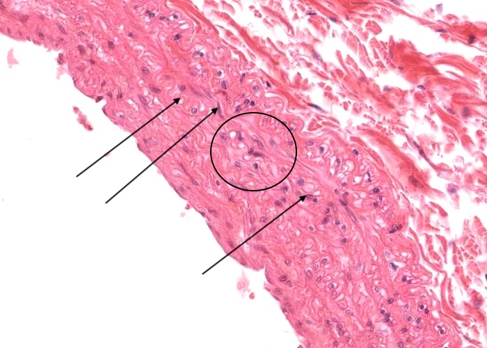
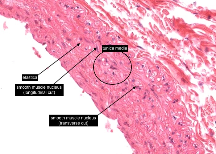
The wall of a pulmonary artery is thinner (relative to the lumen diameter) than that of a corresponding systemic artery because the pulmonary blood pressure is lower.
The tunica media in this image has smooth muscle sectioned both transversely (look for a nucleus with a circular profile) and longitudinally (look for a nucleus with an elongated profile) and there are extensive "wiggly" fibres of elastica - the pulmonary artery is classifed as being an "elastic artery". The expansion and recoil of this vessel due to the elastica maintains pulmonary arterial pressure relatively constant during the cardiac cycle.
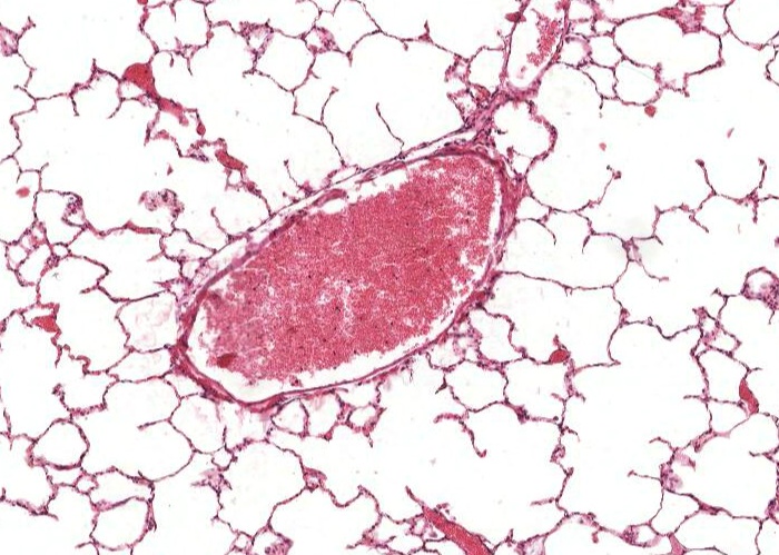
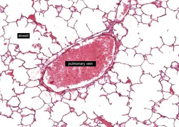
A pulmonary vein can be confidently identified if you view the section some distance away from the large air passageways and look for an isolated large vessel surrounded by alveoli.
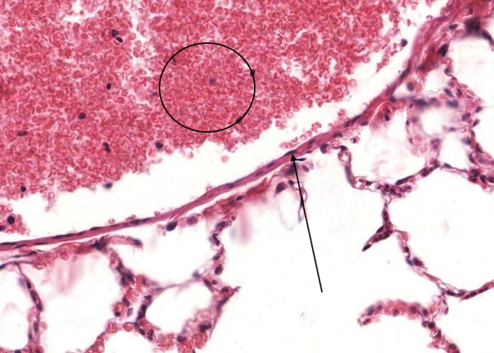
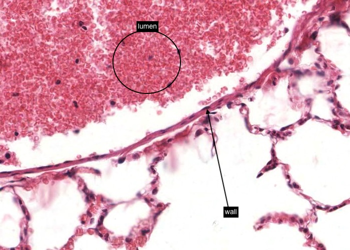
If you have examined the histological section of the monkey lung (H&E) you would have noticed that the pulmonary vein has an extremely thin wall and a wide lumen diameter.
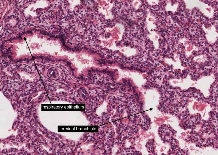
A bronchiole supplies a pulmonary lobule. Make sure you are familiar with the lobular organization of the lung. This image is of a larger bronchiole because if you look very carefully you can see some areas of ciliated epithelium - it will change from pseudostratified, ciliated epithelium to simple, ciliated, columnar epithelium more distally i.e. further down the air passageway. Goblets cells may or may not be present now. Cartilage and serous-mucous glands are absent in bronchioles but there will be smooth muscle in the wall - quite thick in most places.
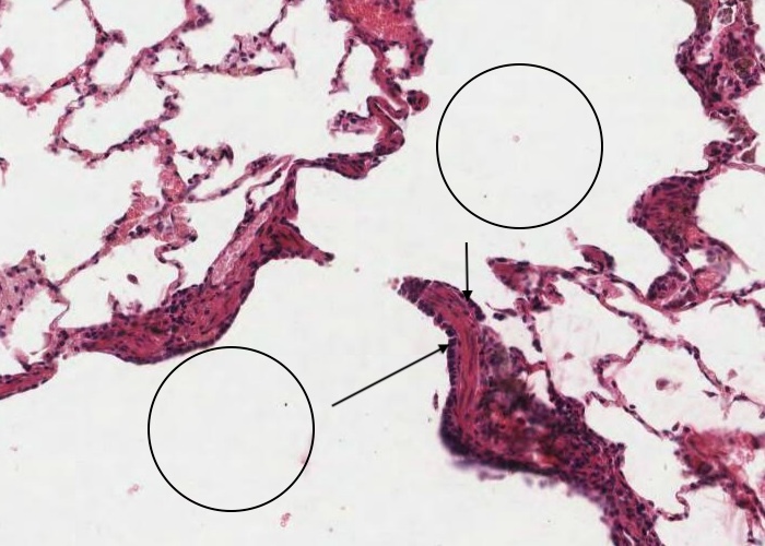
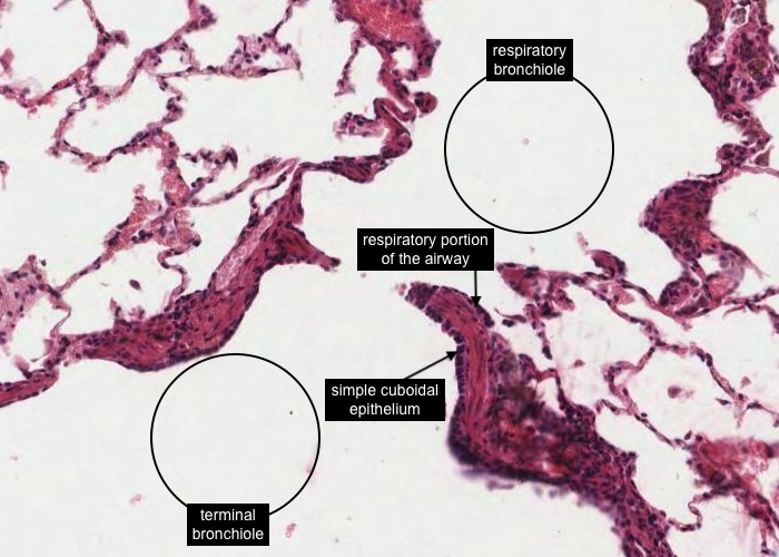
As you are aware bronchioles are the distal, increasingly branching, airways have no supporting cartilage, and are the passageways where typical respiratory epithelium will end. Terminal bronchioles are the smallest bronchioles and are concerned with air conduction only.
Each terminal bronchiole exhibits simple cuboidal epithelium; the ciliated cells disappear and are replaced by "Clara cells". The epithelium begins to flatten where they join the distal respiratory portion of the airway - initially a respiratory bronchiole then the alveoli.
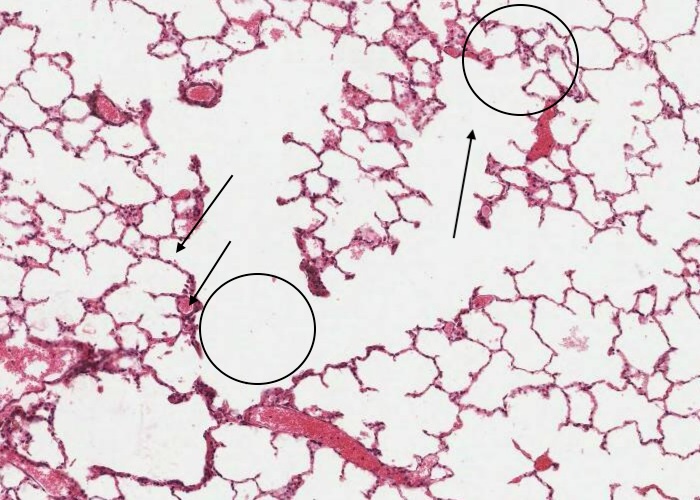
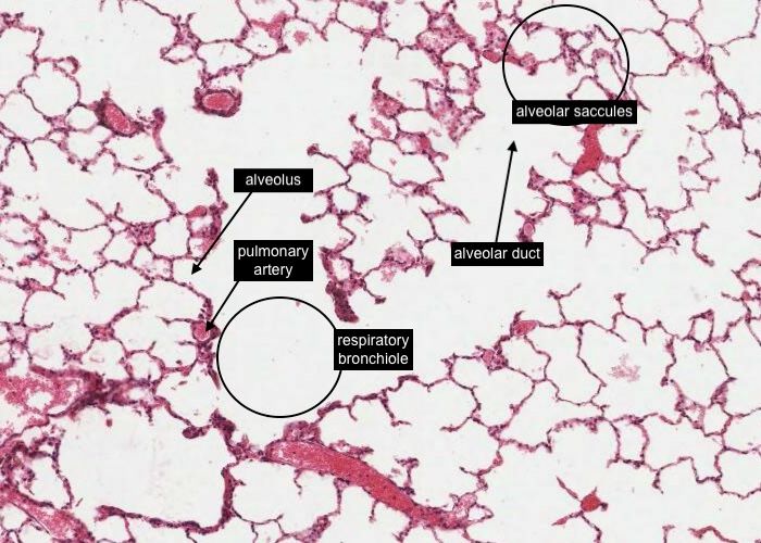
A respiratory bronchiole often has small branches of the pulmonary artery nearby. Typically, the two run next to one another with an alveolus extending from the portion of the wall of the bronchiole some distance from the artery. The respiratory bronchiole will open into the alveolar duct and then alveolar saccules (clusters of alveoli).
An alveolar duct is particularly difficult to comprehend, yet these ducts are a most important structural entity of the lung. Only they and alveolar sacs can significantly expand or contract during inspiration or expiration of air. Thus volume change is an accommodation of these ducts.
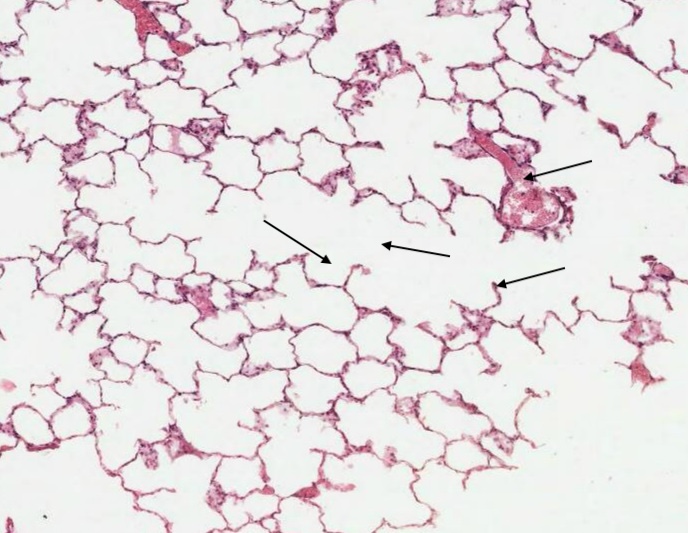
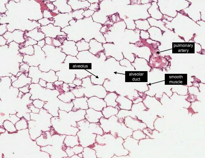
To pick out an alveolar duct it is necessary to understand that the alveolus itself is not part of the walls of the duct but an outpocketing from the wall. The wall itself is composed of little more than minimal bands of smooth muscle overlain by a low epithelium.
Often a section through the wall shows only a single smooth muscle cell cut transversely as a bulge at the end of an alveolar wall. The blood vessels near these alveolar ducts are probably the terminal branches of the pulmonary artery.
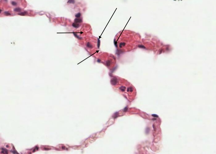
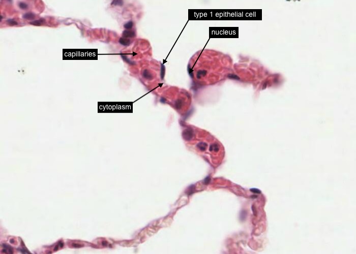
The inter-alveolar wall (air - blood barrier) is formed of two extremely attenuated sheets of epithelium enclosing many capillaries and very little connective tissue. The many elongated nuclei belong largely to type I epithelial cells or the nucleus of an endothelial cell. It is not possible for you to distinguish between these two cell types but in this image I think I may be able to see a tiny amount of cytoplasm adjacent to what I think is the type I cell and it appears to be more adjacent to (and bulging into) the lumen of the the air space than the nucleus of the endothelial cell which I reckon is more internal i.e. within the space of the capillary?
The two other cell types associated with the inter-alveolar wall are not visible in this image.
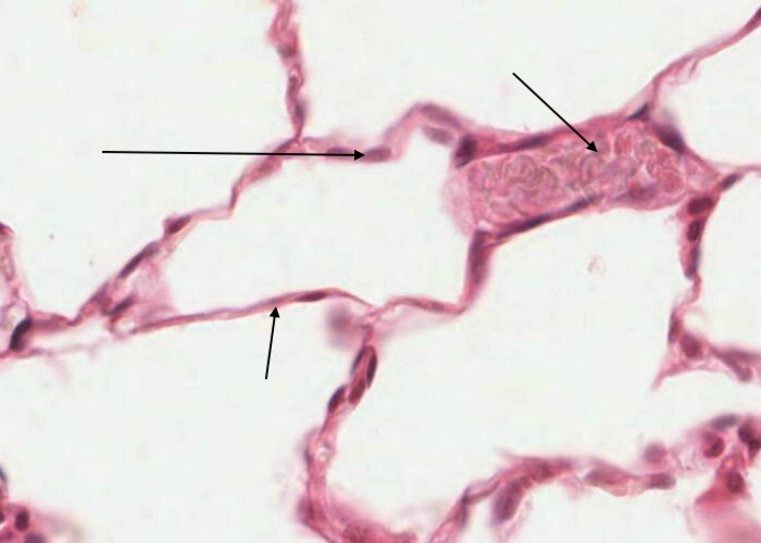
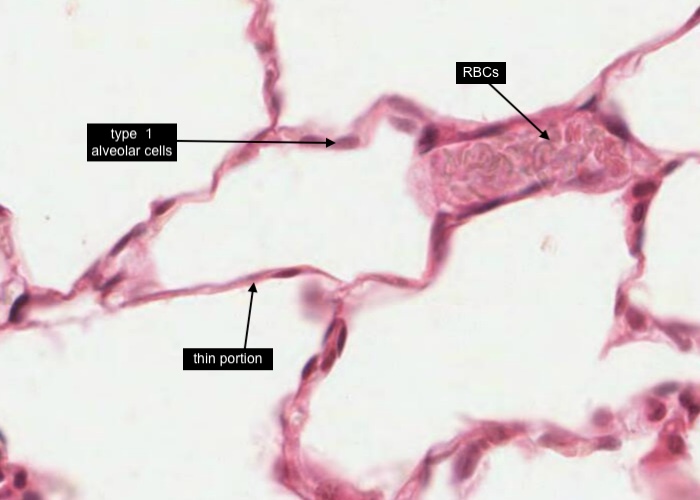
Type I alveolar cells (often also called type I pneumocytes) are very thin, squamous cells and line almost all the surface of the alveolus. They are easier to see in what is often referred to as the "thin portion" of the alveolar septum. A large capillary full of red blood cells is nearby.
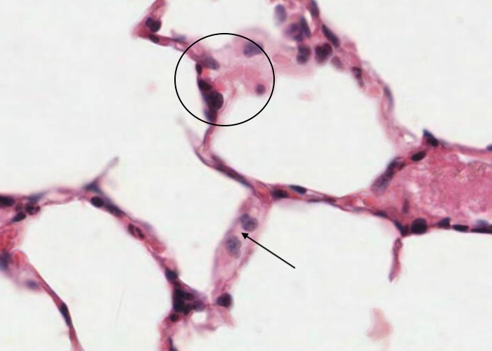
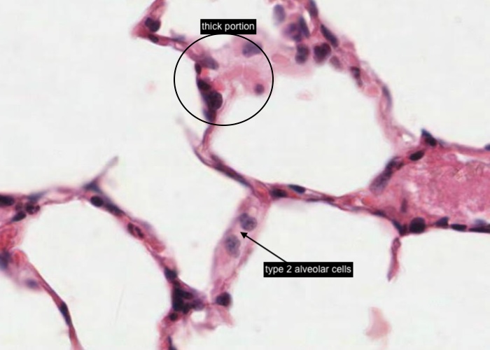
Type II alveolar cells (often referred to as type II pneumocytes or septal cells) are cuboidal and usually occur at the corners where two interalveolar walls come together (often referred to as the "thick portion" of the alveolar septum). They produce surfactant. Picking out these type II cells in H&E sections is not easy so be philosophical if they elude you.
macrophage?
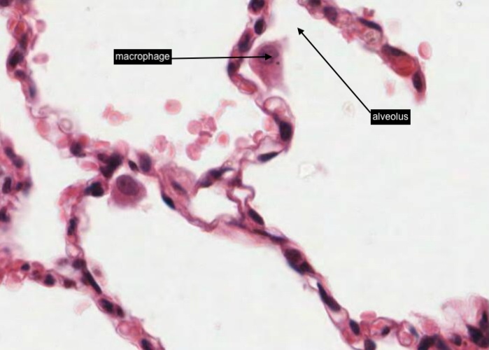
An alveolar macrophage can be found anywhere but I can find them best on the surface of the air space in the alveolus. Once laden with dust they like to retire to a comfortable patch of connective tissue and just relax. Can you see the other one?
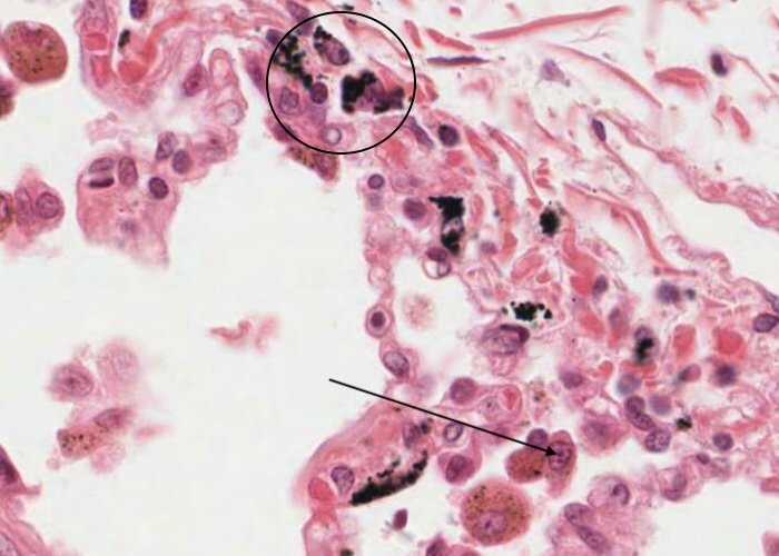
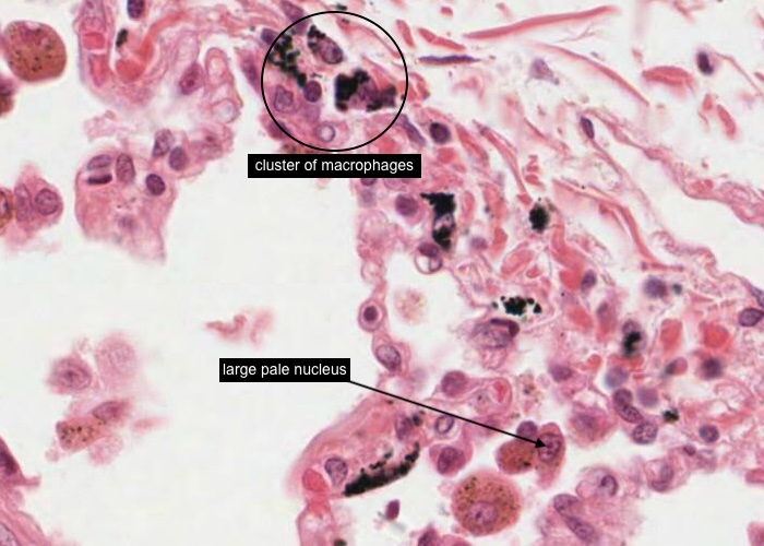
You cannot miss seeing the clusters of macrophages, by noticing the black dust particles that they have ingested. Examine several of the clusters and note that they are located in seams of connective tissue. This is where macrophages permanently retire after they engorge themselves with indigestible particles. Many of the macrophages have taken up so many dust particles that the only detail you can see is their cell boundary.
Other cells have taken up less opaque materials allowing you to see that they are rounded up cells with a large pale nucleus.
The lung develops in the fetus in much the same manner as a gland. We really should think of the lung as a gland - which secretes CO2.
An outpocketing from the foregut (endoderm) grows at the tip first as the trachea and then branches into bronchi and later bronchioles.
Branches of the pulmonary artery are easy to find as they travel with a bronchus and/or a bronchiole. You may be think why have I labelled a bronchus at such a low magnification with so much confidence - it's because I examined the histological section (as you should) and saw a region of developing cartilage in the wall!
Remember that a branch of the pulmonary vein tends to lie in the periphery between lobules. Using them as a guide, and the shrinkage artifacts that emphasize the small amount of interlobular connective tissue, see if you can find or imagine lobules.
The visceral layer of pleura is clearly visible.
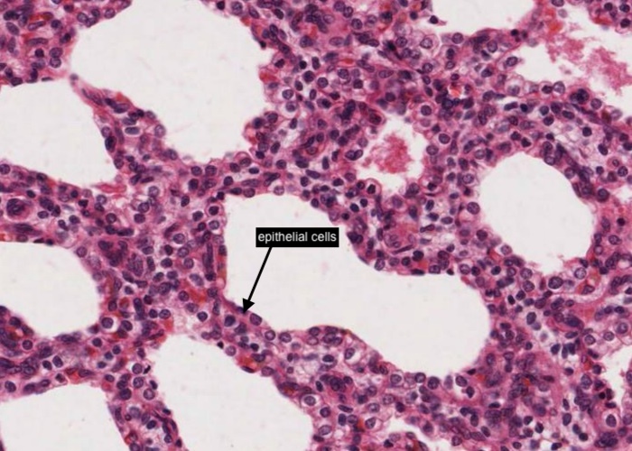
The epithelial cells are cuboidal. When alveoli form, these cells will stretch out to be very squamous. Here and there you may see some cilia.
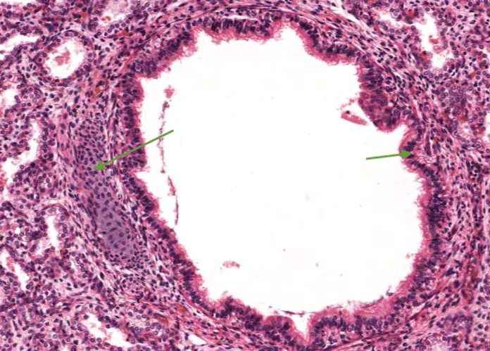
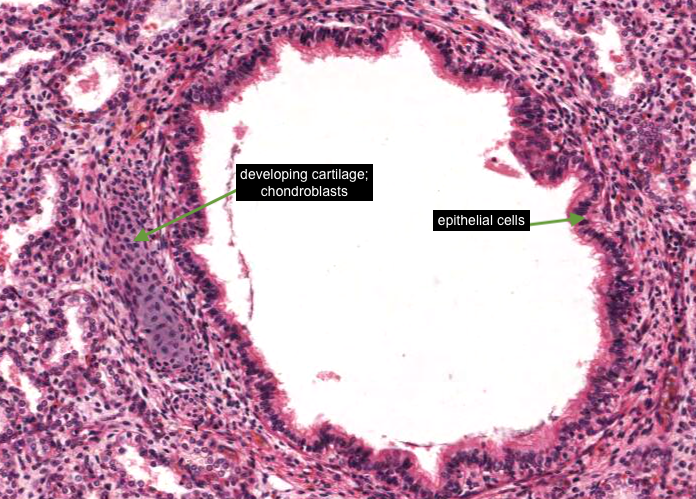
The less abundant bronchi are recognizable by their developing cartilage - so the collections of nuclei you see are those of chondroblasts. Here and there you can see some cilia on the epithelial cells of the developing bronchus.
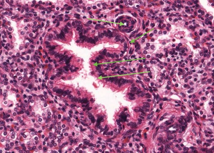
The obvious ducts you see at low power are bronchioles. The "respiratory epithelium" has cells with cilia on their apical surfaces. Maybe there is a hint of a few smooth muscle cells begining to surround the bronchiole wall? The tiny vessel-like structure at the middle-top border of the bronchiole is probably an terminal branch of a pulmonary artery - so a pulmonary arteriole?
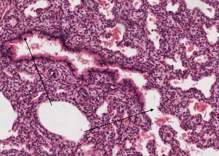
Well you know now that the obvious duct you see at low power (particularly when you see it has a "respiratory epithelium" lining) is that of a bronchiole - oh ..... provided you see no evidence of developing cartilage. If you follow this duct it is obviously going to continue as a terminal bronchiole (cuboidal epithelium) then a respiratory bronchiole that will finally branch into alveolar ducts but their alveoli will have not yet formed.