A&P Lab exam 2
1/154
There's no tags or description
Looks like no tags are added yet.
Name | Mastery | Learn | Test | Matching | Spaced | Call with Kai |
|---|
No analytics yet
Send a link to your students to track their progress
155 Terms
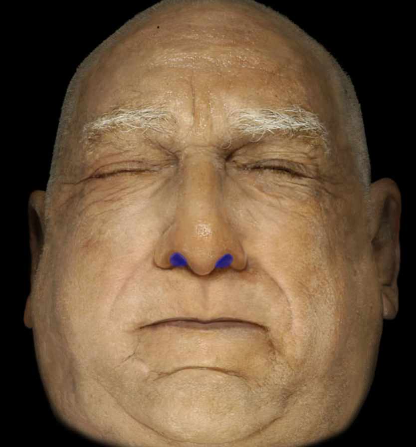
Identify
External nares
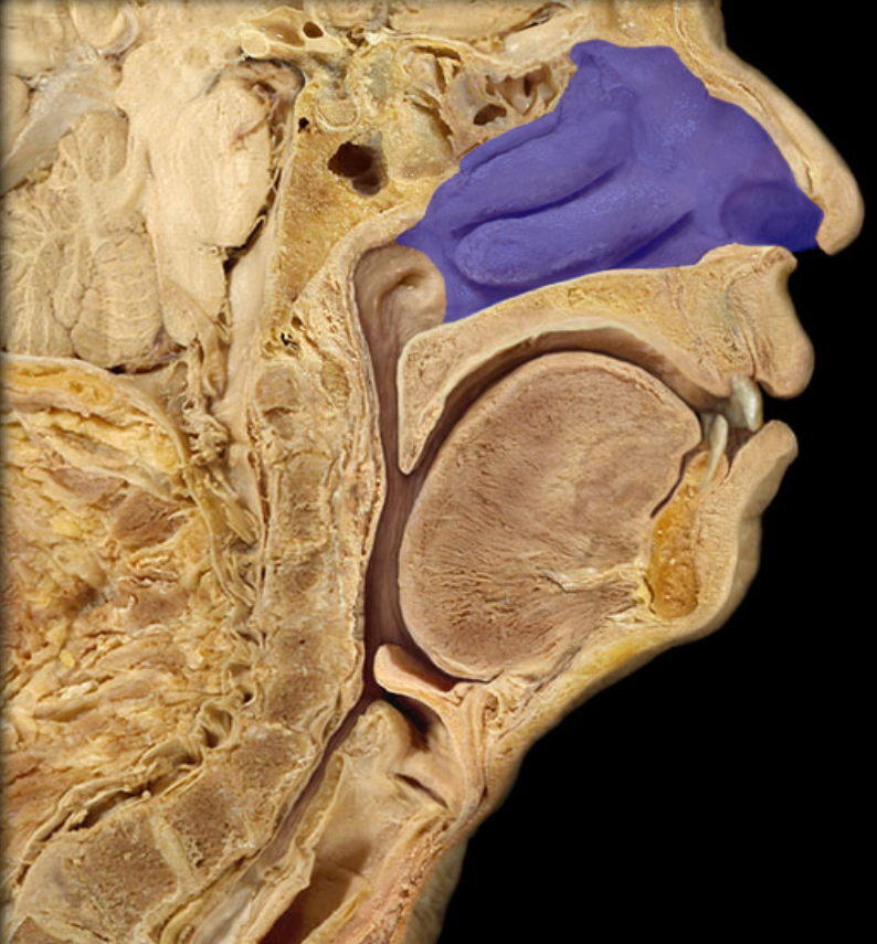
identfiy
Nasal cavity
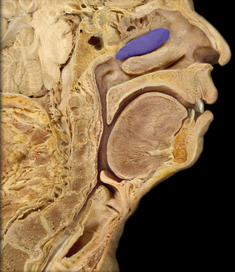
This is one of three what?
Nasal conchae
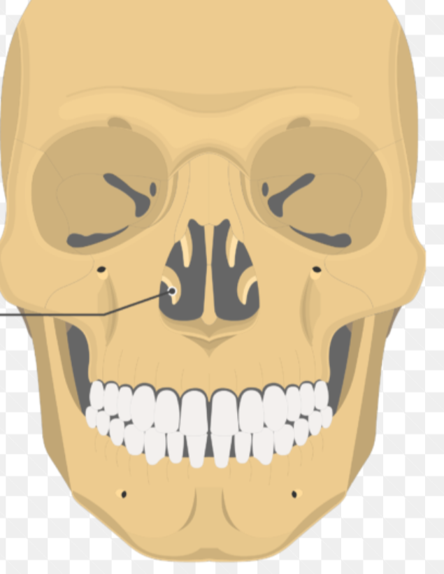
Identify the m arked structure
nasal conchae
Identify
Nasal septum
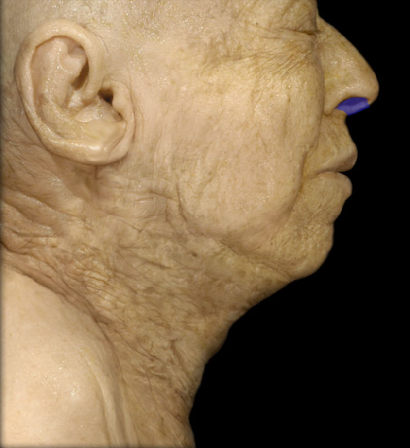
Identify this structure
Frontal sinus
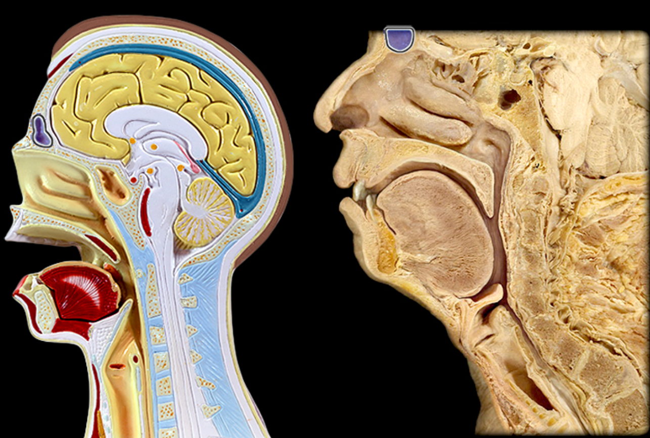
Identify
Sphenoidal sinus
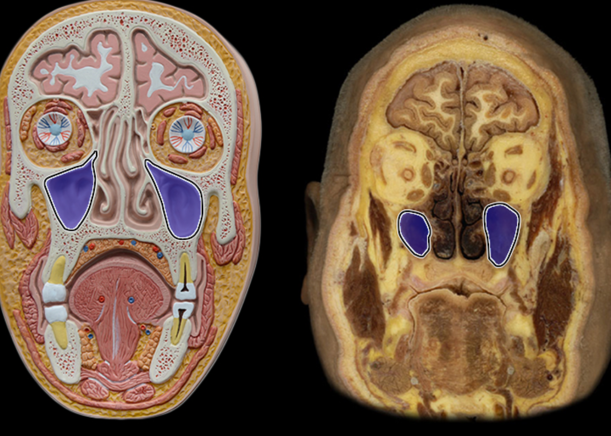
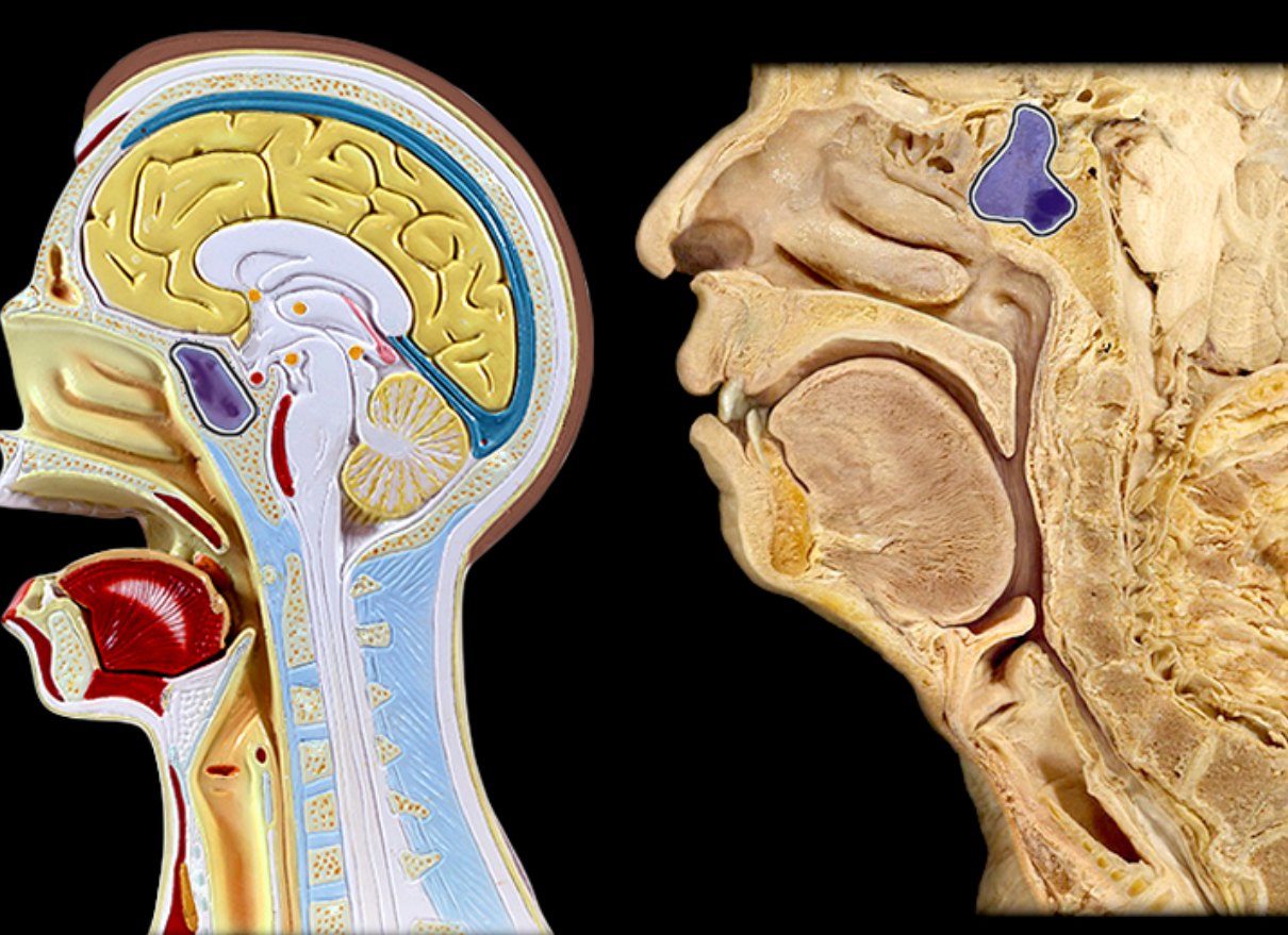
Identify
Maxillary sinus
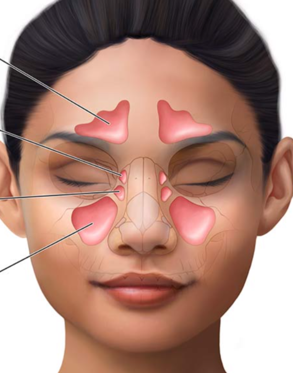
Identify the sinuses superior to inferior
Frontal, sphenoid, ethmoid, and maxillary
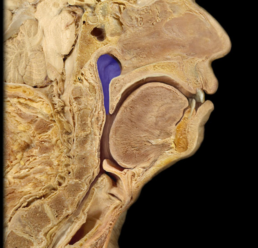
Identify
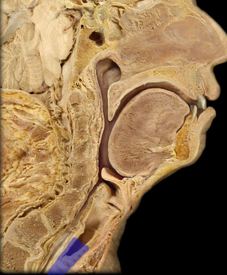
Nasopharyns
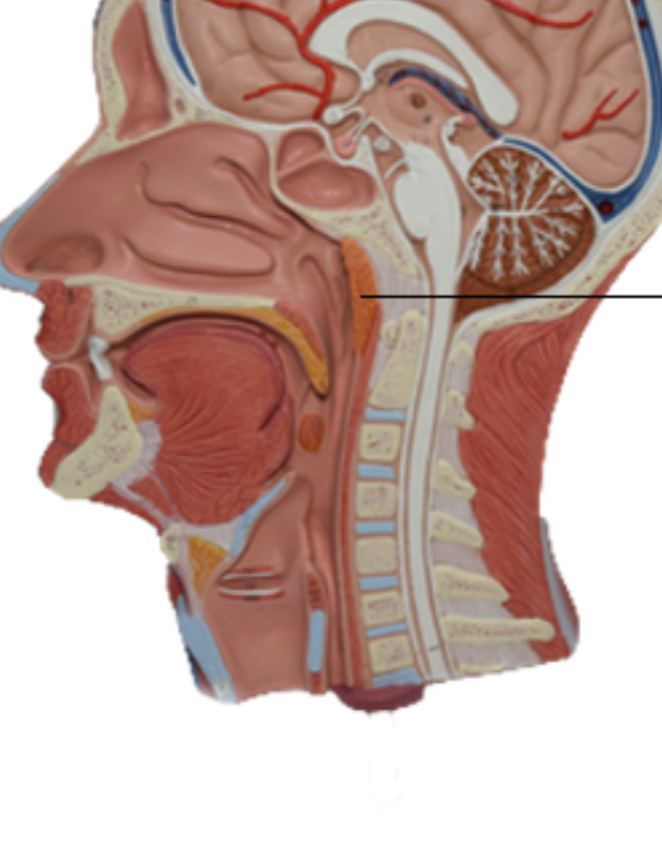
Identify
Pharyngeal tonsil (adenoids)
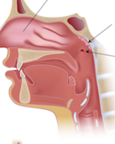
Identify the three structures
Nasal cavity, adenoids/pharyngeal tonisls, and auditory (eustachian) tubes
Identify
Oropharynx
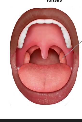
ID
Palatine tonsils
Identify from supior to inferior
Palatine tonsils, lingual tonsils
Describe the location of the pharyngeal/adenoids, palatine, and lingual tonsils
Pharyngeal/adenoids - at the end of the nasal cavity, Palatine - on either lateral side of the throat/mouth, lingual - on back of throat
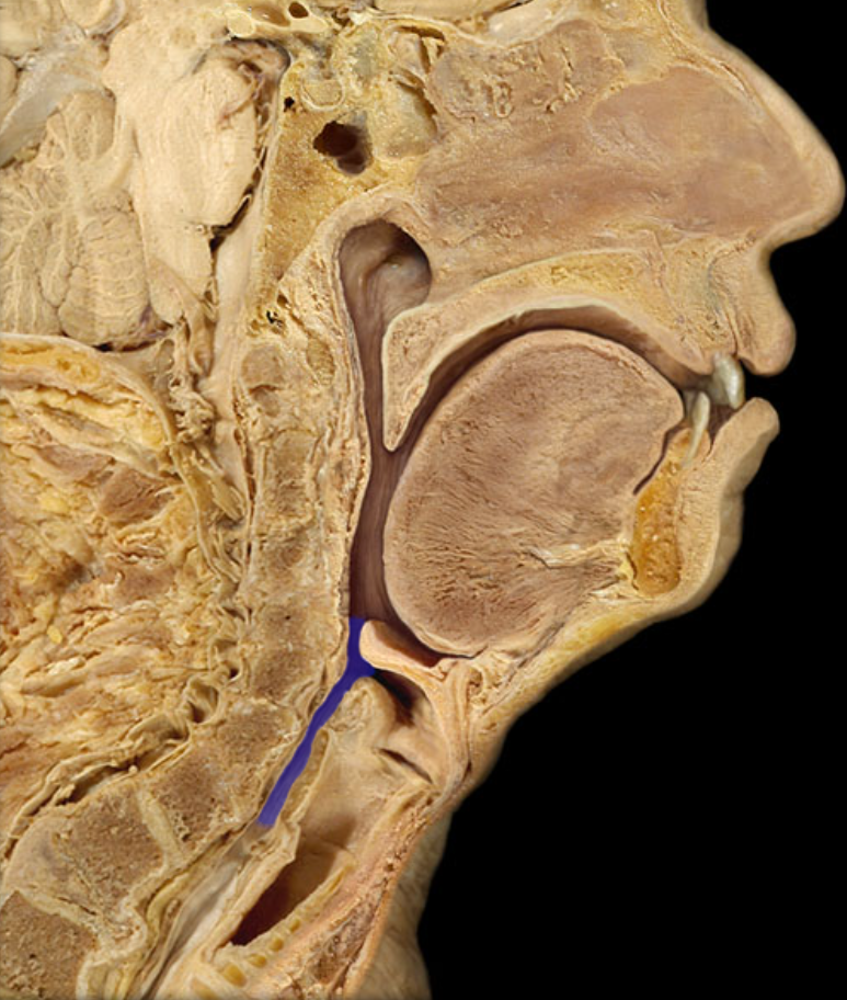
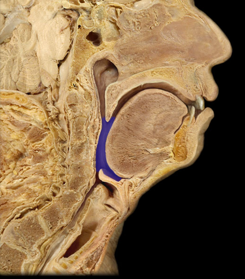
ID
Laryngopharynx
ID
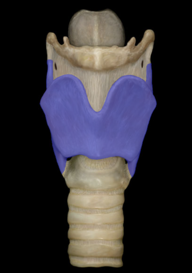
Larynx
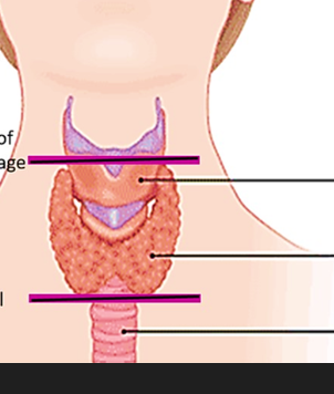
ID from superior to inferior
Thyroid cartilage, thyroid gland, trachea
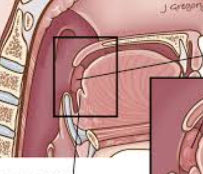
ID
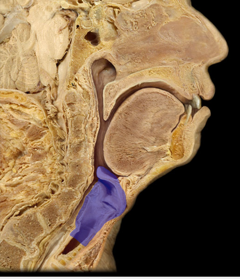
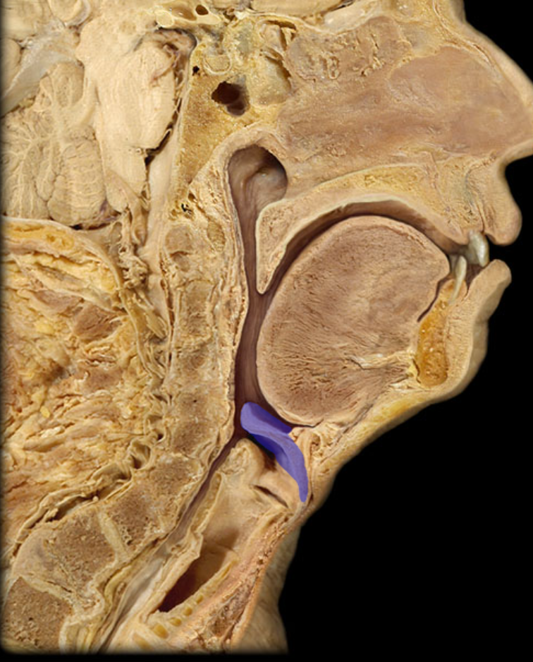
Epiglottis
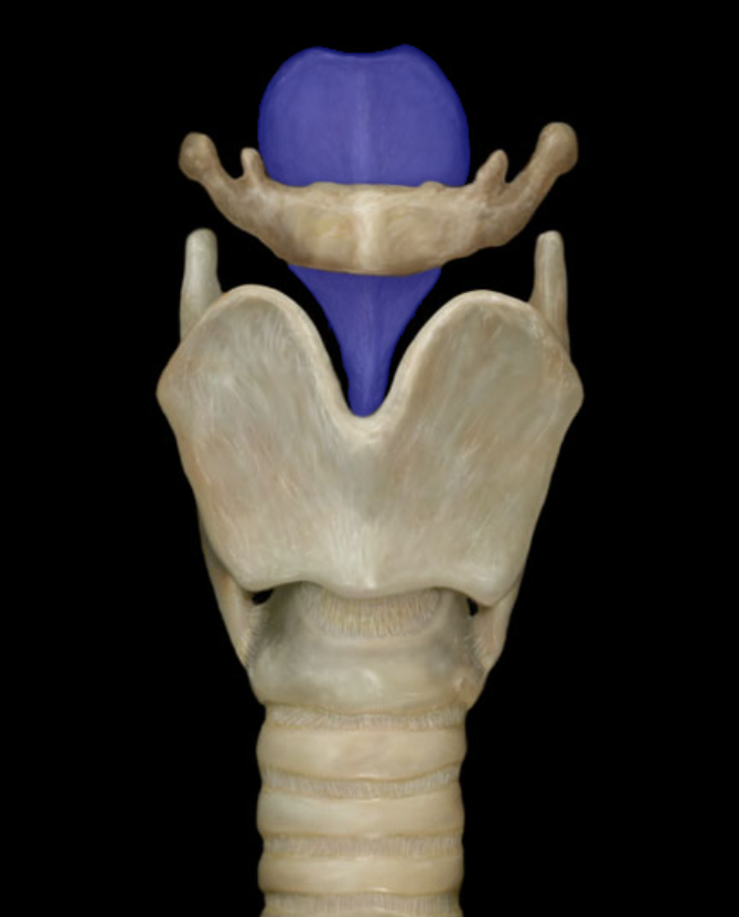
ID
Epiglottis
ID
Thyroid cartilage
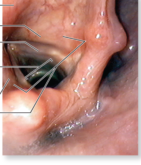
Describe the Glottis
The vocal cords and space between them
ID
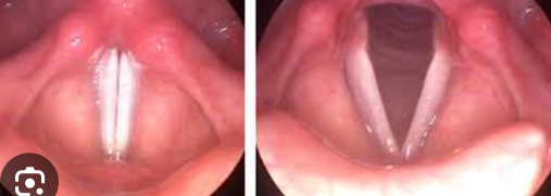
Vocal folds
What is pictured
View of larynx, to the left is the glottis and vocal folds
ID
Trachea
ID
Trachea
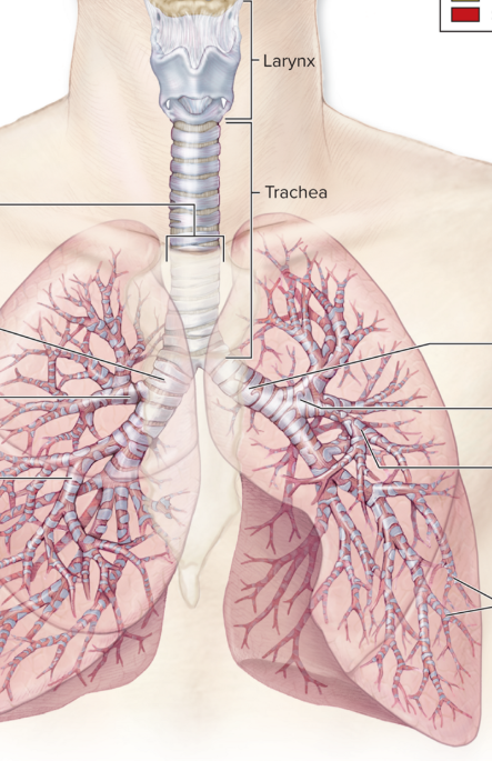
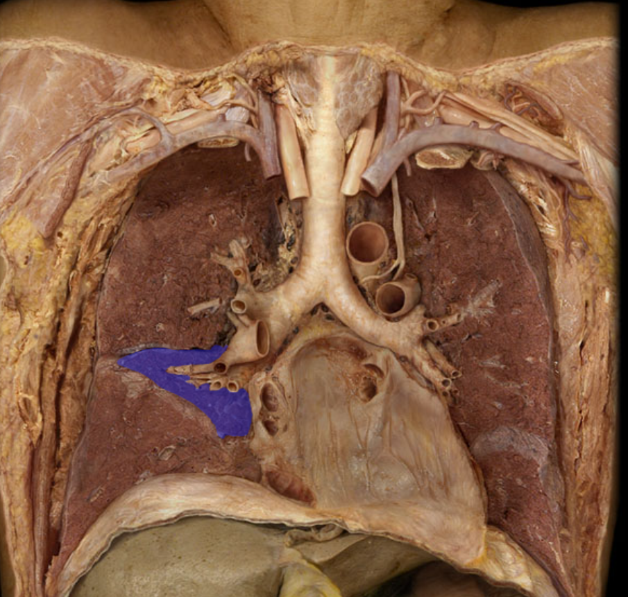
ID
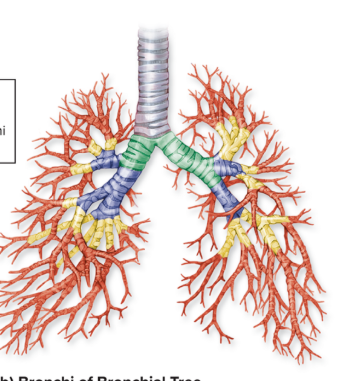
Bronchial tree
How many primary bronchi are there?
2 right and left
How many secondary bronchi are there
3 on right, 2 on left for each lobe of the lungs
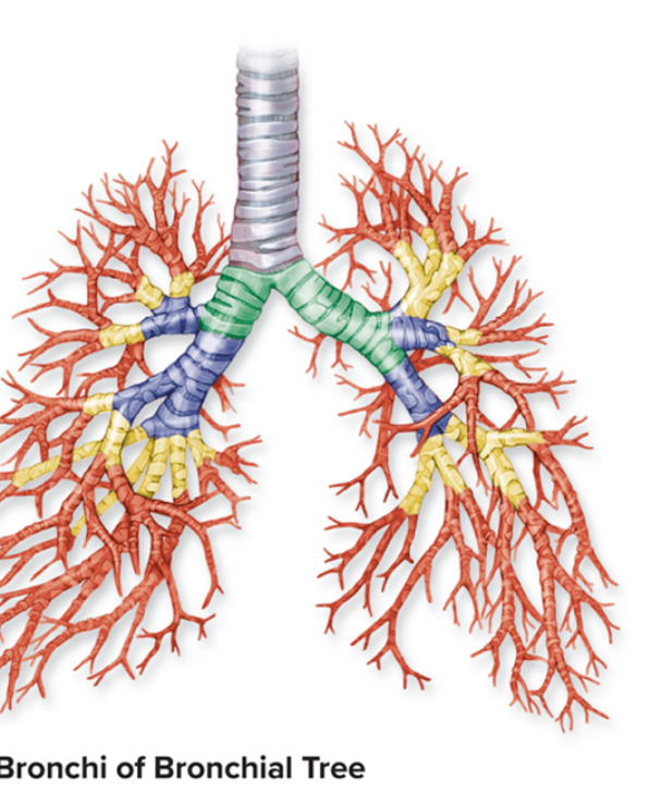
Identify what type of bronchi each color represents
Green primary, blue secondary (lobar(, yellow tertiary, red is smaller bronchi and then bronchioles
What are the bronchioles with alveoli off of them called?
Respiratory bronchioles
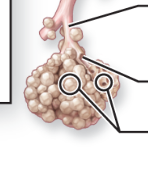
Identify superior to inferior
Respiratory bronchioles, alveolar duct, alveoli
What is the layer of alveolar cells, capillary cells, and the fused basement membranes of the capillary endothelium and alveolar epithelium called?
Respiratory membrane
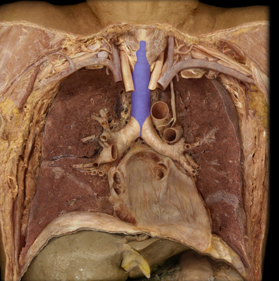
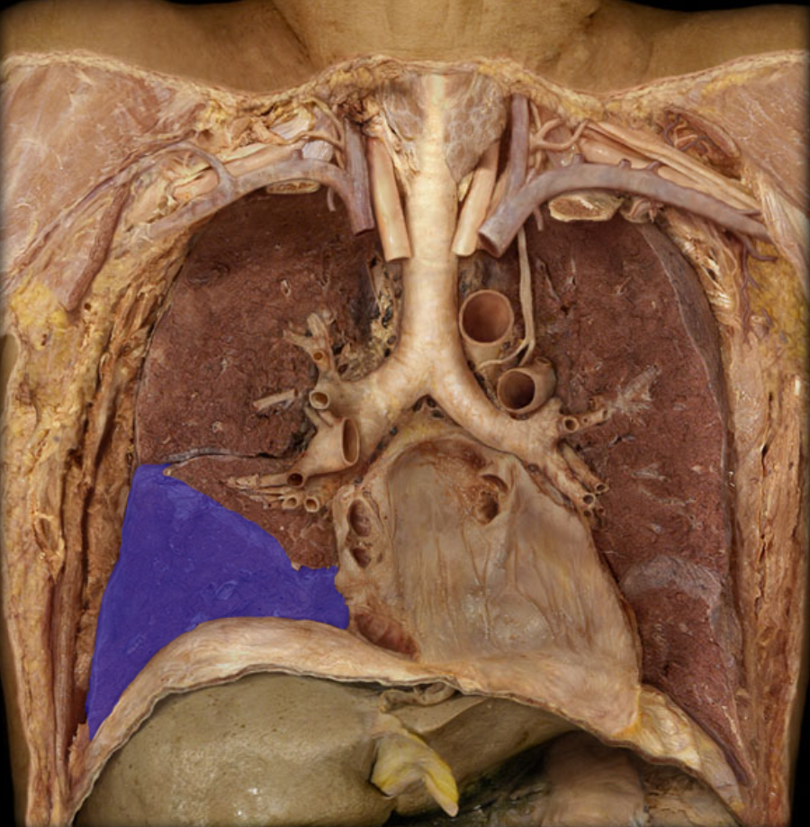
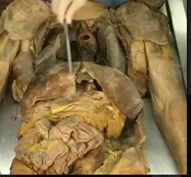
ID the structure in between the lungs
Mediastinum, holds the heart as well
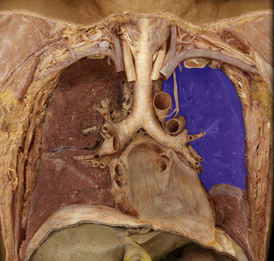
ID
Superior lobe of left lung
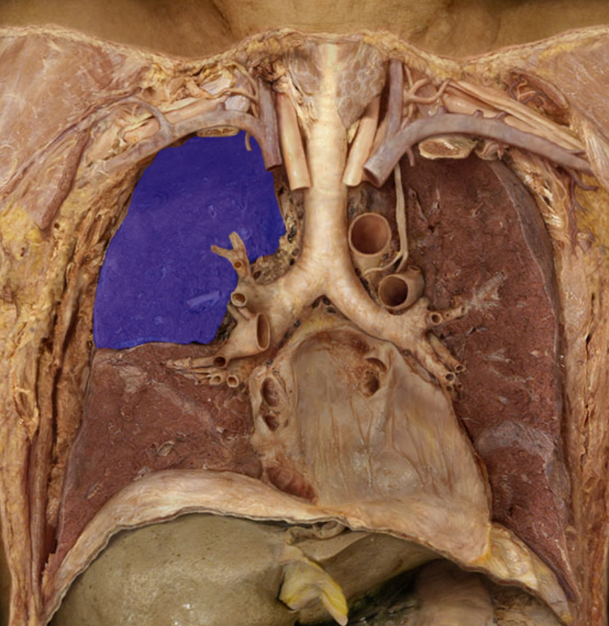
ID
Superior lobe of right lung
ID
Inferior lobe of left lung
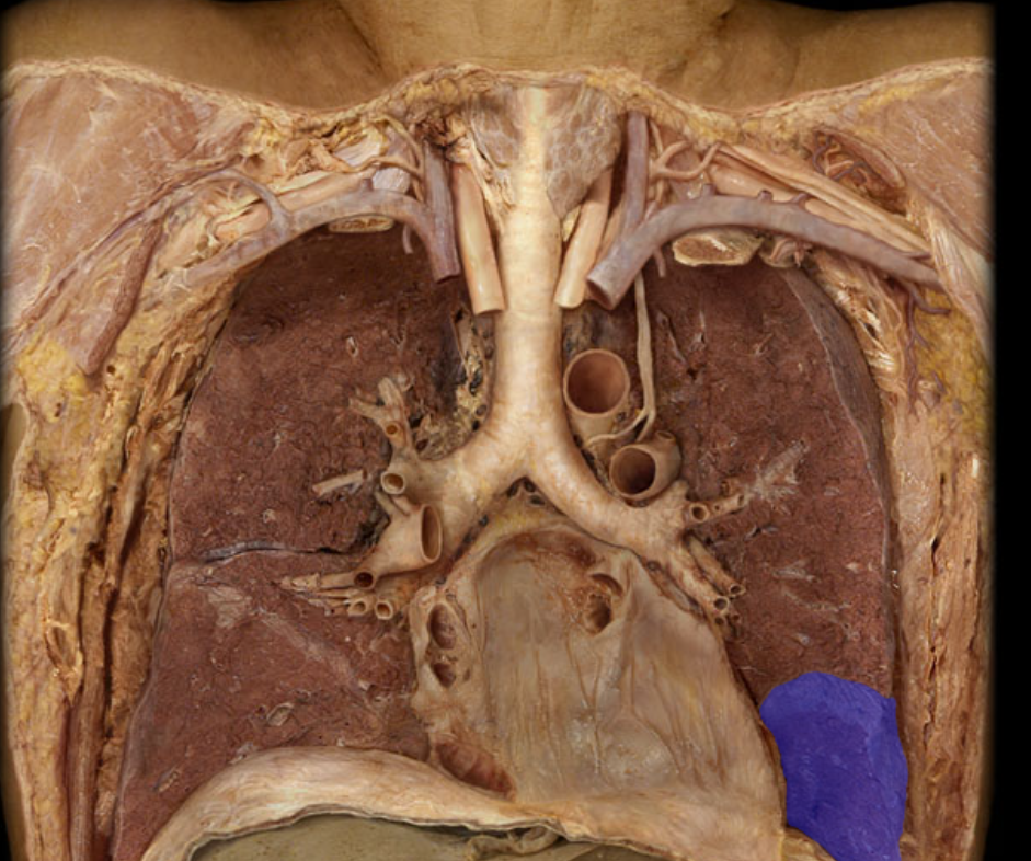
ID
Inferior lobe of right lung
ID
Middle lobe of right lung
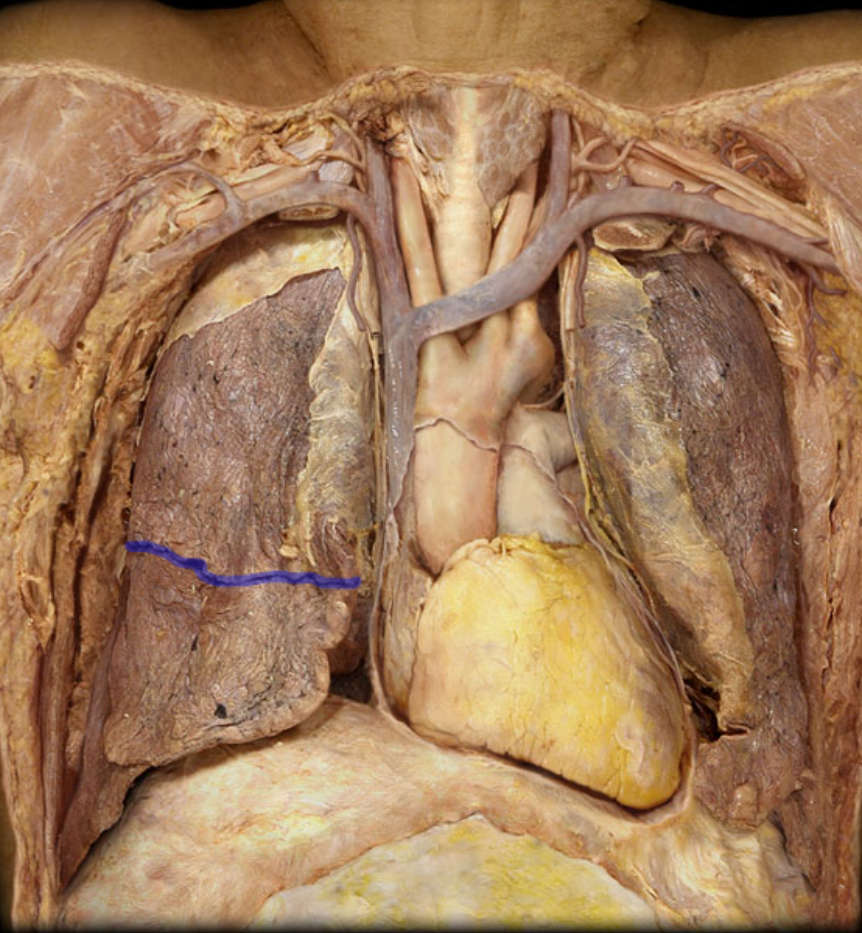
ID
Horizontal fissure of right lung
ID
Oblique fissure of right lung
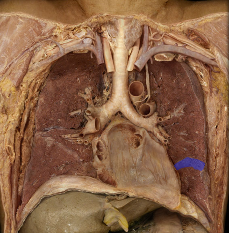
ID
Oblique fissure of left lung
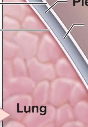
ID from superior to inferior (top three)
Parietal pleura, pleural cavity, visceral pleura,
ID
Diaphragm
ID
Diaphragm
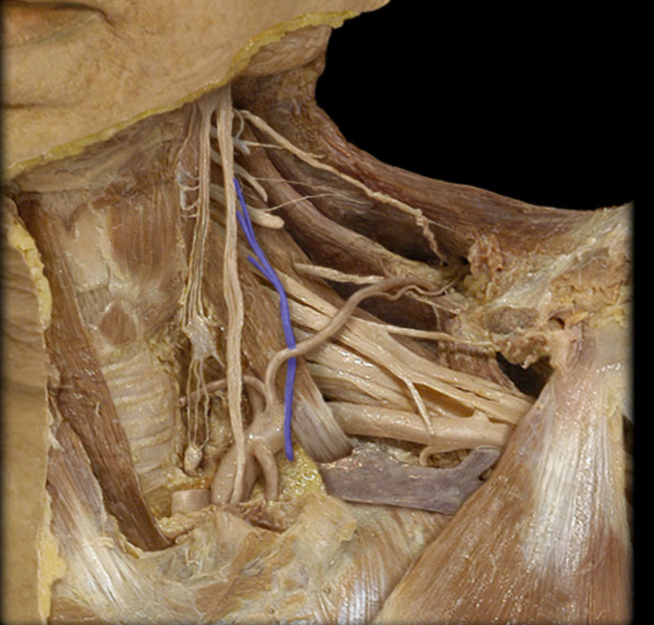
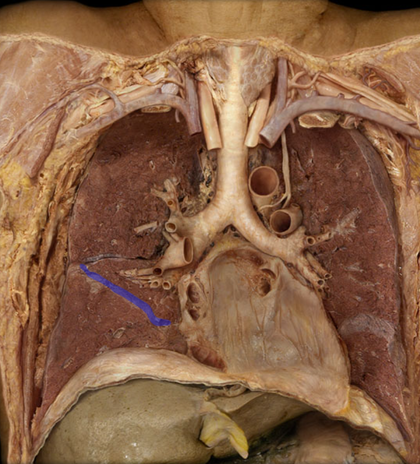
ID
Phrenic nerve
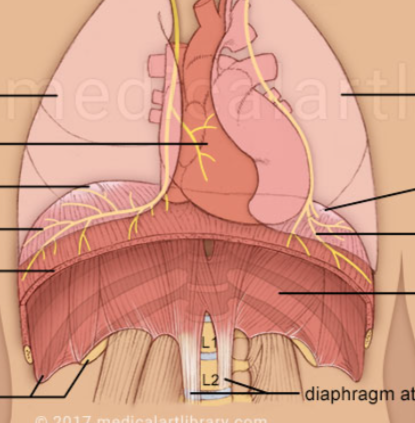
What nerve is seen innervating the diaphragm
Phrenic nerves
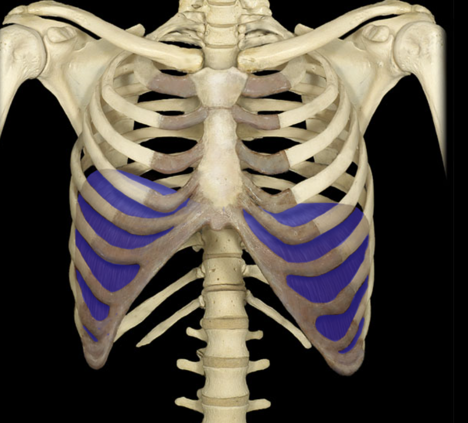
Which intercostal muscles are angled TOWARDS the head
Internal intercostals
Which intercostals are angled AWAY from the head?
External intercostals
What innervates the intercostal muscles?
Intercostal nerve
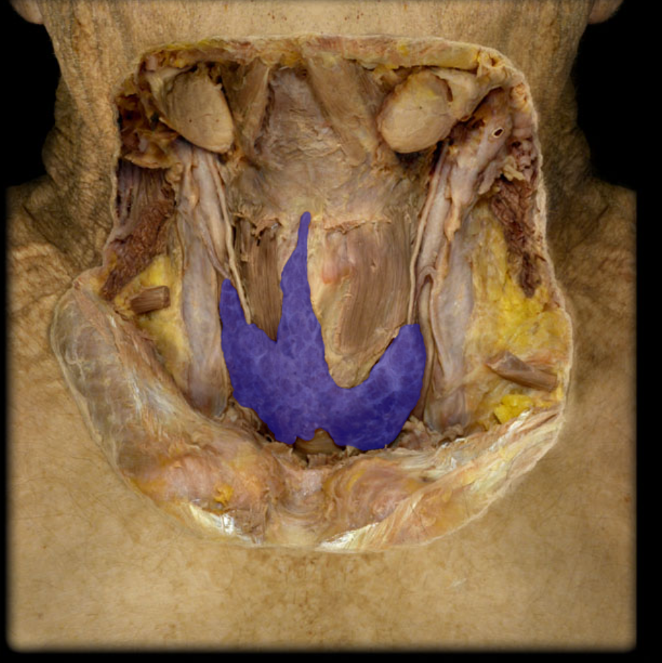
ID
Thyroid gland
What are the hard little lumps on the back of the thyroid gland called?
Parathyroid gland
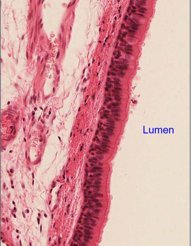
Identify the cell type nand where
Pseudostratified ciliated columnar epithelium, in trachea
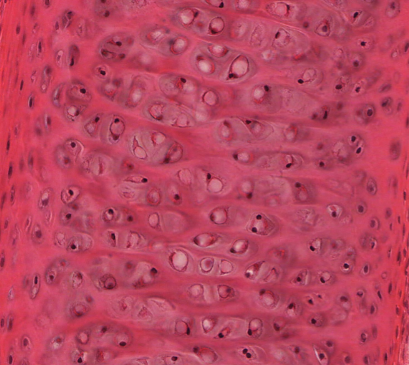
Id the tissue, and where
Hyaline cartilage, in trachea
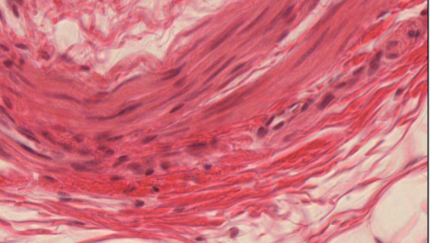
ID the tissue type and where
Smooth muscle in trachea
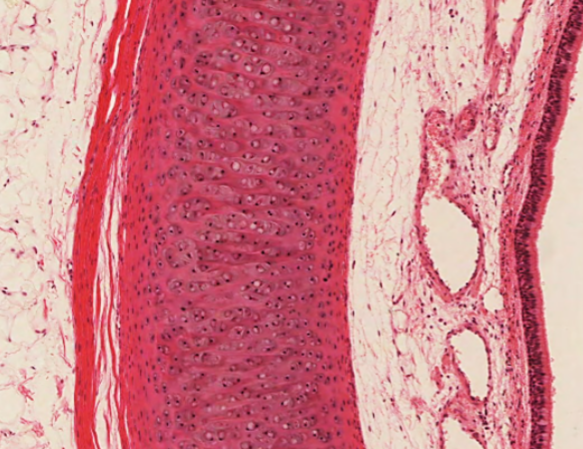
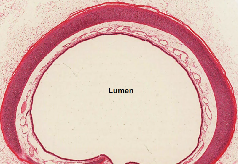
ID the stained redish pink structure
Tracheal cartilage or tracheal rings
Describe and point out the location of the trachealis muscle
White section, c shaped ring deep to hyaline ring
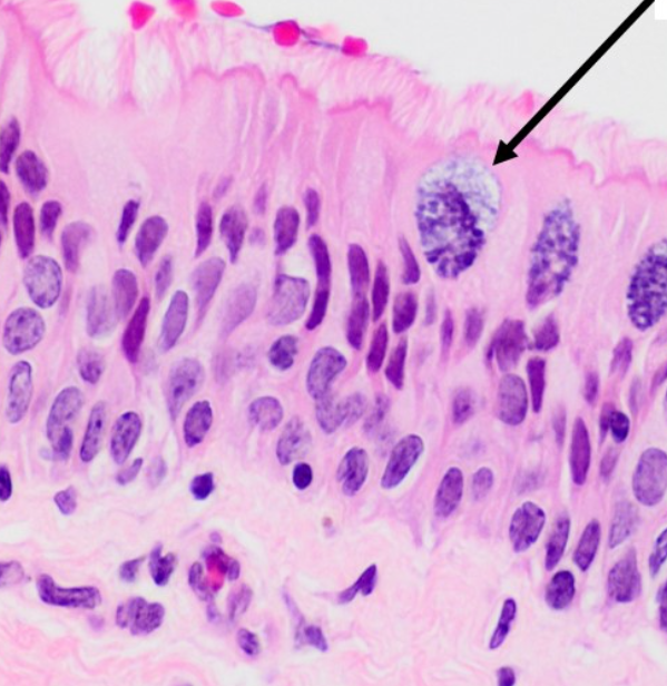
ID the structure
Goblet cells (stained)
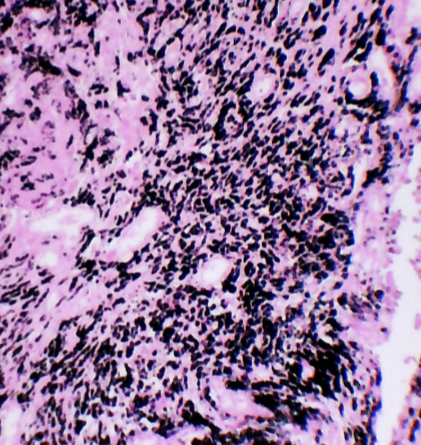
ID this condition
Coal dust/black lung
ID this organ histologically
Lung
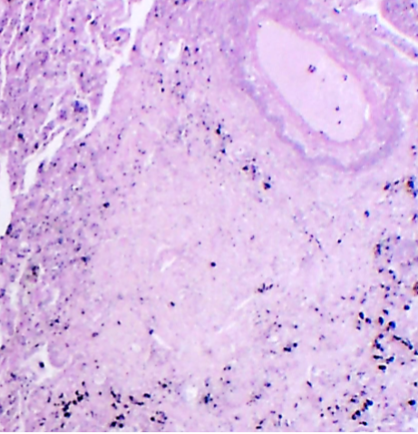
ID this condition, note important features
Tuberculosis, hard bulbs (the rounded tubercle visible in upper right corner)
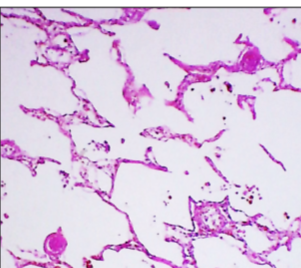
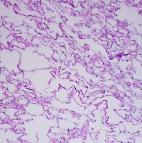
ID this condition and its identifitying features
Emphysema, enlarged airspaces and shortness of breath - destruction of alveoli
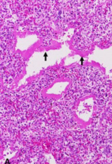
ID this condition and note its cuase and primary features
Neonatal pumonary distress syndrome, thick hyaline membranes because of alveolar collapse from a lack of surfactant
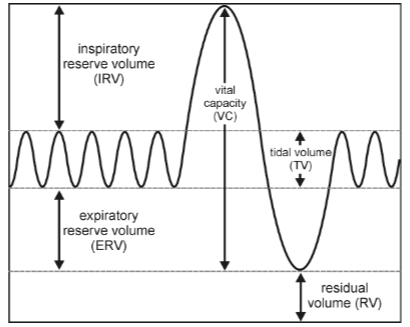
What is the volume of air beathed in and out without conscious effort, and what is its volume
Tidal volume, about 500 ml
What is the additional volume of air that can be inhaled with maximum effor after a normal inspiration, and what is the average value ml
Inspiratory reserve volume, about abt 3000 ml
What is the additional volume of air that can be forcibly exhaled after normal exhalation, +avg value
Expiratory reserve volume, 1100 ml
What is the total volume of air that can be exhaled after a maximum inhalation +avg ml
Vital capacity, 4600ml
What is the volume of air remaining in the lungs after maximum exhalation, and why +ml
Residual volume, the lungs can never be completely emptied, abt 1200ml
What is the VC+RV?
Total lung Capacity
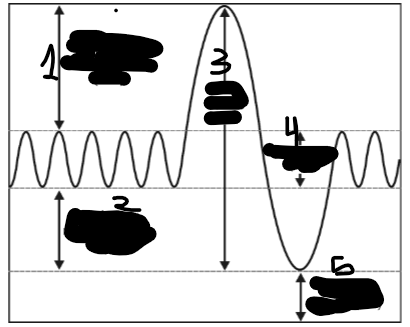
Locate the ERV, IRV, TV, RV, and VC
2, 1, 4, 5, 3
TC= Blank +IRV +ER
TV
blank = VC +RV
TLC
During hyperventilatiion what happens to blood pH
Raises ph
Hypoventilation does what to blood pH
lowers it, less CO2 is expelled
What happened when you blew into the alkaline water with the alkaline indicator?
It turned clear again as you added CO2 to the water and it lowered the pH (added H+ ions)
What happened after excercise to make it take less time to turn the indicator clear?
Excercise increased rate of breath which raised the pH meaning it raised the pH of the solution faster
Define (and list the organs involved in) the gastrointestinal tract/digestive tract
Tube from oral cavity to anus. Includes oral cavity, pharynx, esophagus, stomach, small intestine, and large intesting
List the accesory digestive organs
Teeth and tongue, salivary glands, pancreas, digestive glands, liver, and gallbladder
Define digestion
The breakdown of ingested food into smaller components that can be absorbed
Define asorbtion
The membrane transfer of digested molecules, electrolytes, vitamins, and water across the epithelial lining
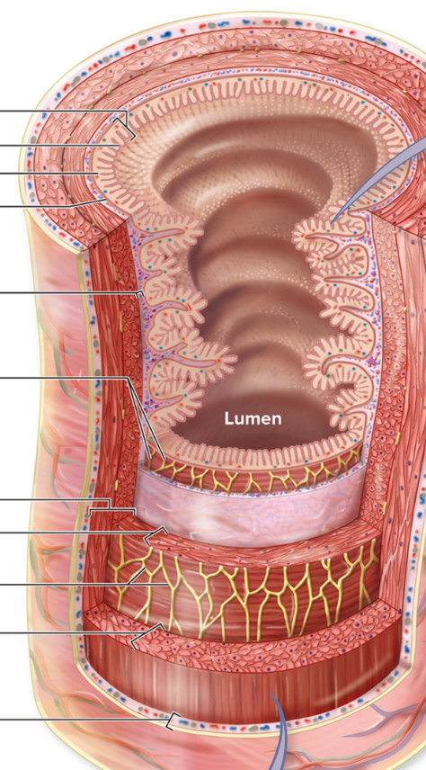
Identify the layers from internal to external
(Lumen) Mucosa, submucosa, muscularis, serosa.
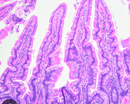
Identify the structures pictured
villi
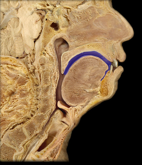
ID
Oral cavity
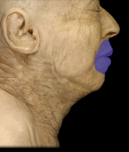
ID
Labia (lips)
a slit-like, horseshoe-shaped space between the inner surface of the lips/cheeks and the outer surface of the teeth/gums
Vestibule
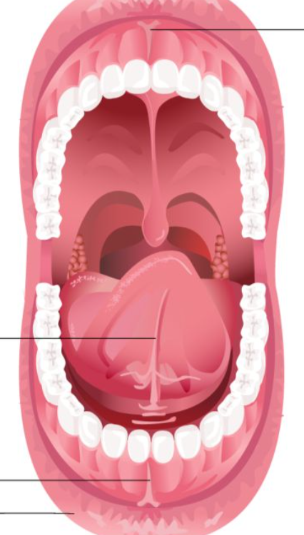
ID superior to inferior
Superior labial frenulum, lingual frenulum, inferior labial frenulum
ID
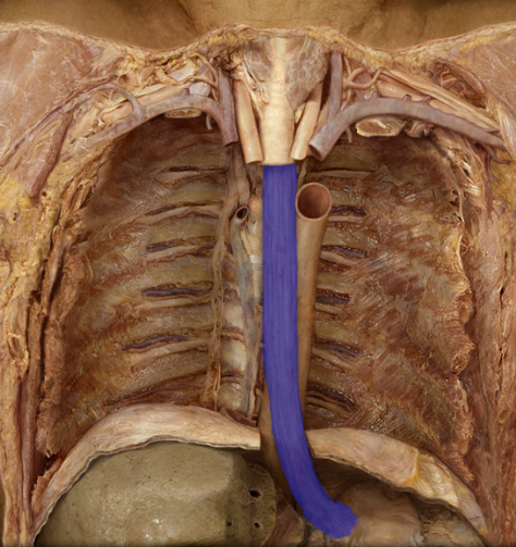
Esophagus
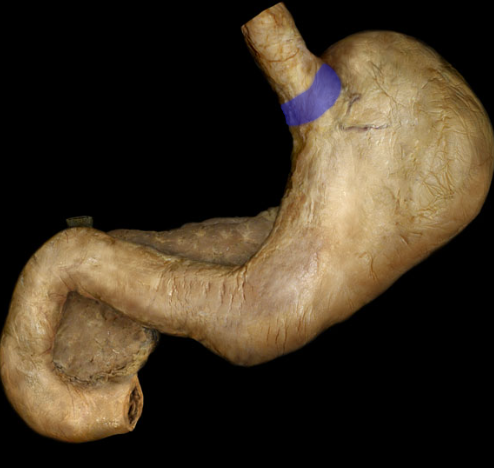
What is the sphincter that connects the stomach and the esophagus
Gastroesophageal (cardiac) sphincter
ID
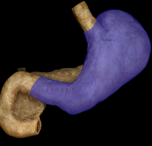
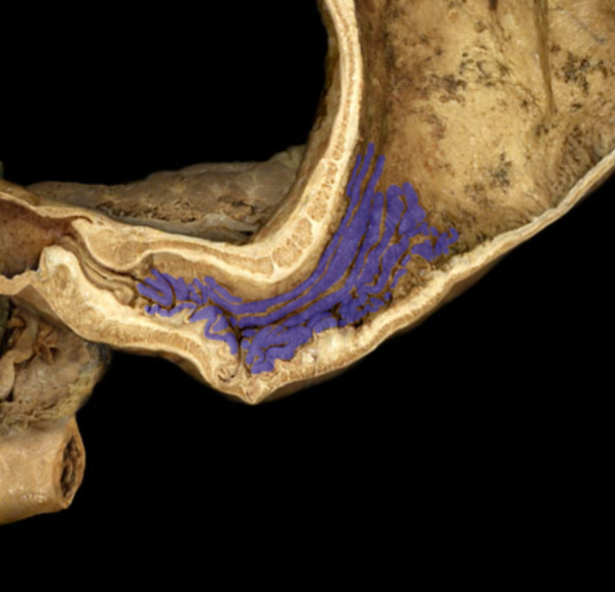
Stomach
ID
Rugae/gastric folds
ID
Cardiac region of stomach
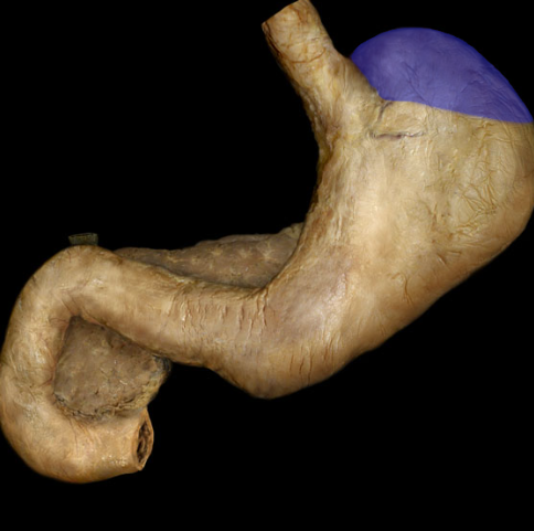
ID
Fundus of stomach
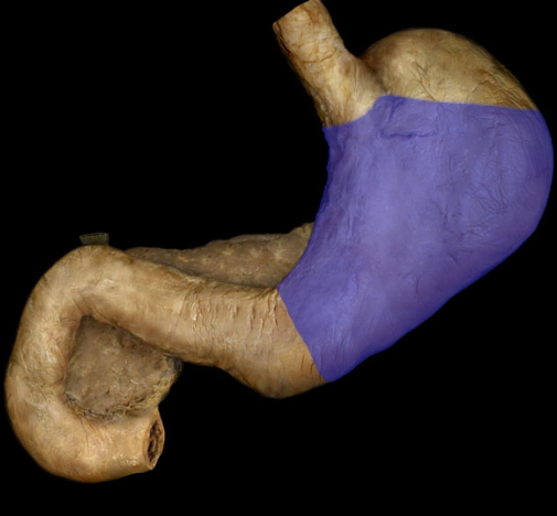
ID
Body of stomach
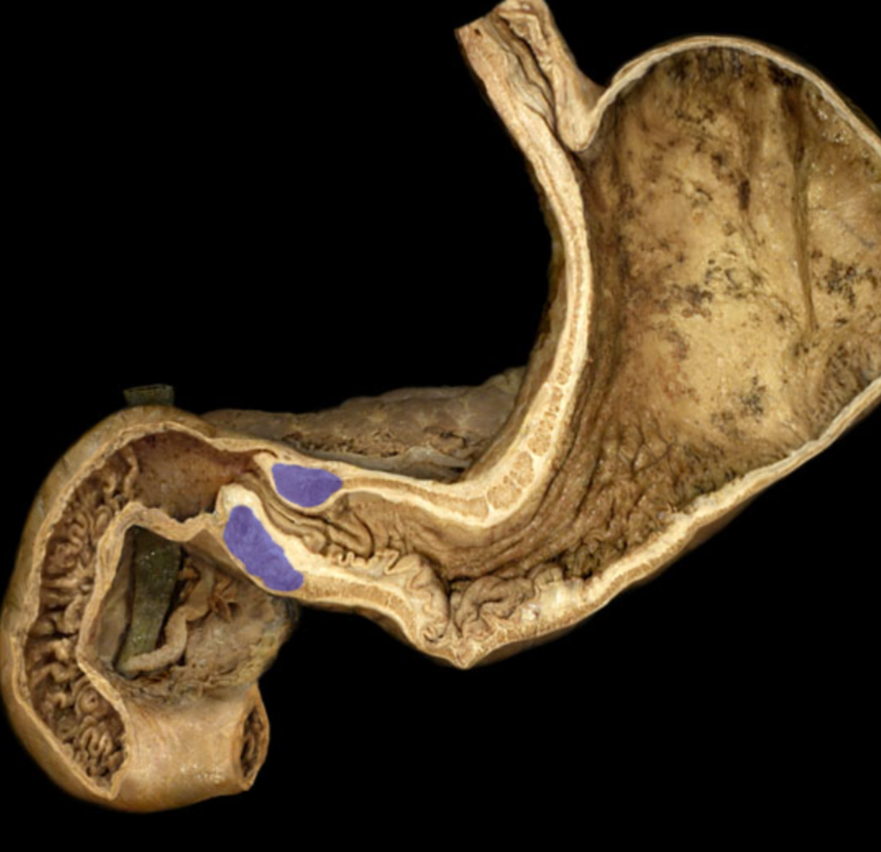
ID this muscle
Pyloric sphincter
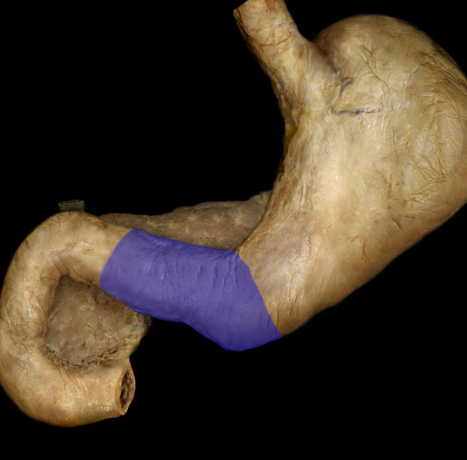
ID
Pylorus (region) - where the stomach goes to sm intestine
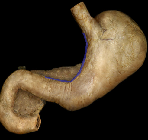
ID
Lesser curvature
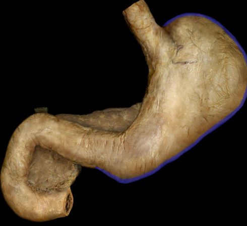
ID
Greater curvature
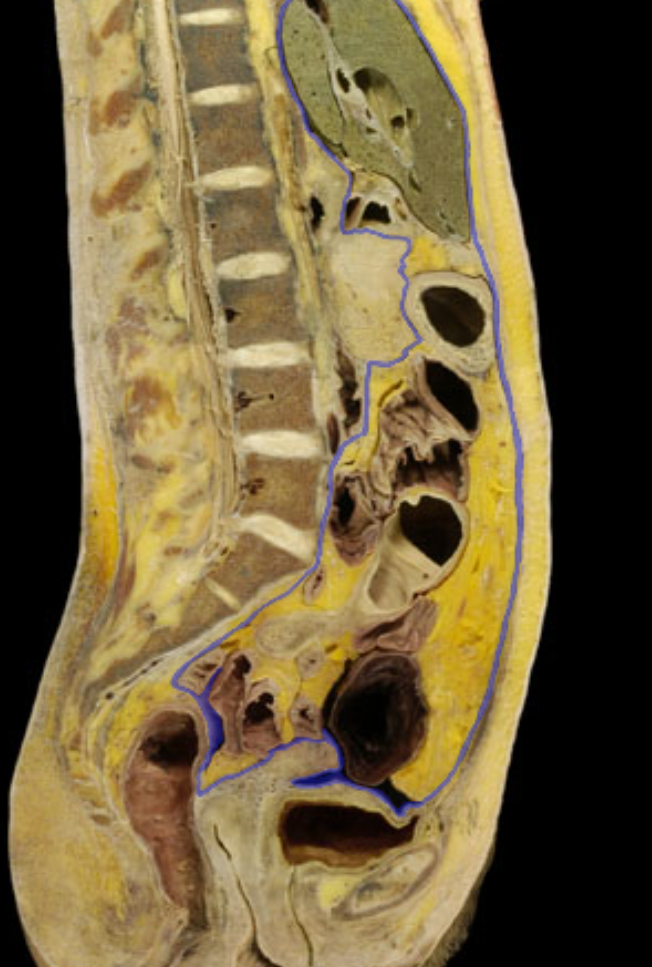
ID
Parietal peritoneum
ID
Visceral peritoneum