BIOL 2052 - Ionic basis of the membrane potential and synaptic integration
1/27
There's no tags or description
Looks like no tags are added yet.
Name | Mastery | Learn | Test | Matching | Spaced |
|---|
No study sessions yet.
28 Terms
neurons
functional unit of the nervus system
generate and transmit electrical impulses
allows motor control, sensory processing and proprioception
recap of physiology of an action potential
the sodium potassium ATPase is electrogenic and generates the membrane potential
3 sodium ions pumped out for every 2K+ ions in
the membrane potential is a measure of charge separation across the membrane
membrane acts as a capacitor —> charge capacitance will be bigger in bigger cells and smaller in smaller cells
each ions charge affects the membranes voltage by 16 nanovolts
the concentrations do not flip, just the charge separation becomes sufficiently larger to create an action potential
movement of ions
overall electrochemical gradient moves Na in an K out
RMP is -70mV due to permanently expressed leak channels which allow K+ in
when the inside becomes ore positive Na is repelled so it doesnt keep moving in but reaches an equilibirum
at some point K ions will equilibrate (at a little lower than -70mV)
sodium ions outside the cell are attracted to the potassium ions inside the cell
typical ion concentrations
ION | INSIDE | OUTSIDE |
Na+ | 15 mM | 145 mM |
K+ | 150 mM | 4 mM |
Cl- | 10 | 110 mM |
the nernst equation to calculate equilibrium potential
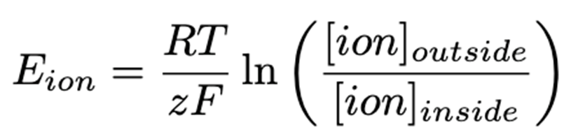
units in V
F is faradays constant - 96485
z is ionic charge
T is temp in K
R is the gas constant
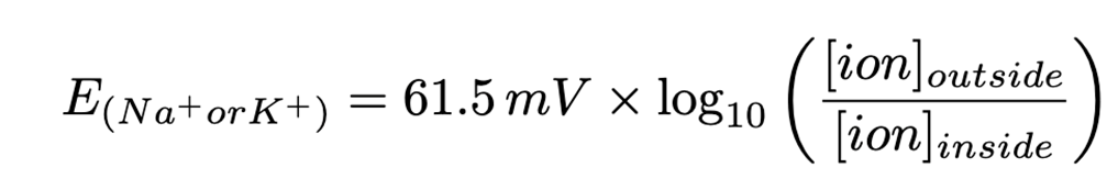
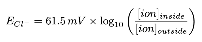
this simplifies to
goldman hodgkin katz equation
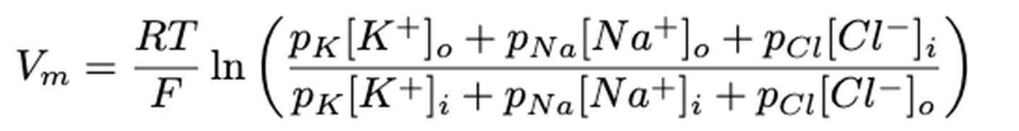
for neurons at rest pK:pNa:pCl is 1:0.05:0.45
useful for when the concentrations change with time and determining membrane concs at a particular moment
summary of control of action potentials
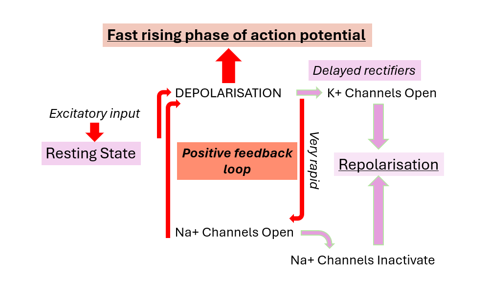
firing of neurons
neurons fire 10 times/s but some ultra rapid ones can fire up to 200x/s
sodium is responsible for the AP which was proved by hodgkin and hatch in squid axons as decreasing the sodium concentration decreases the depolarisation of the membrane
as you decrease the sodium conc the AP also moves to later as the concentration gradient is less steep
how can we measure neuronal activity
EXTRACELLULARLY
local field potential
electroencephalogram
INTRACELLULARLY
action potentials
passive/active electrical properties
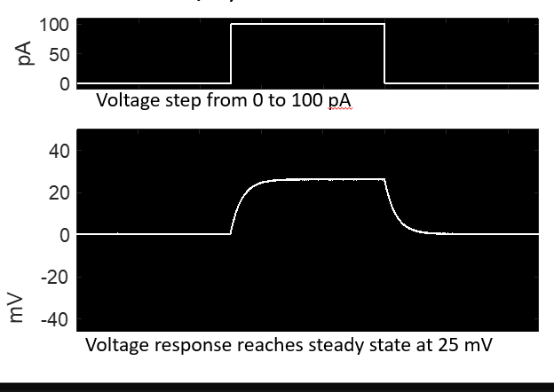
inserting a voltage into an axon will give a response
ohms law states that V=IR where current is in amps and R is in ohms
once the threshold for activation is reached an action potential is formed which is an active property of an action potential
passive membrane properties
the voltage effect of the membrane current takes time due to membrane capacitance - this is called the time constant
the voltage effect of the membrane current decreases with distance from the injection site (the length constant )
the voltage change as a response to current injection depends on the cells resistance (input resistance determined by V=IR)
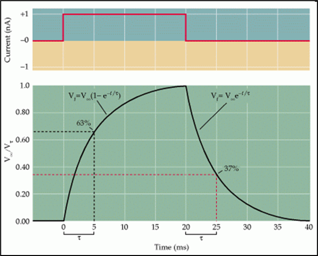
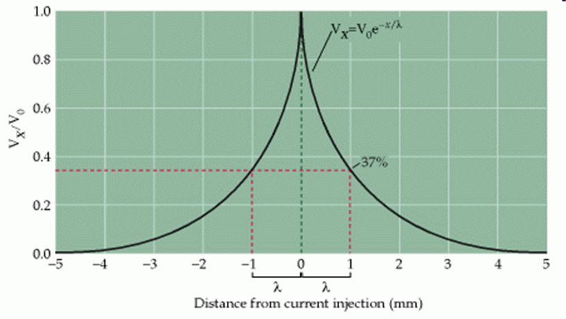
exponential equations can be used to model behaviour of many membrane properties
we can refer to the constant extracted from these equations to qualify the properties of neurons
example of synaptic integration - dentate gyrus
the dentate gyrus is involved in the generation of new neurons
new neurons have different properties to the old neurons:
NEW
small
high resistance
low capacitance
require less input to fire
OLD
low input resistance
high capacitance
requires more input to fire
if you multiply the capacitance and the resistance, this is a direct measure of how slow the cell will be at integrating inputs
capacitance
the ability of membranes to store charge
capacitors and resistors in parallel (which act as a model axon) has only passive properties, whilst actual axons have passive and active properties
how can we dissect the contribution of different currents to neuronal activity
use of a voltage clamp to keep a set voltage
you can then figure out which ions are flowing through the axon at set voltages so can tell at which voltage sodium channels are activated
however, this experiment was run in squid axxons which differ from our brains as squid axons have slower sensory processing
propogation of signal
delay in stimulation and change in current due to the membrane acting as a capacitor
as the membrane becomes charged the current flows through the capacitance path
gradually, more current flows through the resistance branch (the membrane potential)
when the current is terminated the current through the capacitance branch flows through the resistance branch until the membrane potential returns to baseline
this is represented by the “wave shaped” response
as the input moves through the axon the change in the membrane potential will become smaller and smaller
summary of passive vs active responses of neurons
PASSIVE
delayed response caused by capacitance of membrane
attenuation across distance
ACTIVE
excitatory input
na channels and K channels opening
does not attenuate over distance
active properties - the action potential
usually, when current is injected the membrane depolarises with the magnitude of the injected current unless the injected current surpasses the threshold for activation, then an AP is generated which doesnt attenuate over distance
this is an active property of neurons
cells with a high resistance are more likely to surpass threshold
the active properties will depend on the type of ion channel expressed and the specific localisation of these within cells
active properties of neurons
bought about by ion channels —> either voltage gated or ligand gated
ion channels have multiple subunits which must be activated which is why sometimes in equations why factors are raised to the power of 4
patch clamping
allows us to study behaviour of ion channels
a pipette is connected to an amplifier is used to isolate a patch of membrane from a cell and measure the current
when the pipette is brought to the membrane it forms a bond which prevents the flow of ions - called a giga-ohm seal
once the seal is formed the membrane patch can either be cut off or left attached
to cute the cell - apply more suction
when electrical potential is passed across the membrane the ion channels open and close and the amplifiers record the current
pipette amplifiers are able to measure the current and clamp the voltage to keep it the same or they are able to keep it at the same current and record the voltage
this info can be used to determine the resistance of the membrane
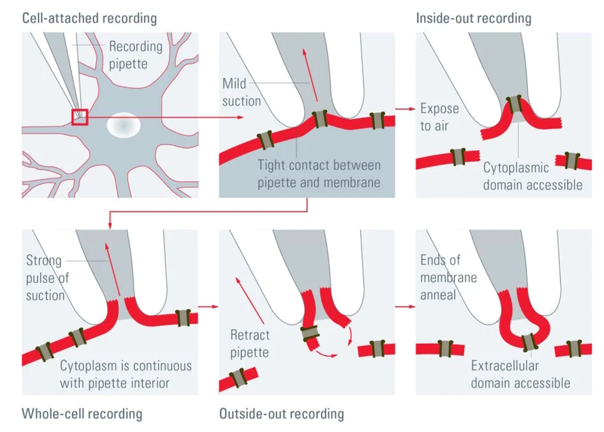
PATCH CLAMPING REQUIRES:
clean glass
air pressure
membrane
giga-ohm seal
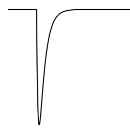
ion current plotting: +ve in
entry of positive current into the cell causes depolarisation
this causes an inwards deflection
the negative current exits the cell
for example: glutamate receptors
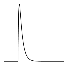
ion current plotting: -ve in
entry of negative ions
exit of positive current
hyperpolarisation of the cell causes an outward deflection
for example: the GABA receptor
how do ligand gated ion channels encode electrical activity
the electrical activity is dictated by the opening of ion channels
current flowing through receptors = conductance x driving force

glutamate receptors have huge driving force and so the number of ion channels is equal to the current through the plasma membrane
GABA has a smaller driving force because were closer to the reversal potential to chloride (RMP)
however, the membrane potential always changes so we need to consider synaptic integration
synaptic integration
neurons receive synaptic inputs and the resulting currents are summed by cells
the net effect of the inputs modifies the output of the neuron
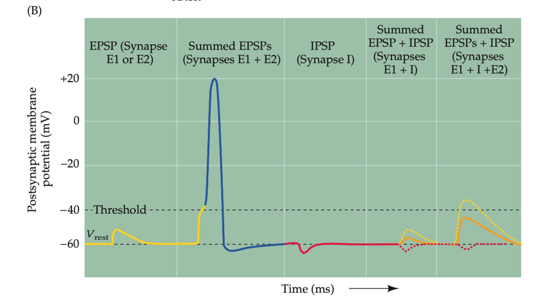
EI and I will give a smaller input as the result of EI is cancelled by I
EI and E2 combined with I is not enough to generate an AP due to the inhibitory effects of I
is synapses are more active and the connections stimulated by experience, studying etc, then the synapse expresses more AMPA receptors on the post synaptic membrane
many receptors are ion channels
all or none response
the larger the voltage/ further the voltage is from 0 the larger the current as the larger the driving force
experiments often conducted in absence of magnesium but this is not physiological
NDMA receptors are slow acting, AMPA receptors are fast acting
influence of magnesium - NDMA and AMPA receptor relationship
physiological conc of Mg2+ is 1000um
at low concentrations of magnesium it causes flickering of voltage as magnesium blocks NDMA receptors —> total block at high concentrations
activation of NDMA receptors is caused by the activation of AMPA receptors and at resting membrane potential the NDMA receptors will be blocked by magnesium
AMPA receptors activated by glutamate —> when the cell is depolarised the positive charge of the cell will repel the magnesium ion and will allow flux of cations and Ca2+ which will cause downstream effects
therefore, NDMA receptors are voltage dependent and act as coincident detectors
for them to be activated depolarisation and glutamate bound
Mg2+ is expelled at around -50mV and
plasticity
cells that fire together wire together
as you continue to stimulate an axon, the excitatory post synaptic potential amplitude increases from a stable baseline
receptor activation of AMPA and NDMA receptors results in synaptic plasticity
continued activation will result in more AMPA receptors
strengthening of synaptic function associated with growth of synaptic spine and increased cytoskeleton
back propogation of action potentials
is you generate action potentials in the soma it can be detected in the dendrites dur to the back propagation of the action potential
this does not mean that the AP can travel in both directions it can just travel to parts of the cell that haven’t experienced the action potential
GABA receptors
when GABA receptors are activated it produces an inhibitory input
causes these leaky channels which allows Na+ into the cell but not enough to cause an AP due to integration of inhibitory input