Comprehensive Unit 1 Exam Study Guide - Anatomy and Physiology I - BIO 1121
1/156
There's no tags or description
Looks like no tags are added yet.
Name | Mastery | Learn | Test | Matching | Spaced | Call with Kai |
|---|
No analytics yet
Send a link to your students to track their progress
157 Terms
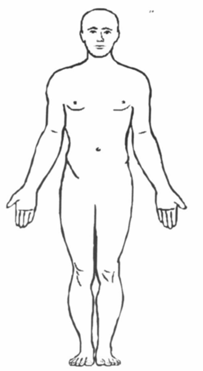
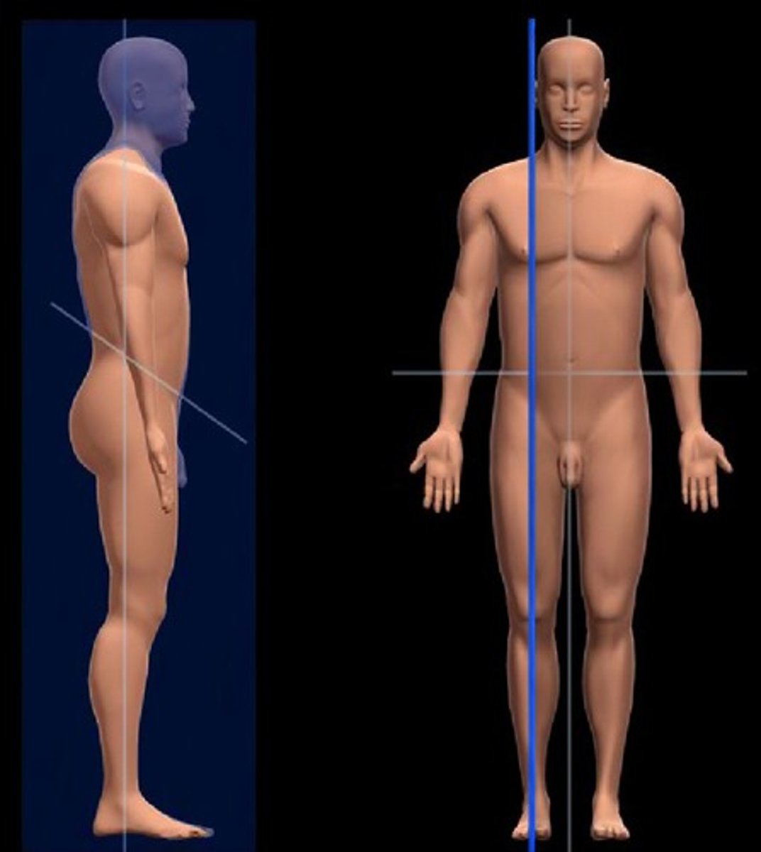
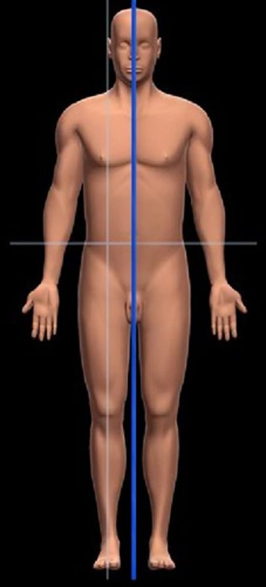
Anatomic position
- Standing erect
- Facing forward
- Palms forward
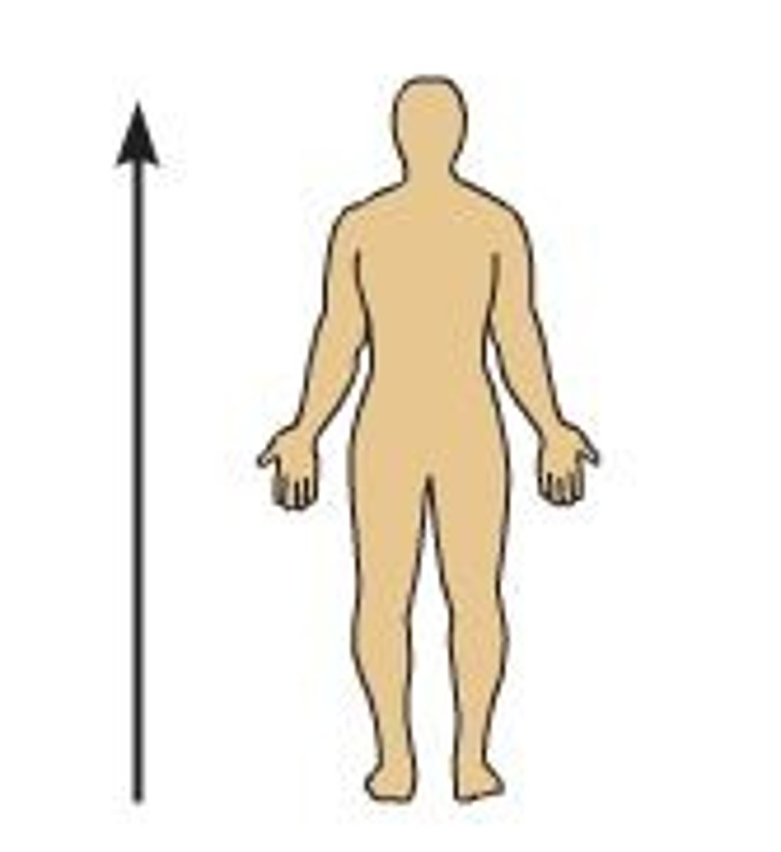
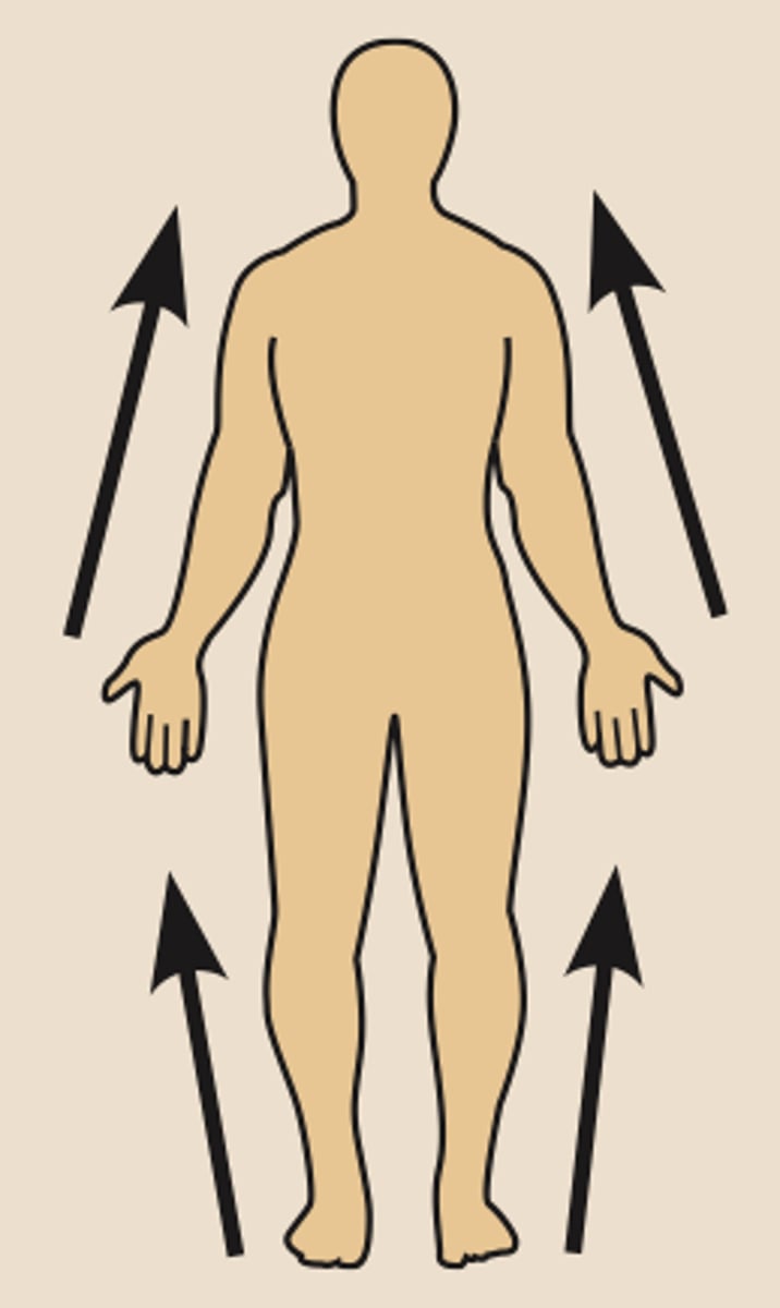
Superior (directional term)
above
Ex) The forehead is superior to the nose.
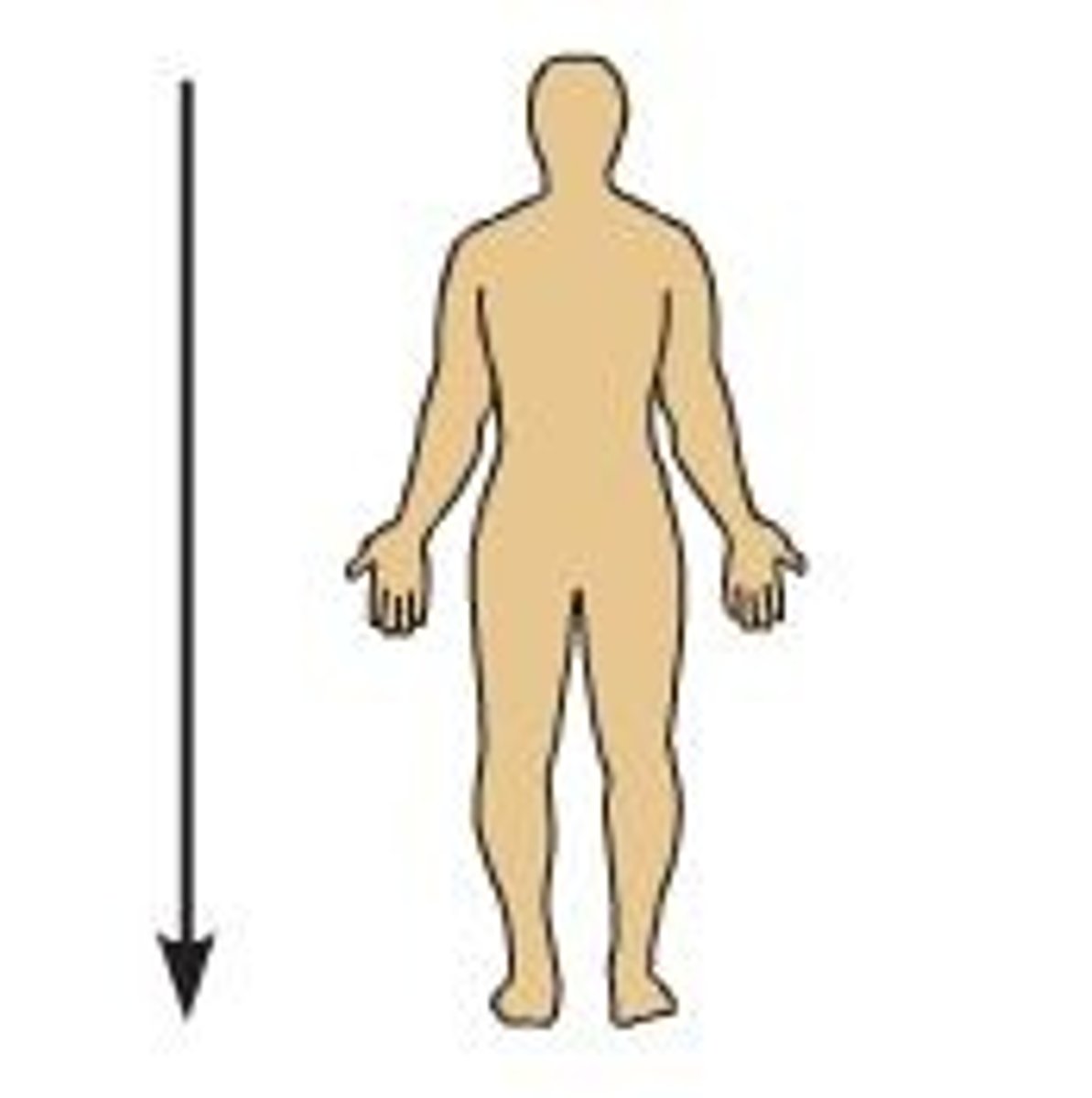
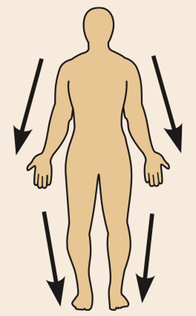
Inferior (directional term)
below
Ex) The nose is inferior to the forehead.
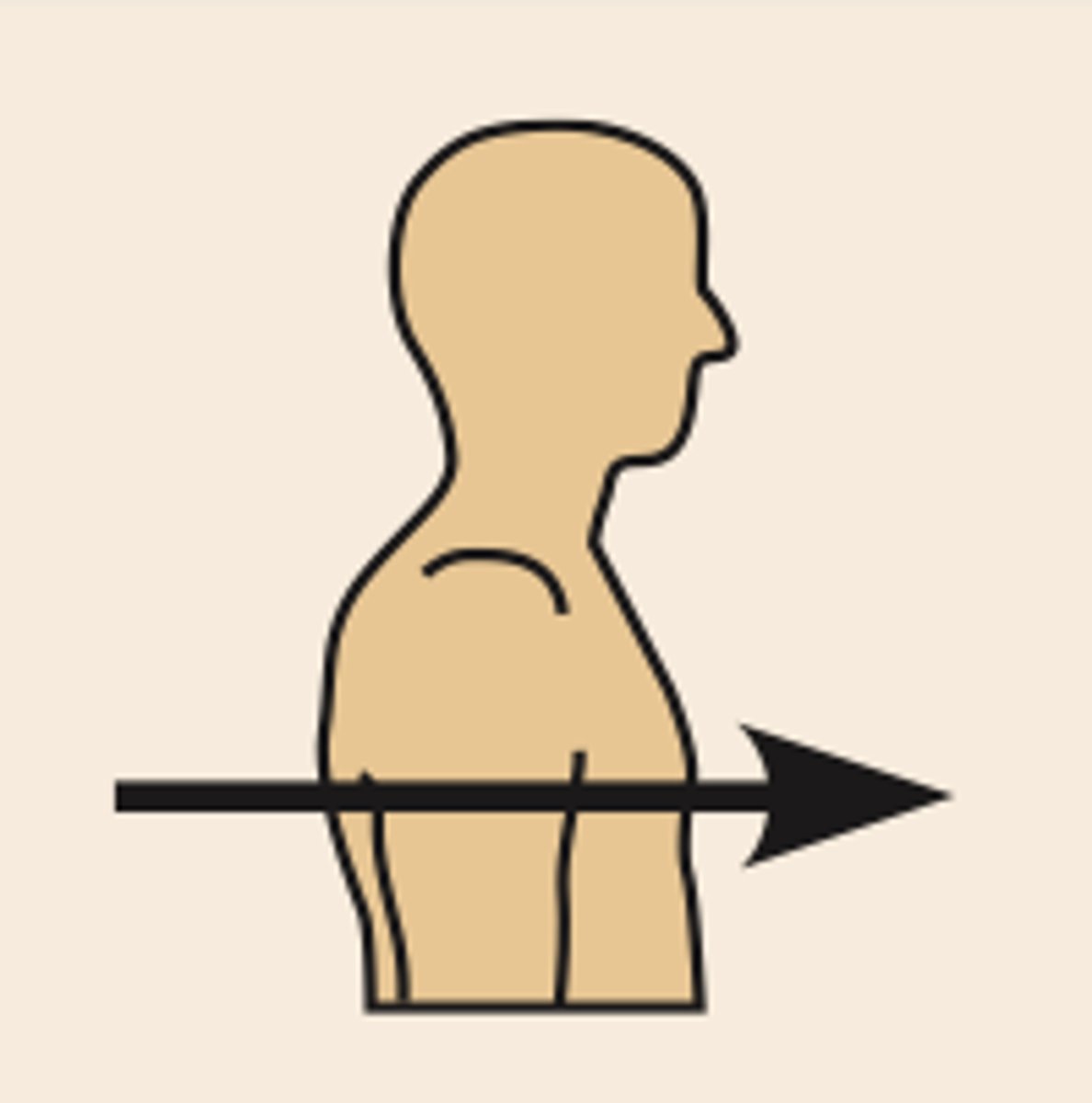
Anterior (directional term)
Toward or at the front of the body
Ex) The breastbone is anterior to the spine.
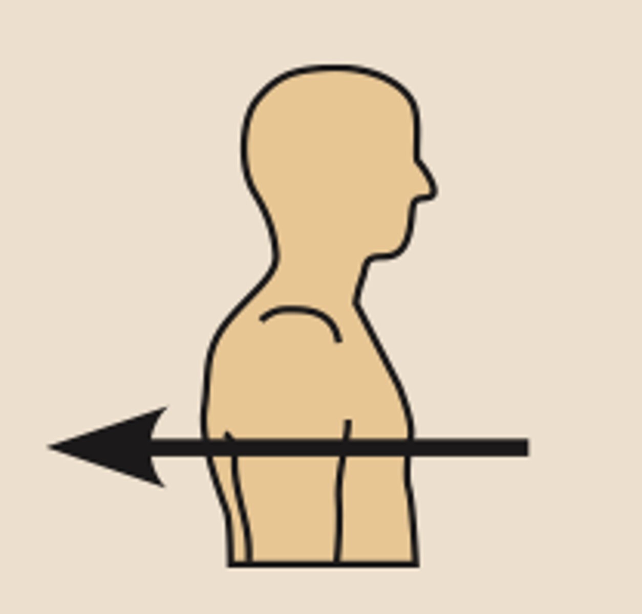
Posterior (directional term)
Towards the back or at the backside of the body - Behind
Ex) The heart is posterior to the breastbone.
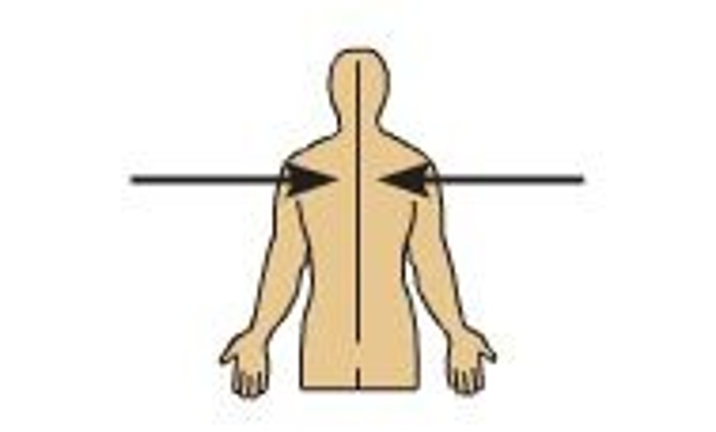
Medial (directional term)
Toward or at the midline of the body
Ex) The heart is medial to the arm.
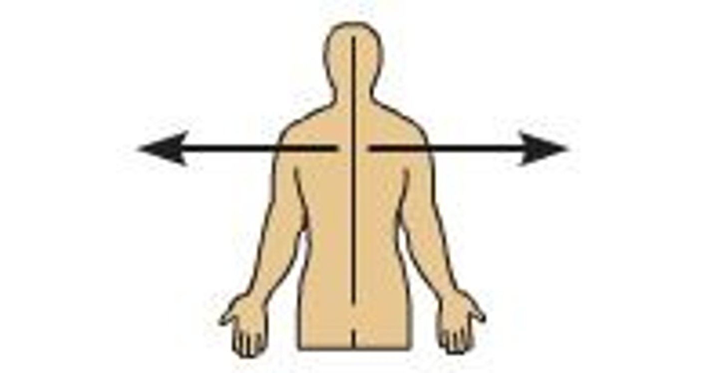
Lateral (directional term)
Away from the midline of the body
Ex) The arms are lateral to the chest
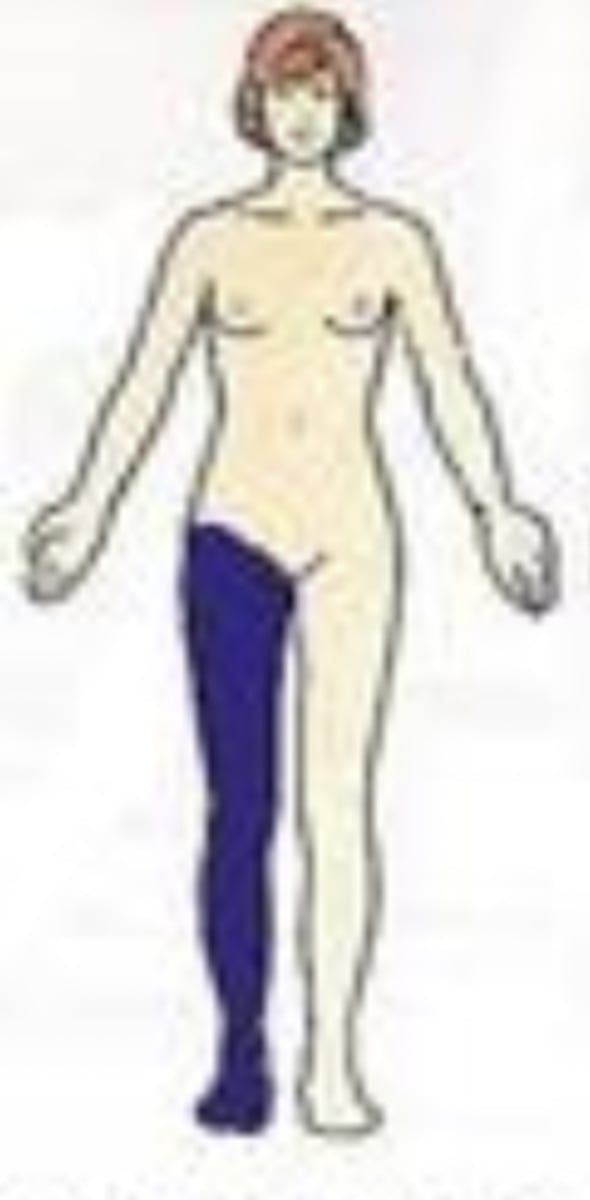
Distal (directional term)
Away from the point of attachment
Ex) The knee is distal to the thigh.
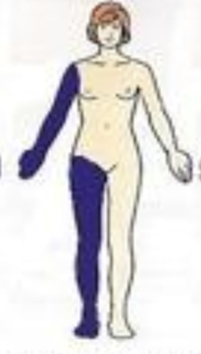
Proximal (directional term)
Toward the point of attachment
Ex) The elbow is proximal to the wrist.
Superficial (directional term)
Toward or at the body surface
Ex) The skin is superficial to the skeleton.
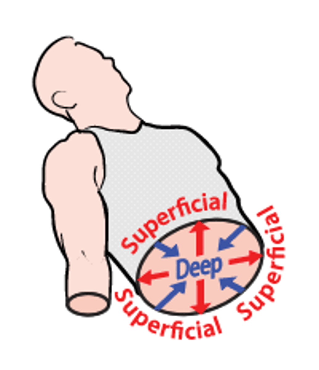
Deep (directional term)
Away from the surface of the body
Ex) The lungs are deep to the rib cage.

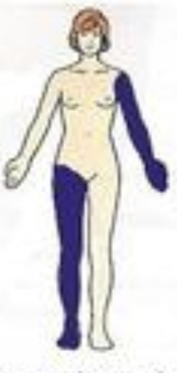
Bilateral (directional term)
On both sides of the body
Ex) The eyes are bilateral structures.
Unilateral (directional term)
On one side of the body only
Ex) The liver is a unilateral organ.
Ipsilateral (directional term)
Structures found on the same side of the body
Ex) Right arm is ipsilateral to the right leg
Contralateral (directional term)
Structures found on opposite sides of the body
Ex) The right hand is contralateral to the left leg.
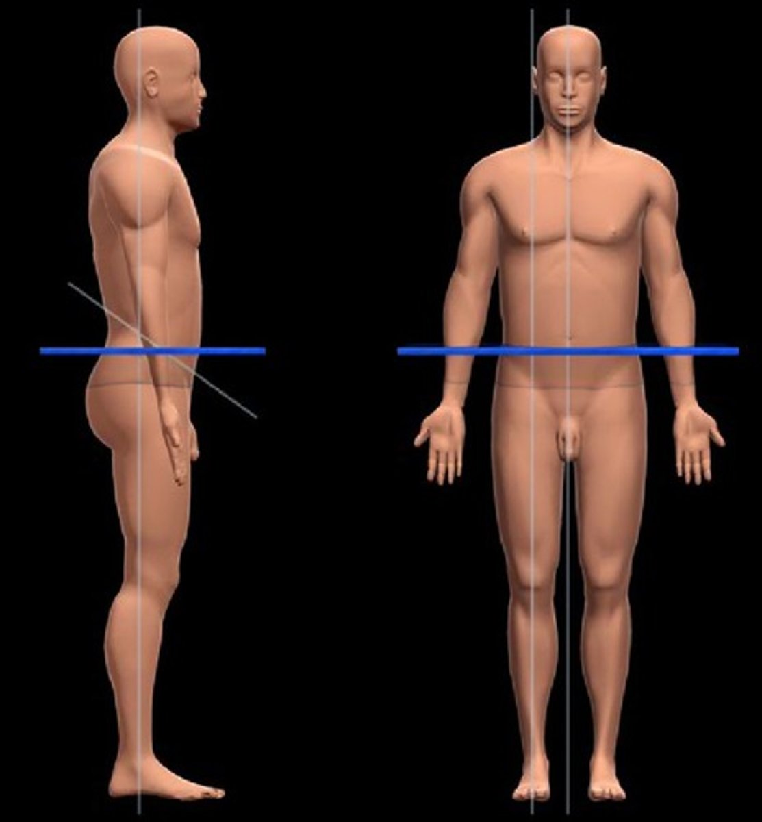
Frontal Plane
Divides the body into front and back portions (anterior and posterior).
Also known as a coronal plane
Sagittal Plane
A vertical plane that divides the body (or an organ) into right and left parts.
Midsaggital Plane
Divides the body into equal right and left halves.
Also known as a Median Plane.
Transverse Plane
Divides the body into superior and inferior parts
Also known as a Horizontal Plane or a Cross Section.
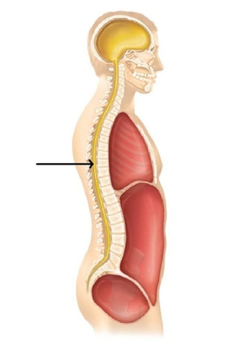
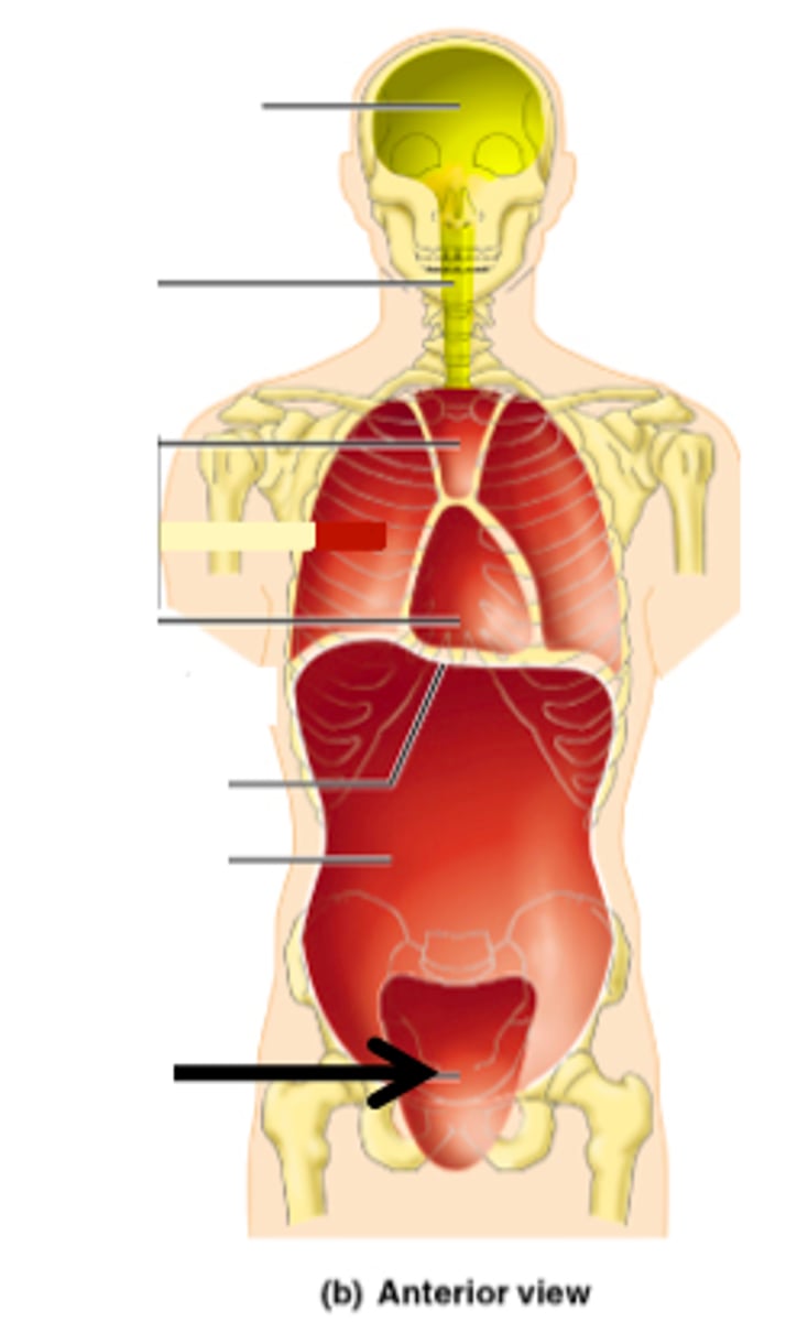
Dorsal Cavity
Contains the brain and spinal cord.
This cavity is further subdivided into the cranial and vertebral cavities.
Also known as the posterior body cavity.
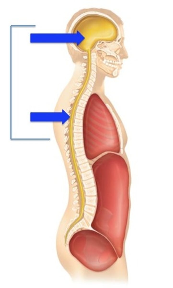
Cranial Cavity
Contains the brain
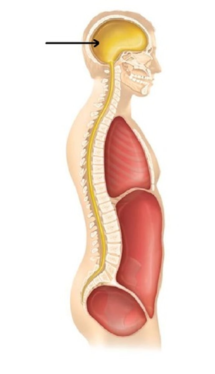
Vertebral Cavity
Contains the spinal cord.
Also known as the Spinal Cavity.
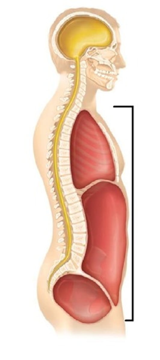
Ventral Cavity
Made up of the thoracic cavity and the abdominopelvic cavity separated by the diaphragm.
Also known as the anterior cavity.
Thoracic Cavity
Located superior to the diaphragm. This cavity is divided into three sections.
1. left pleural cavity - contains left lung
2. mediastinal cavity - contains trachea, esophagus, and pericardial cavity (which contains the heart)
3. right pleural cavity - contains right lung
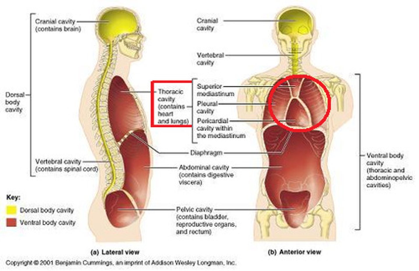
pleural cavity
contains the lungs
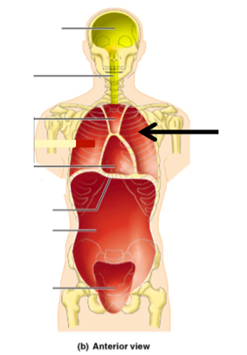
mediastinal cavity
The cavity between the lungs that contains the heart, trachea, esophagus, and thymus gland.
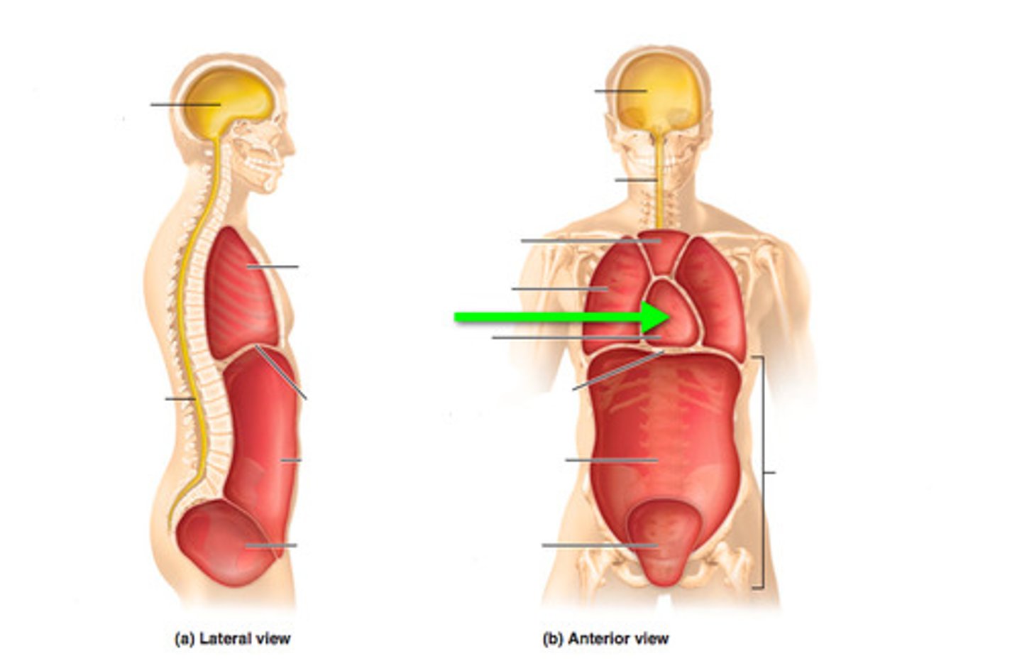
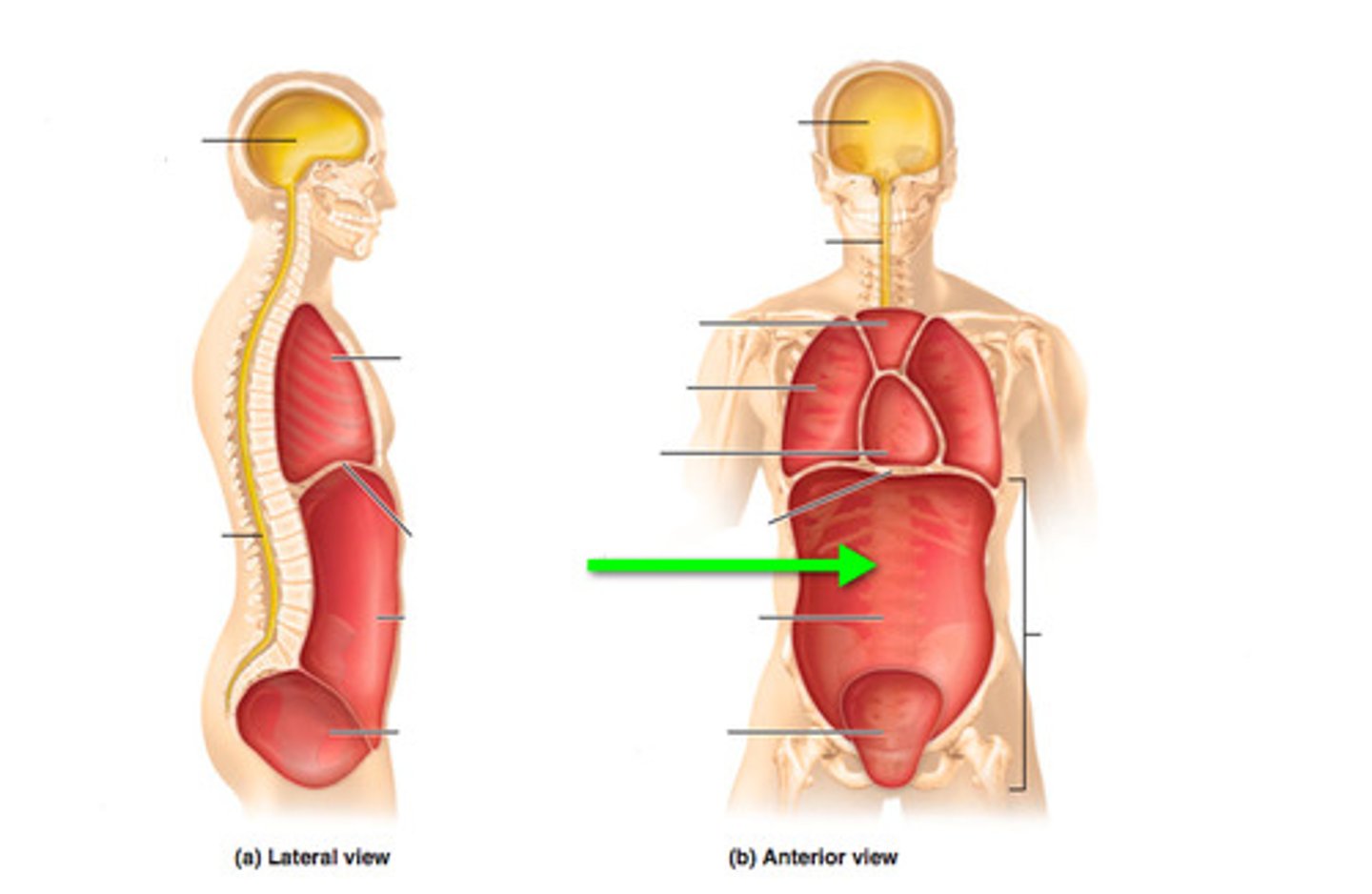
Abdominopelvic Cavity
Contains abdominal and pelvic cavities.
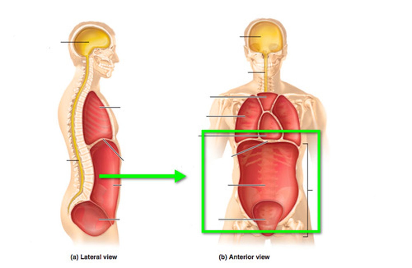
Abdominal Cavity
Contains the stomach, intestines, spleen, and liver, and other organs.
Pelvic Cavity
Contains the urinary bladder, portions of the large intestine, and internal organs of reproduction.
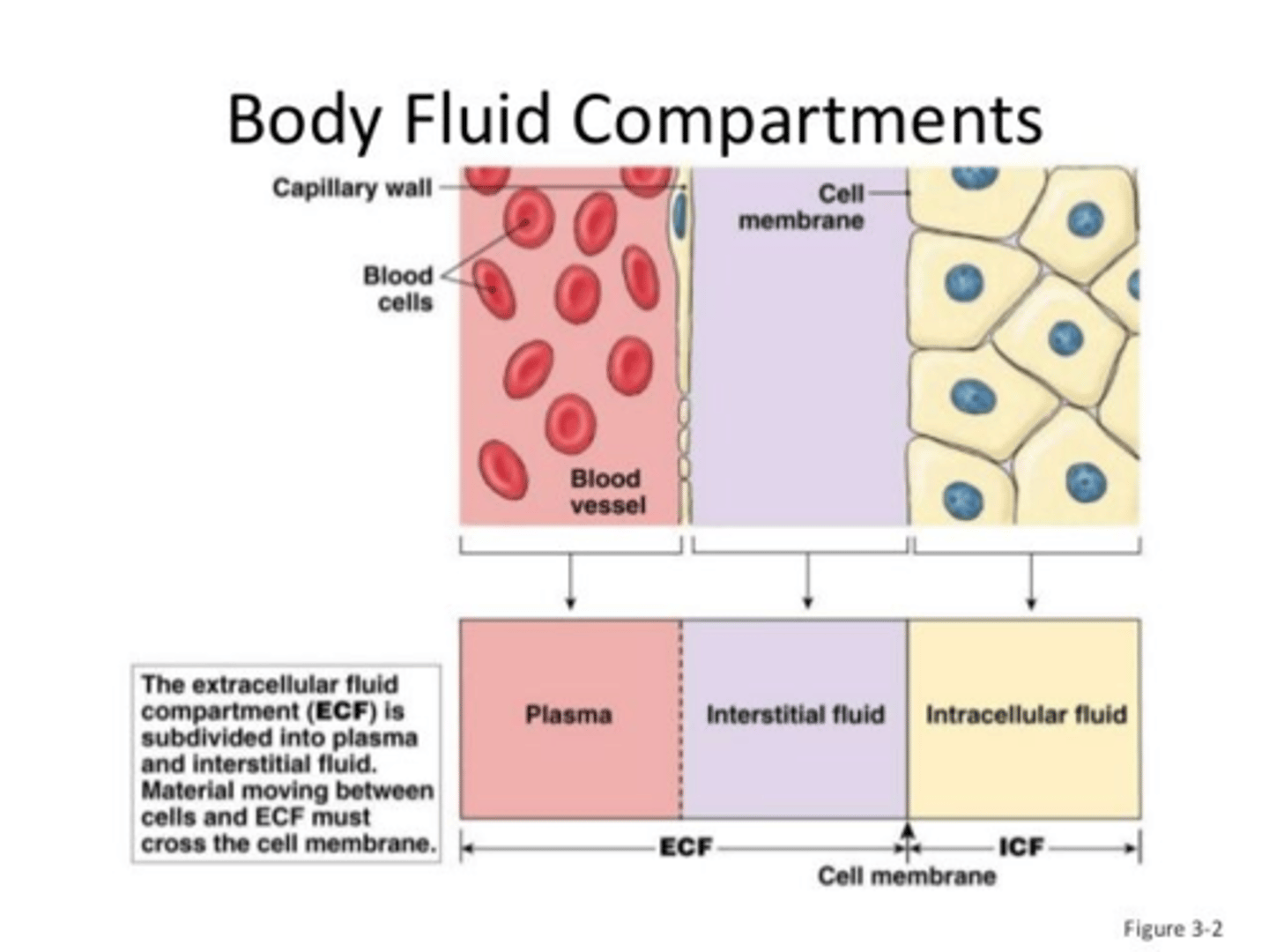
Intracellular fluid (ICF)
fluid inside cells

Extracellular fluid (ECF)
Fluid outside the cells; includes intravascular (i.e. blood plasma) and interstitial fluids.
Homeostasis
The process by which organisms maintain a relatively stable internal environment.
Ex) body temp, blood glucose, blood pressure, etc.
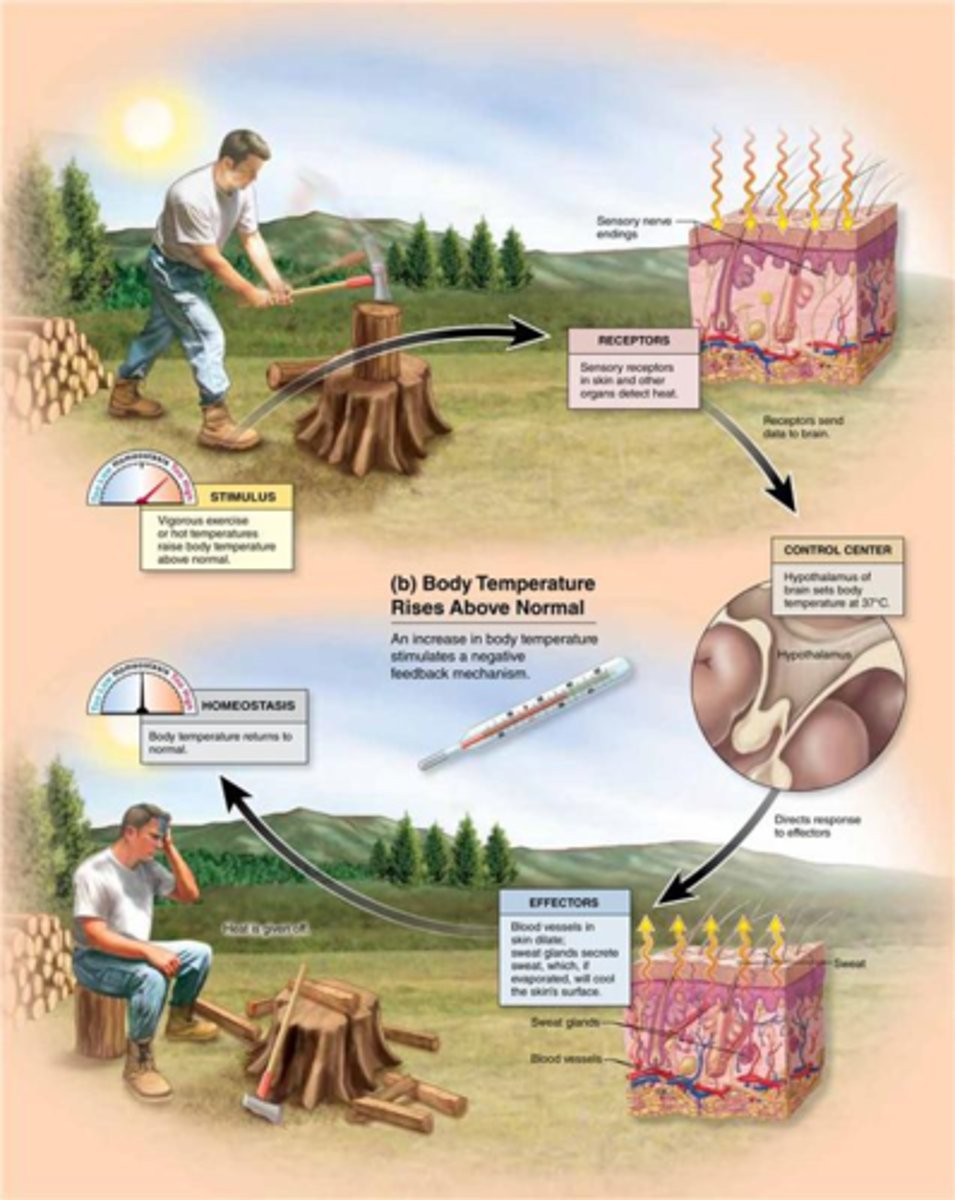
Negative Feedback
A type of regulation that responds to a change in conditions by initiating responses that will counteract the change. Maintains homeostasis.
Three components used in a negative feedback system
1. Sensory Receptor
2. Control Center
3. Effector
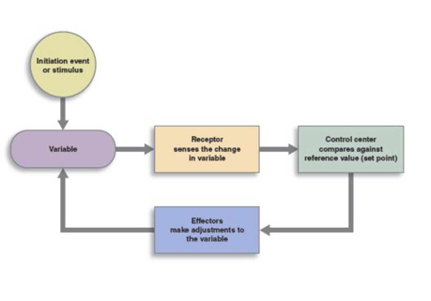
Stimulus (negative feedback loop)
A stimulus is a change in the external environment causing internal changes away from normal.

Sensory Receptor (negative feedback loop)
Monitors internal environment and responds to changes (stimuli).
An afferent pathway relays messages to the control center.

Control Center (negative feedback loop)
Analyzes the information received from afferent pathways and determines the response.
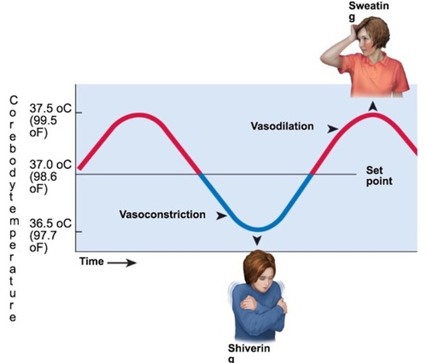
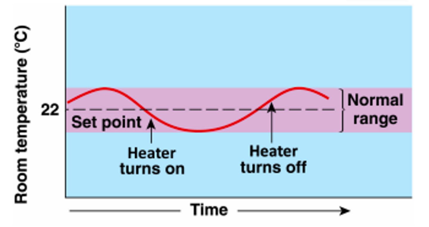
normal set point
The level at which a variable is normally maintained.
Effector (negative feedback loop)
Provides the means for the regulatory center's response (output).
An efferent pathway relays messages from the regulatory center to the effector.

The response (negative feedback loop)
The result of the negative feedback mechanism causing the initial stimulus to be depressed.

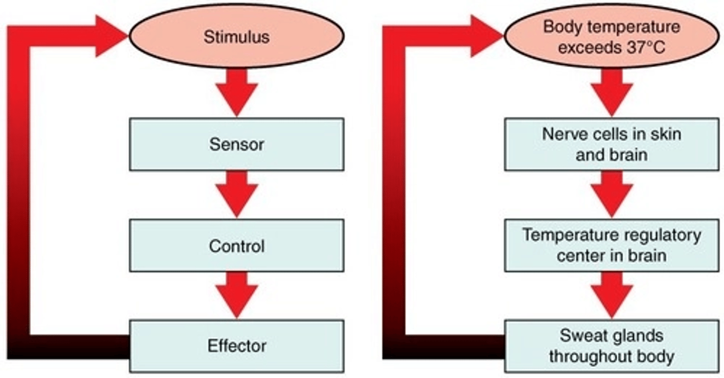
Negative feedback example - body temperature
1. Exercise causes body temperature to increase (too hot)
2. Increased body temperature is detected by thermoreceptors (Sensory Receptor)
3. Thermoreceptors send information to the hypothalamus, a thermostat in the brain (Control Center)
4. Nervous impulses generated by hypothalamus to skin (Effector)
5. Sweating and vasodilation of blood vessels occur
6. Body loses heat due to evaporative and radiative heat losses
7. Body temperature is reduced back down to normal

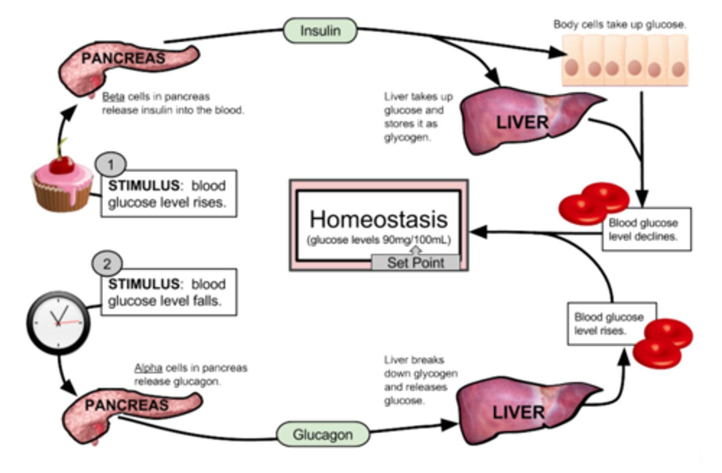
Negative feedback example - blood glucose
When blood sugar rises, receptors in the body sense a change.
The control center (pancreas) secretes insulin into the blood effectively lowering blood sugar levels.
Once blood sugar levels reach homeostasis, the pancreas stops releasing insulin.
Positive Feedback
A type of regulation that responds to a change in conditions by initiating responses that will amplify the change.
Takes organism AWAY from Homeostasis. NOT NORMAL IN THE BODY.
Ex) Birth, ovulation, blood clotting, sodium ion movement during an action potential.
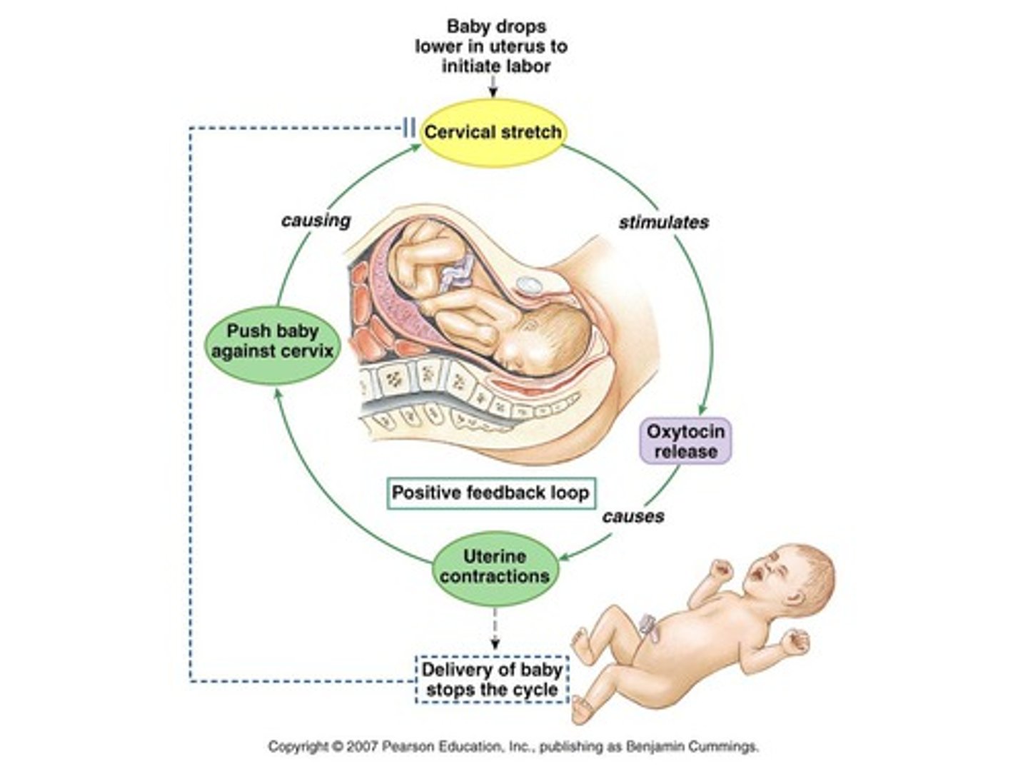
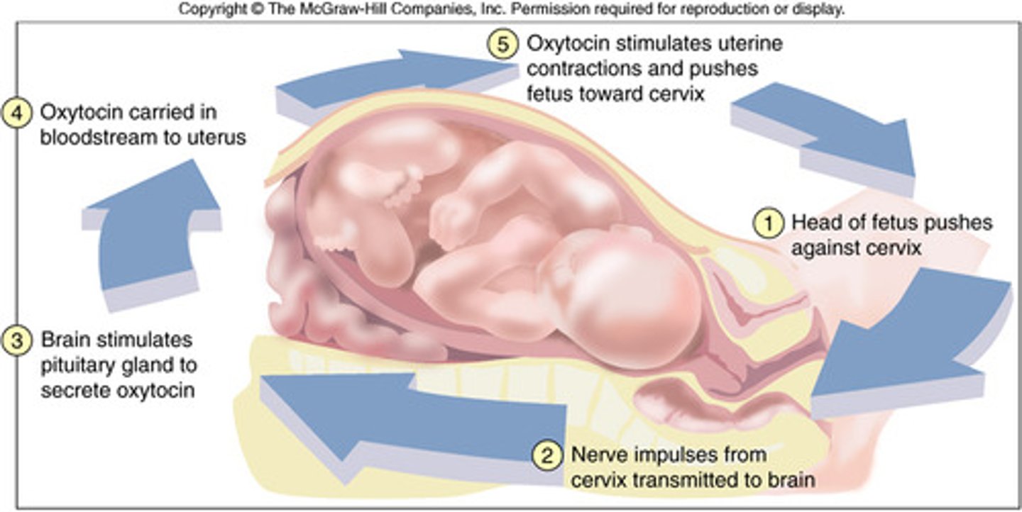
Positive feedback Example - labor
- Pressure: Irritation, stretch of the uterine wall and cervix cause the release of oxytocin
- Oxytocin: Causes uterine contraction which pushes baby into cervix creating more irritation and distension
- Results in: The release of more oxytocin which produces more contractions etc... until the baby is delivered
Active Transport
Energy-requiring process that moves substances from an area of low concentration to high concentration across a cell membrane (Against the concentration gradient).
- Requires Energy in the form of ATP
- Substances move from low concentration to high concentration
- Analogy: Pushing a ball up a hill
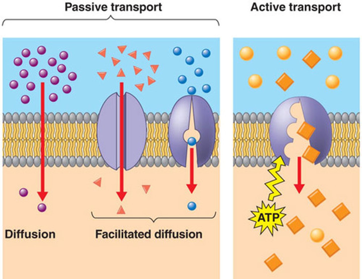
Passive Transport
The movement of substances across a cell membrane without the use of energy by the cell
- No Energy is required.
- Substances move from high concentration to low concentration
- Analogy: Allowing a ball to roll down a hill
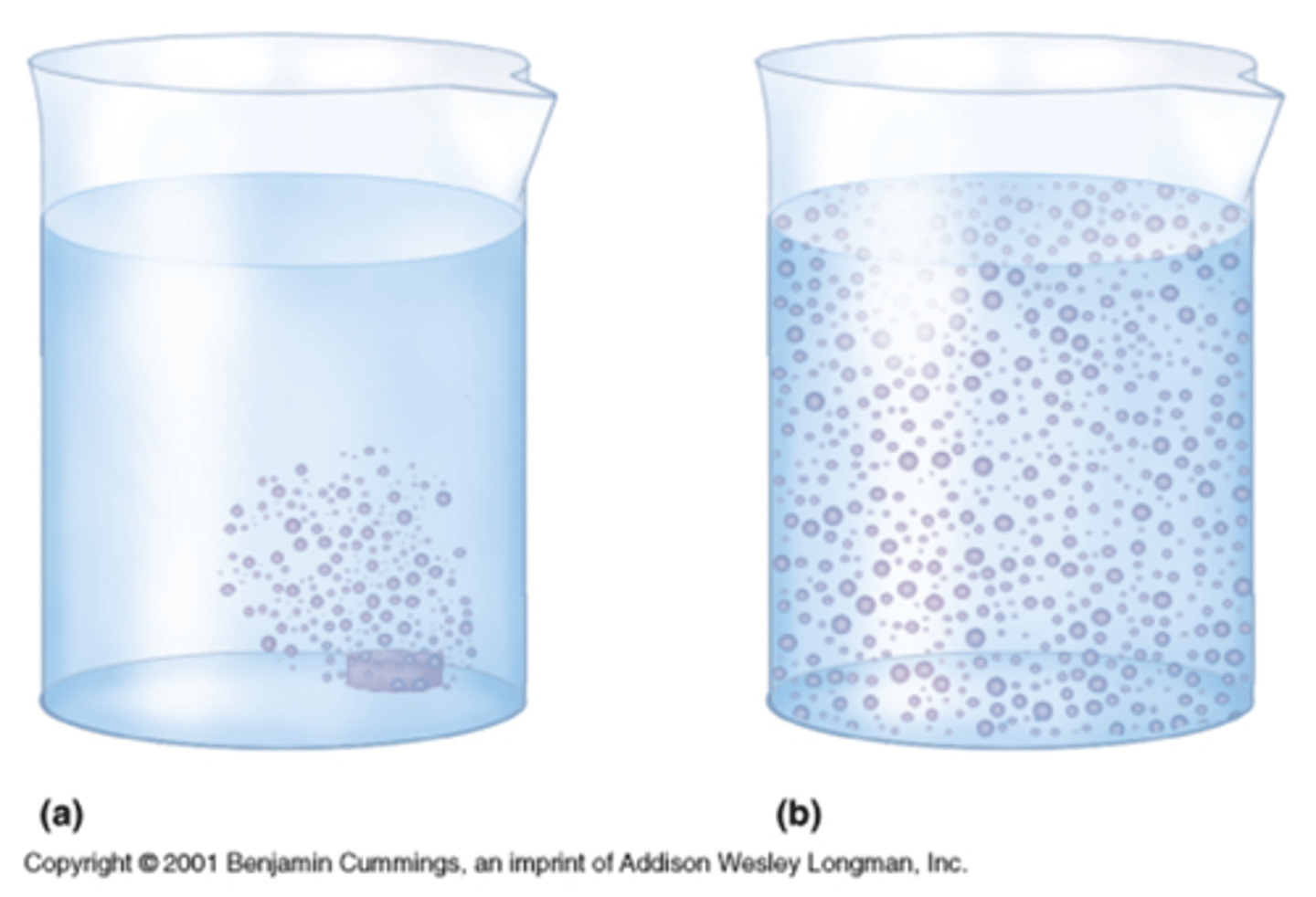
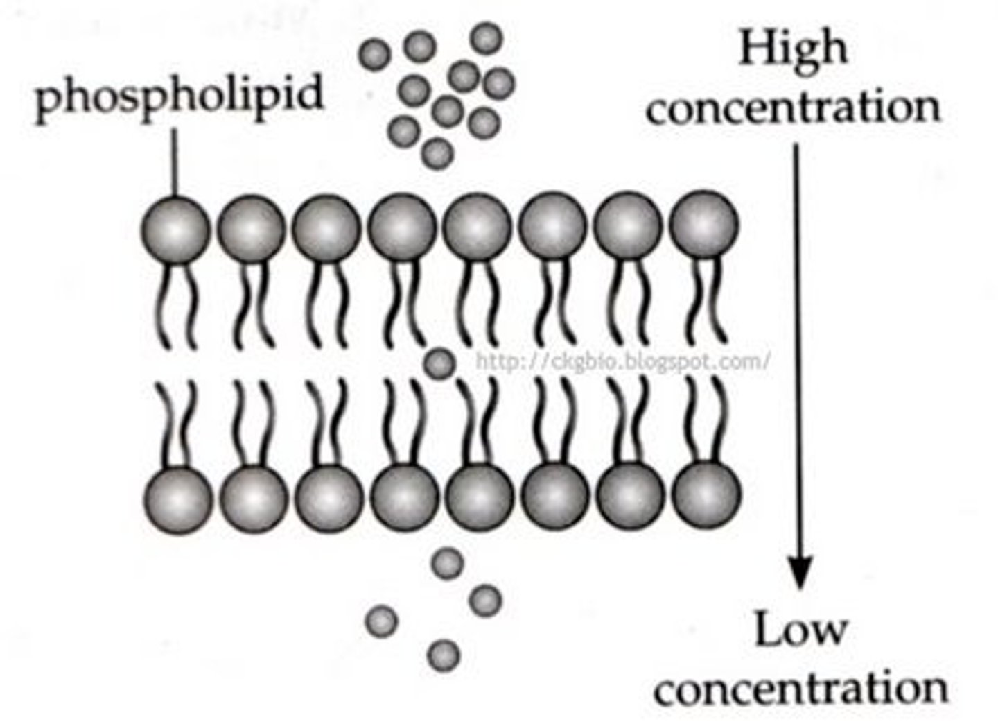
Simple Diffusion
Small substances pass through the cell membrane, from an area of high concentration to an area of low concentration.
Type of Transport - Passive
Non-polar (uncharged) substances move across the phospholipid bilayer
Polar (charged) substances move through a channel or pore in the membrane
Factors Influencing Rate of Diffusion
Molecular Mass - Decreased molecular size/mass increases rate of transport (lighter molecules diffuse faster than heavier ones)
Polarity of the molecule – non-polar (lipid soluble) molecules pass more easily through the cell membrane.
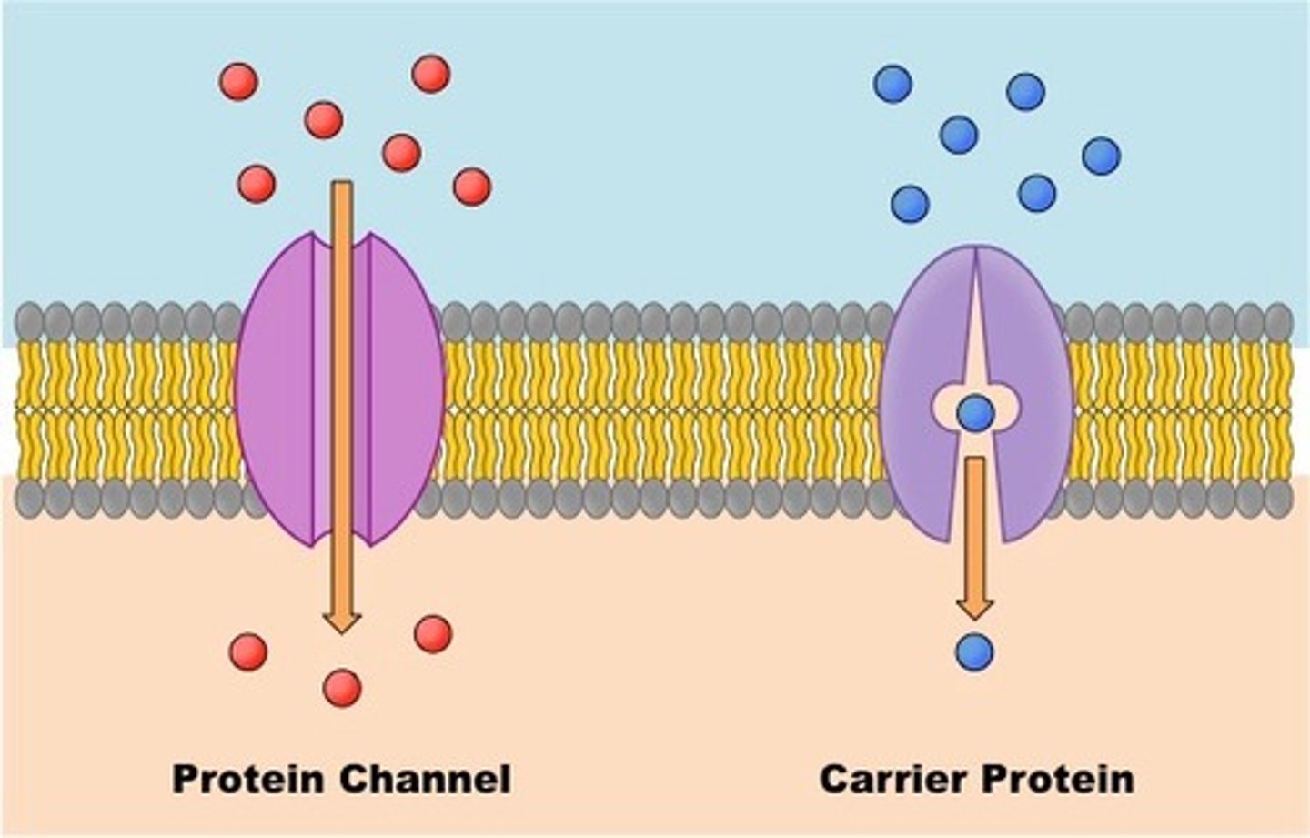
Facilitated Diffusion
The transport of substances through a cell membrane along a concentration gradient with the aid of carrier proteins
Type of Transport - Passive
Is used to transport larger substances into or out of the cell, from an area of high concentration to an area of low concentration
The rate of facilitated diffusion increases with an increased number of carrier proteins (more carriers = faster rate)
Example: The transport of glucose and amino acid from the bloodstream into the cell.
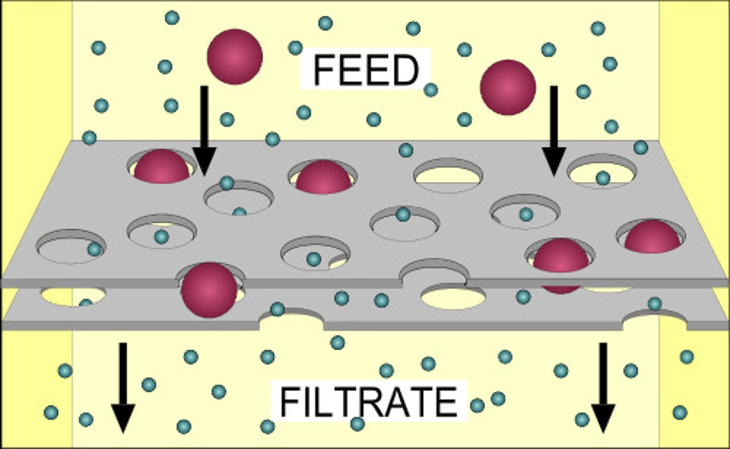
Filtration
Water and solutes are forced through a membrane due to a pressure gradient.
Requires a filter and a pressure gradient
Type of Transport: Passive
Ex. Occurs in the kidney as the first process in producing urine.
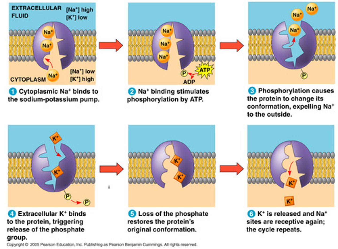
Sodium-Potassium Pump
A carrier protein that uses ATP to actively transport sodium ions (Na+) out of a cell and potassium ions (K+) into the cell.
Type of Transport: Active
Bulk transport
The movement of larger quantities of material across the cell membrane.
Type of Transport: Active
Two types based on direction of transport:
1. Exocytosis = out of cell
2. Endocytosis = into cell
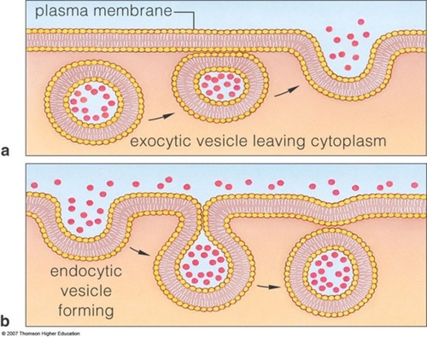
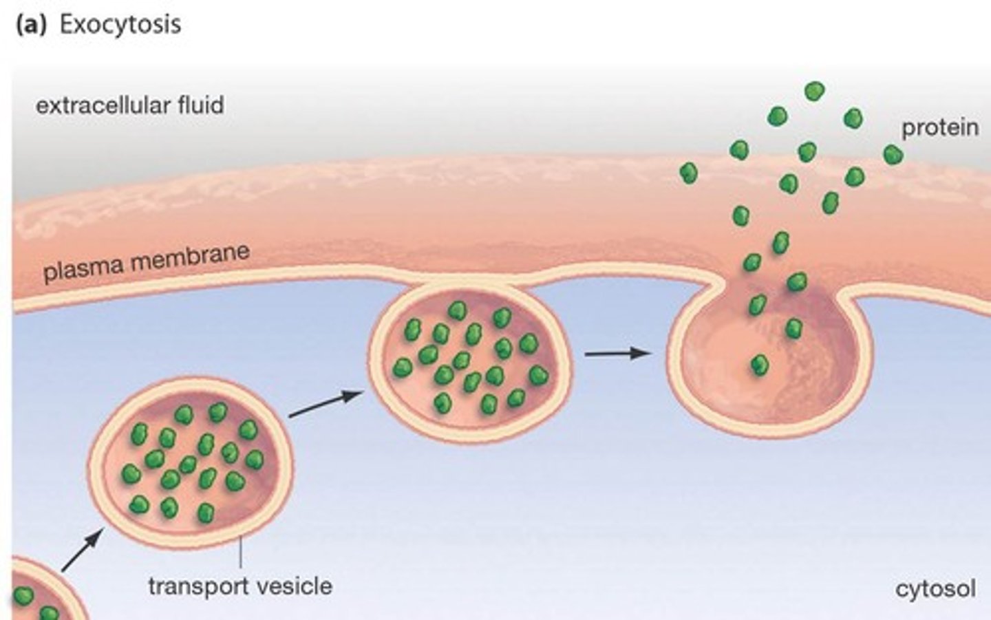
Exocytosis
Substances leave (exit) the cell.
Type of Transport: Active
Example: excrete waste and other large molecules from the cytoplasm to the cell exterior
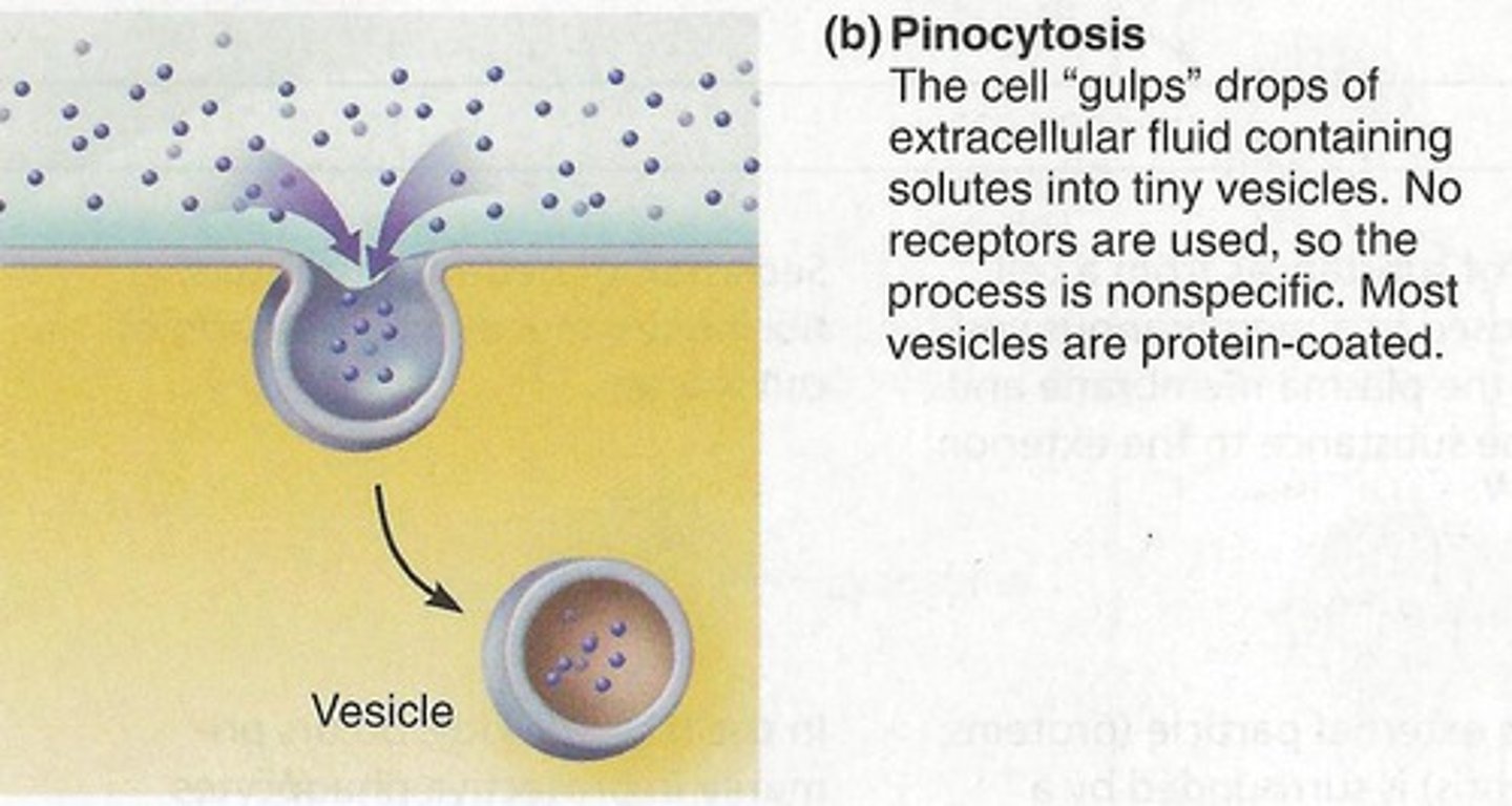
Endocytosis
Defined: process by which a cell takes material into the cell by infolding of the cell membrane.
Type of Transport: Active
Example: leucocytes, neutrophils, and monocytes can engulf foreign substances like bacteria.
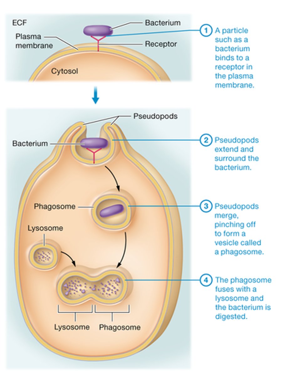
Phagocytosis
Large solutes like bacteria are taken into the cell.
Known as "cell eating"
Type of Transport: Active
Pinocytosis
Fluid and dissolved solutes are taken into the cell. Known as "cell drinking"
Type of Transport: Active
Osmosis
Passive transport of water through a selectively permeable membrane.
The water moves from an area of lower solute concentration to an area of higher solute concentration.
Water concentration is important.
Type of Transport: Passive

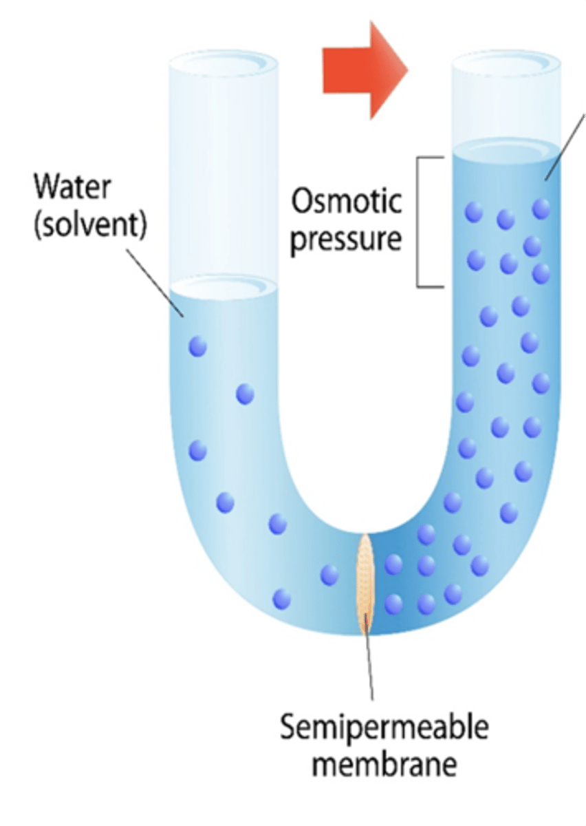
Osmolarity
A measure of the total solute concentration. The greater the solute concentration, the greater the osmolarity.
In this picture, the right compartment has a greater osmolarity than the left compartment.

Osmolarity and Osmosis
In osmosis, water moves toward the area of greater osmolarity (you could say “Water follows salt”)
In this picture, the right compartment has a greater osmolarity than the left compartment and so water moves from left to right.
Tonicity
The ability of a solution to influence the shape or “tone” of a cell.
Tonicity is also a measure of the concentration of non-penetrating solutes in the solution. When the solutes cannot move, water will move to achieve equilibrium.
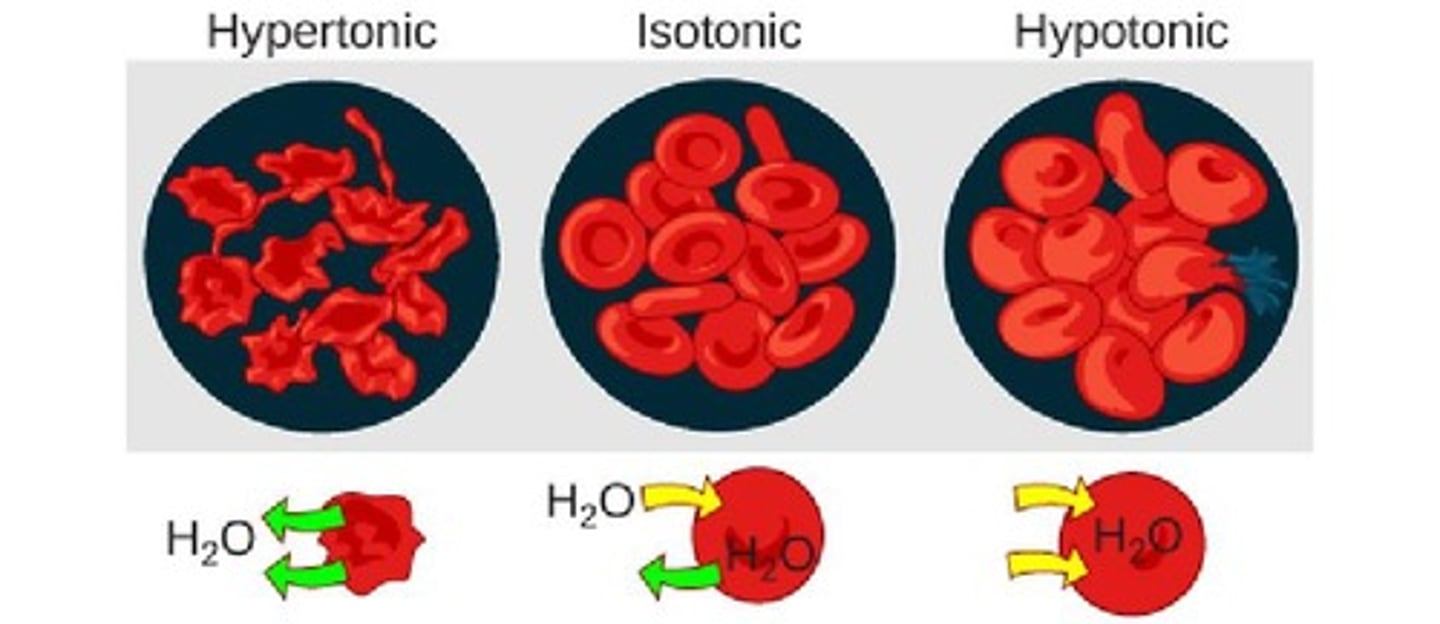
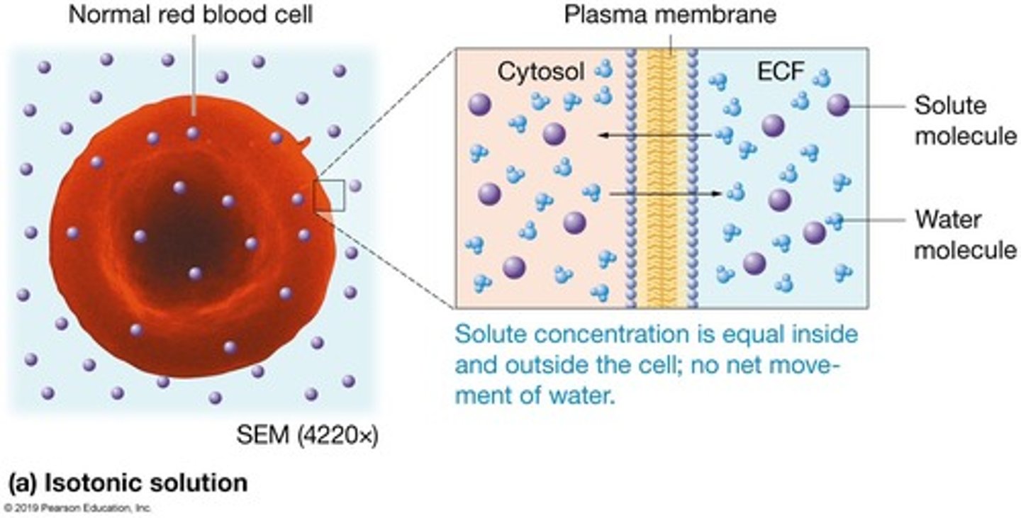
Isotonic
Same concentration of non-penetrating solutes as the cell.
No effect on cell.
These are used as intravenous solutions.
(the prefix iso- means equal)
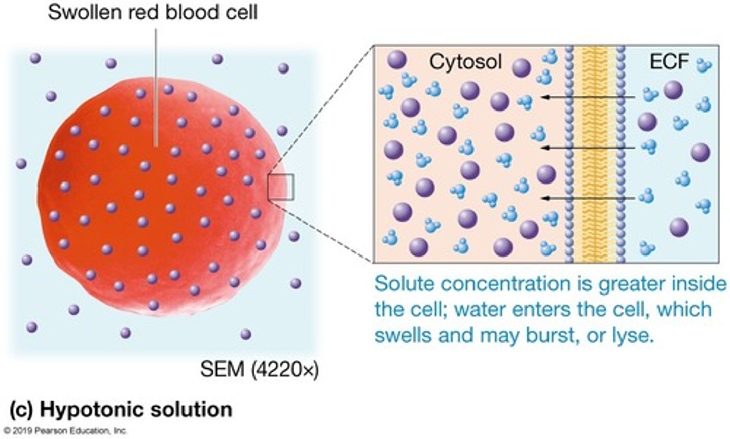
Hypotonic
Lower concentration of non-penetrating solutes than the cell.
Lyses the cell (cell takes up water and bursts).
NEVER given via IV
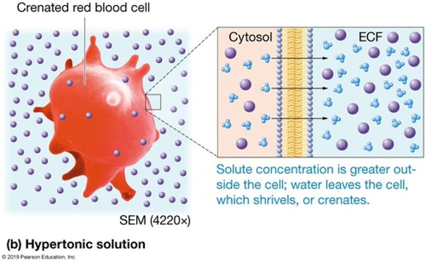
Hypertonic
Greater concentration of non-penetrating solutes than the cell.
Crenates the cell (cell loses water and shrivels).
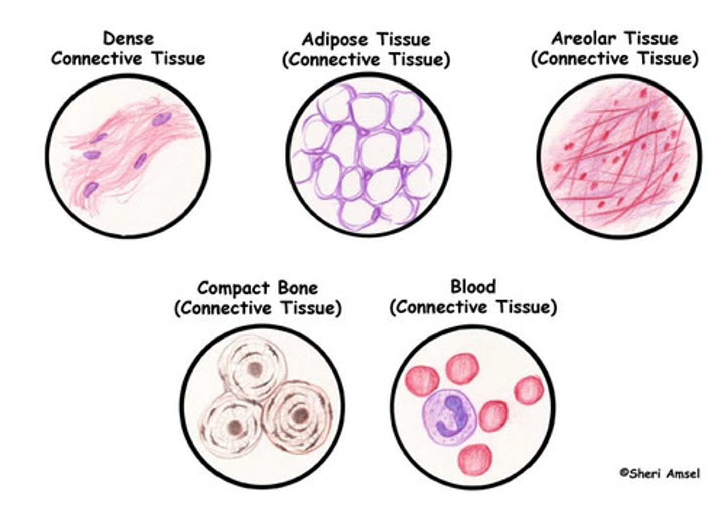
Tissue
A group of similar cells.
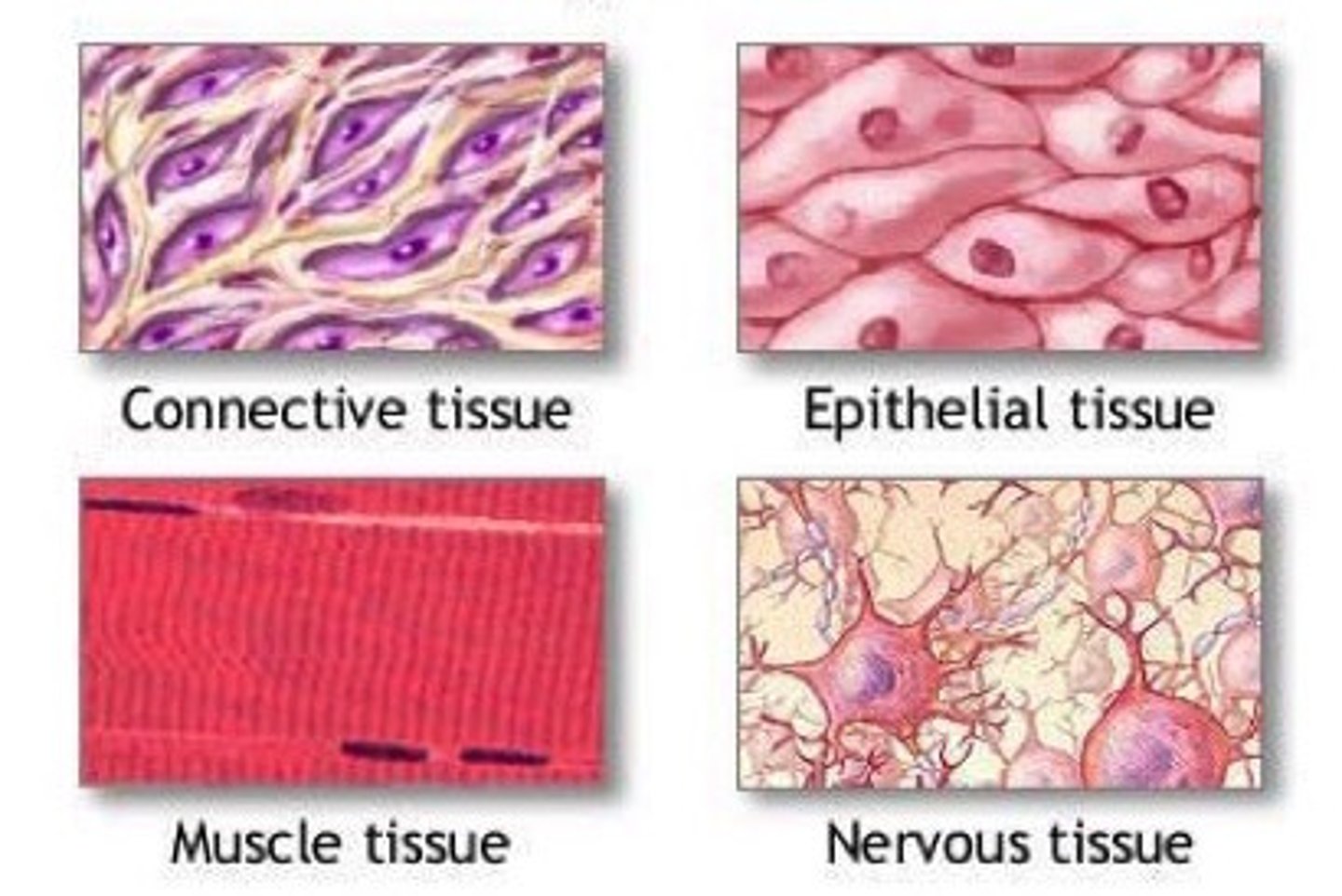
The Four Tissue Types
1. Epithelium
2. Muscle
3. Connective
4. Nervous
Locations of epithelial tissue
Covers the body surface and lines the inner body cavities.
Functions of epithelial tissue
protection, absorption, filtration, secretion
Characteristics of epithelial tissue
avascular, highly regenerative, and very cellular.
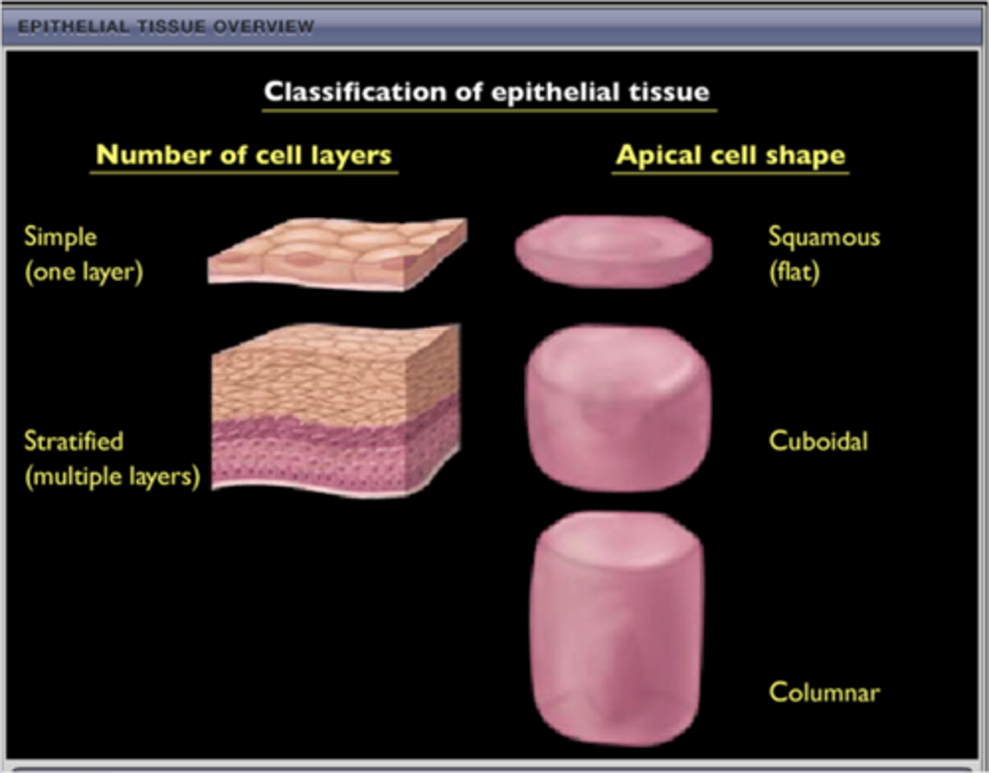
Classification of epithelial tissues
Each type of epithelial tissue has 2 names -
The first name is for the number of cell layers it has (simple or stratified)
The second name is for the shape of the apical cells making up that epithelial tissue (squamous, cuboidal, columnar, pseudostratified, transitional).
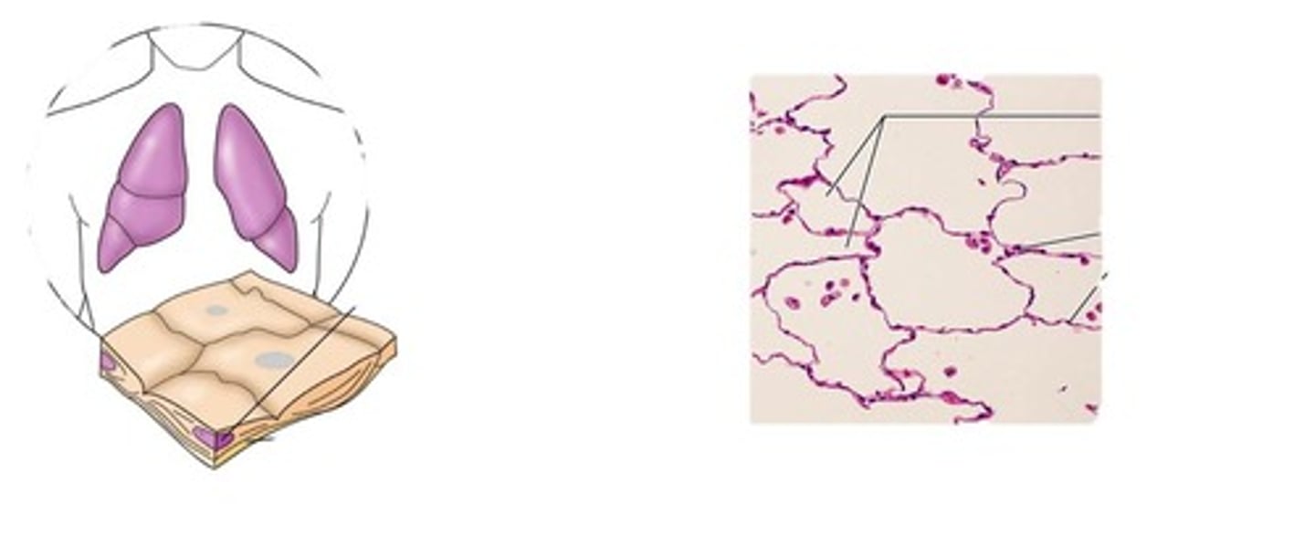
simple squamous epithelium
A single layer of flattened cells
Function: Allows passage of materials by diffusion and filtration in sites where protection is not important.
Location: Kidney glomeruli, air sacs of lungs, the lining of heart, blood vessels, and lymphatic vessels; lining of the ventral body cavity.
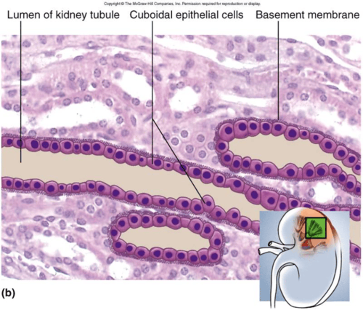
simple cuboidal epithelium
A single layer of cube-shaped cells
Function: secretion and absorption
Location: Kidney tubules; ducts and secretory portions of small glands, ovary surface.
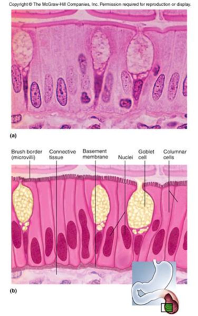
simple columnar epithelium
A single layer of tall cells that fit closely together
Function: Absorption; secretion of mucus, enzymes, and other substances; ciliated type propels mucus (or reproductive cells).
Location: nonciliated type lines most of the digestive tract, gallbladder, and excretory ducts of some glands; ciliated variety lines small bronchi, uterine tubes, and some regions of the uterus.
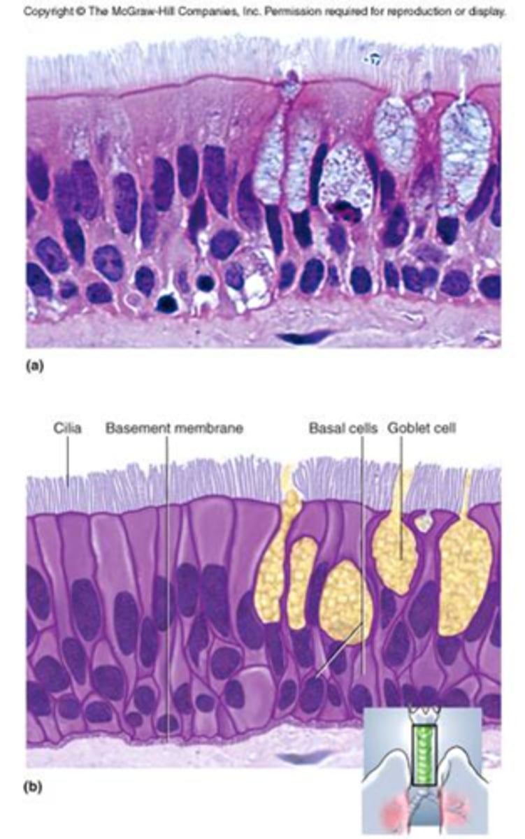
pseudostratified columnar epithelium
Tissue that consists of a single layer of irregularly shaped and sized cells that give the appearance of multiple layers; found in ducts of certain glands and the upper respiratory tract
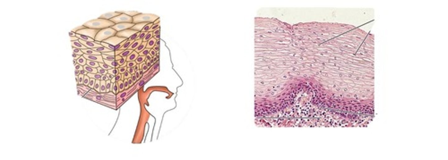
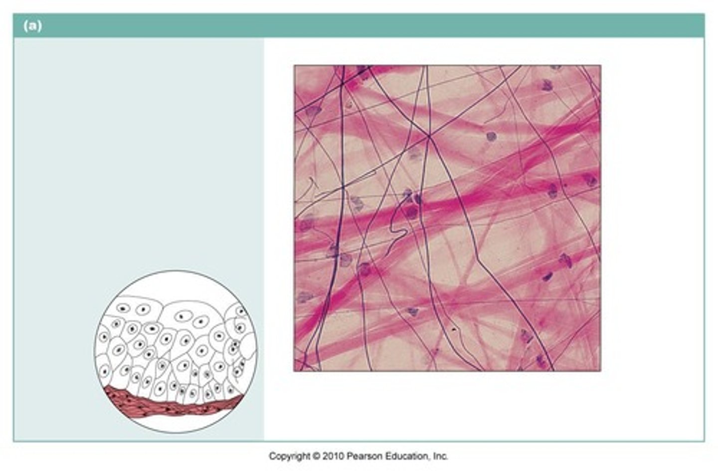
stratified squamous epithelium
Multiple layers of flat cells.
Function: protects underlying tissues in high friction areas.
Location: nonkeratinized type forms the moist lining of the esophagus, mouth, and vagina; keratinized type forms the epidermis of the skin, a dry membrane.
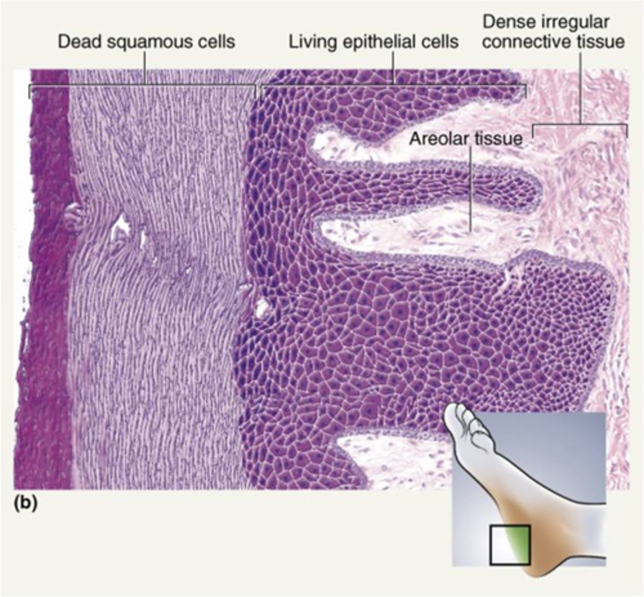
keratinized stratified squamous epithelium
This tissue, consisting of many layers of flat cells, is found in the epidermis of the skin.
The surface cells are dead and are full of keratin (a protein which prevents water loss).
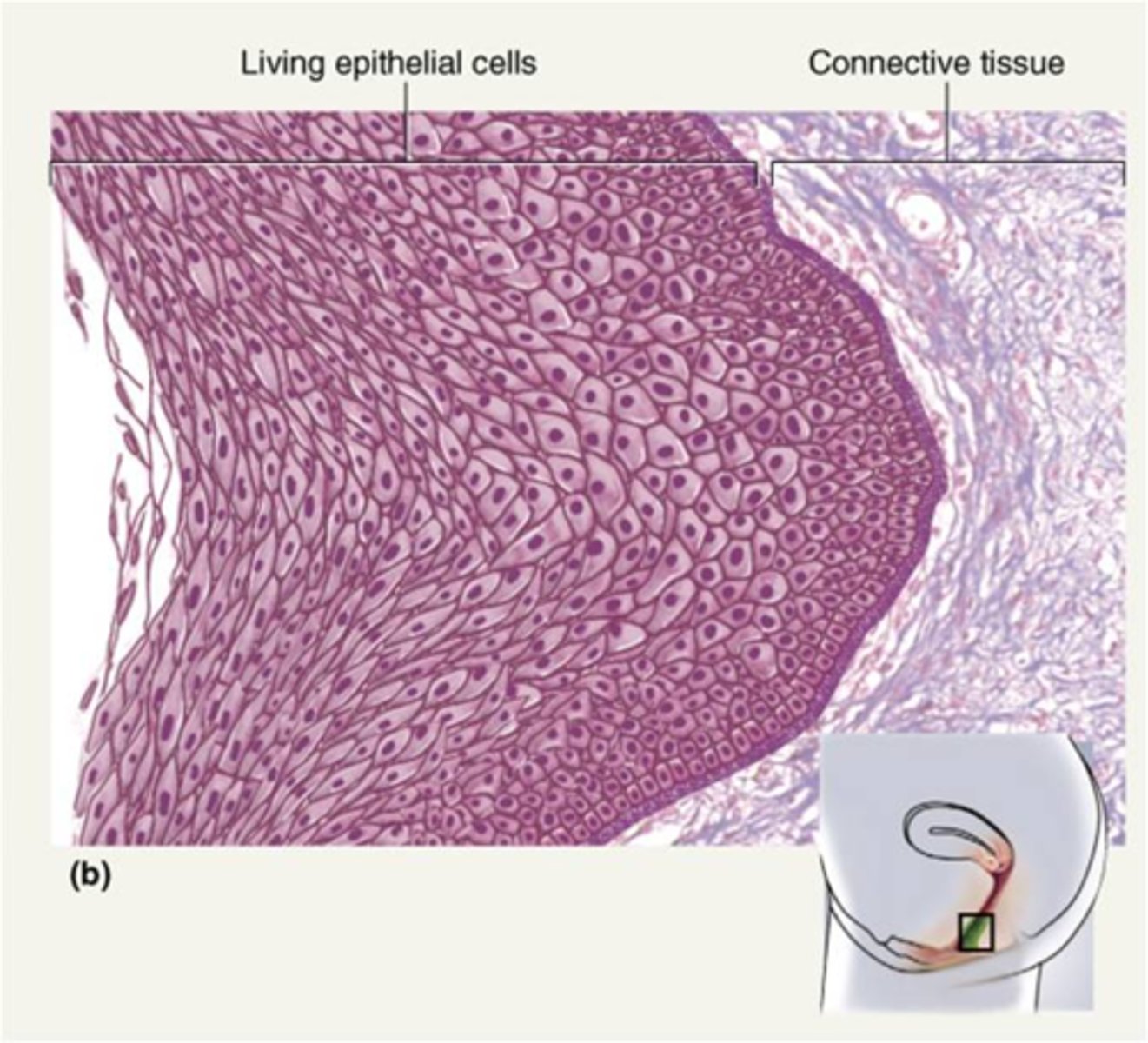
nonkeratinized stratified squamous epithelium
a stratified squamous epithelium that does not contain a superficial layer of dead cells.
All layers including the most superficial layer of cells contain nuclei.
This tissue is good at resisting abrasion but lacks the ability to resist desiccation, so it is found in places where the surface is covered by a layer of mucus.
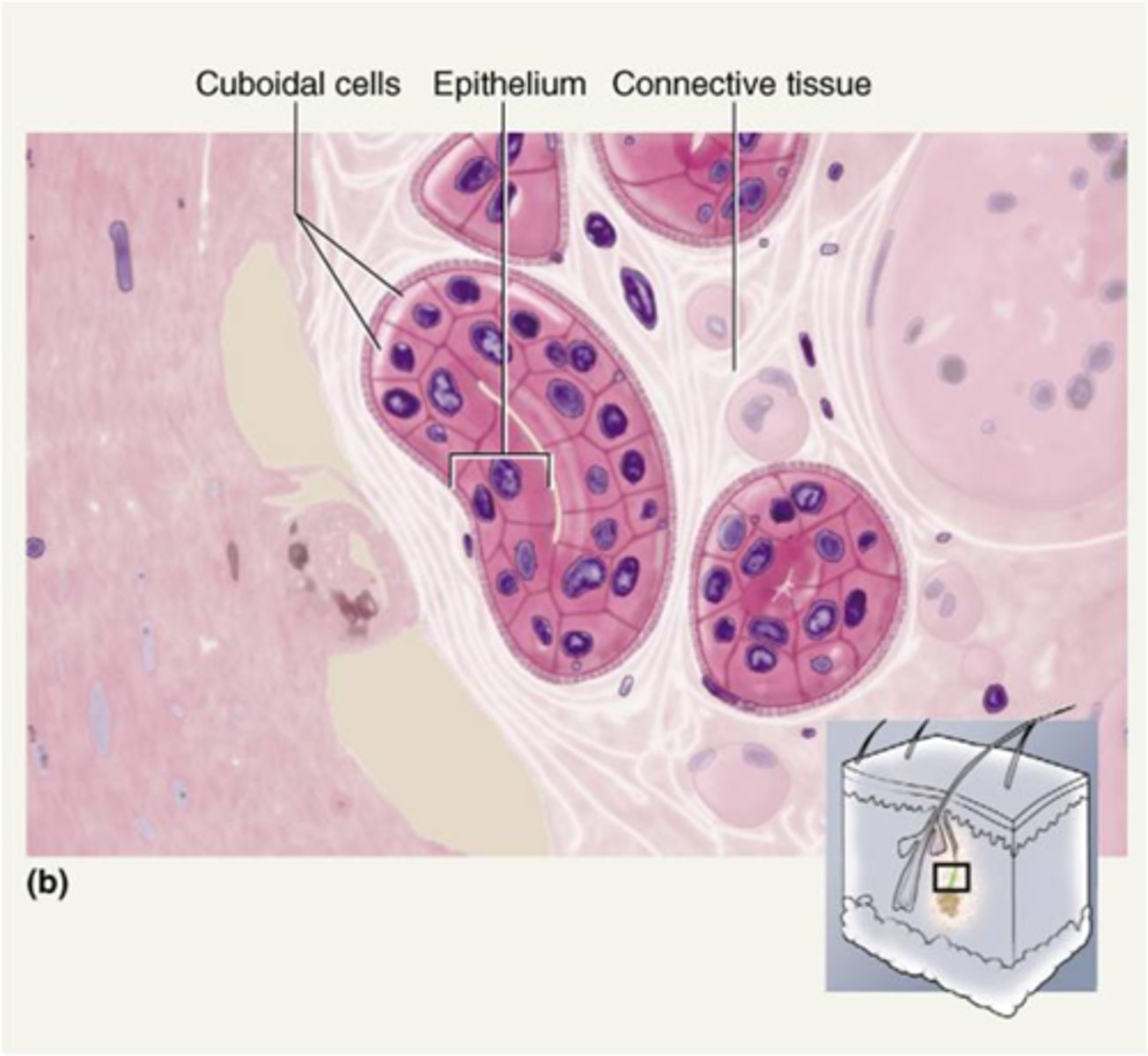
stratified cuboidal epithelium
Multiple layers of cube-shaped cells.
Function: protection
Location: Largest ducts of sweat glands, mammary glands, and salivary glands.
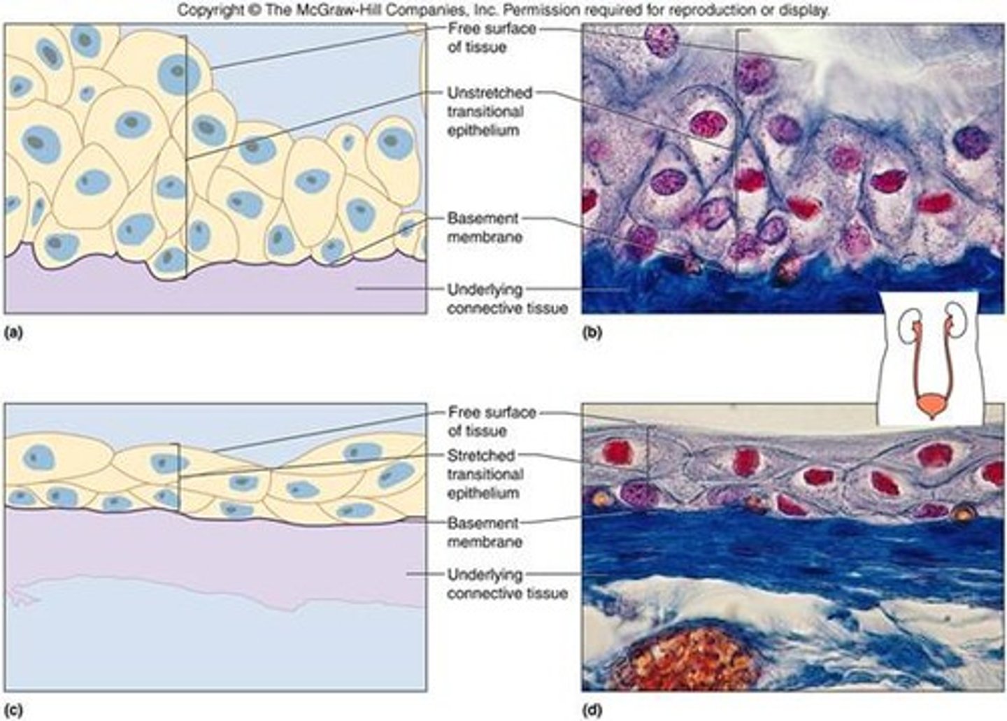
transitional epithelium
Multiple layers of epithelial cells which can contract and expand in order to adapt to the degree of distension needed.
Function: stretches readily and permits distension of urinary organ by contained urine
Location: lines the ureters, urinary bladder, and part of the urethra.
Characteristics of Connective Tissues
Few cells - consists mainly of a nonliving material between the cells, called the extracellular matrix.
The extracellular matrix consists of fibers and a fluid or gel-like ground substance.
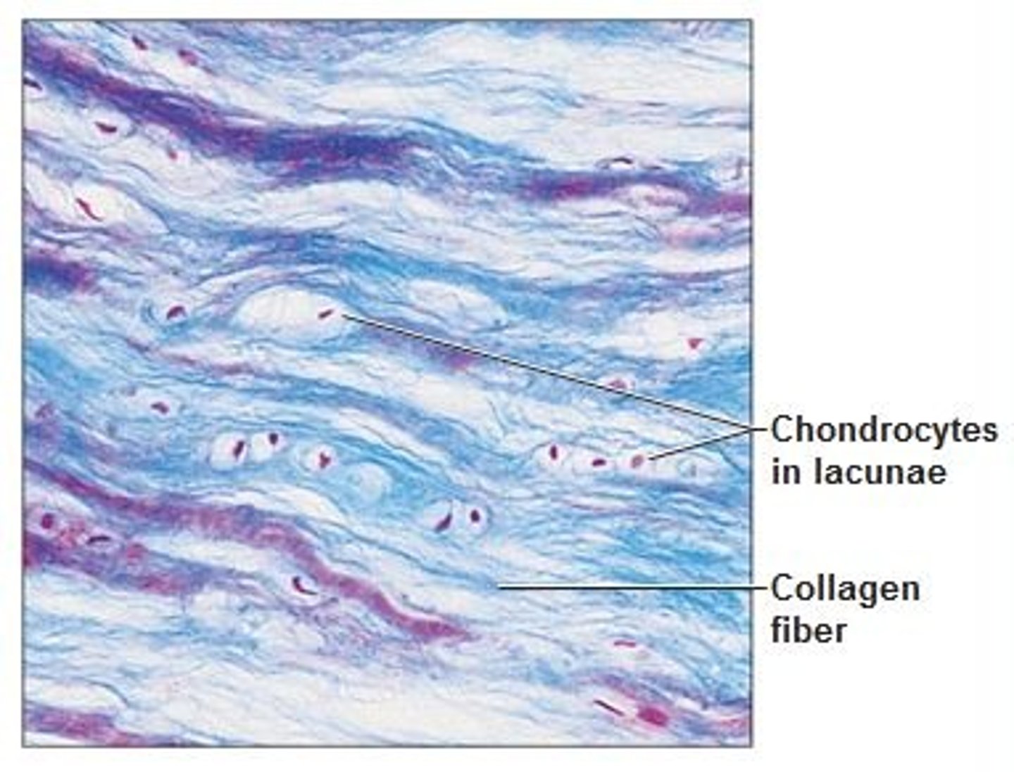
The fibers include collagen fibers (for strength), elastic fibers (for elasticity), and reticular fibers (form networks that support the cells).
The ground substance acts as a medium to allow oxygen and nutrients to diffuse between the cells.
Common origin – they all originate from mesenchyme (embryonic connective tissue) in the embryo.
Functions of Connective Tissues
-Establishing a structural framework for the body
-Transporting fluids and dissolved materials
-Protecting delicate organs
-Supporting, surrounding, and interconnecting other types of tissue
-Storing energy reserves, especially triglycerides
-Defending the body from invading microorganisms
Three types of loose connective tissues
areolar, adipose, reticular
areolar connective tissue
Loose arrangement of fibers and cells in a large amount of ground substance.
Function: Wraps and cushions organs. Composes basement membranes.
Location: Widely distributed under epithelia of body. It is found underlying epithelial tissue, between muscles, and under the skin (in the subcutaneous tissue).
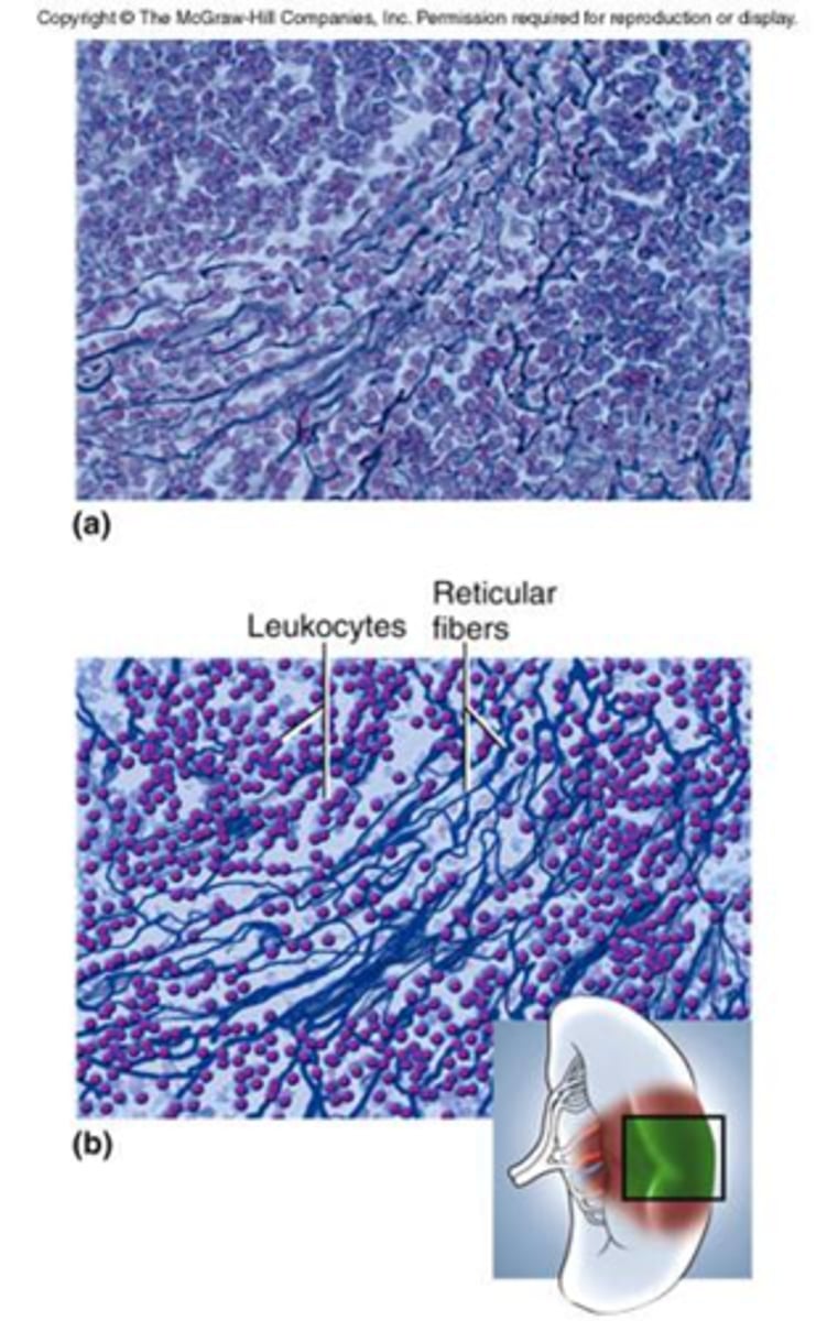
reticular connective tissue
A loose network of reticular fibers and cells.
It is found in the kidney, spleen, lymph nodes, and bone marrow.
It forms the supportive framework (stroma) of those organs.
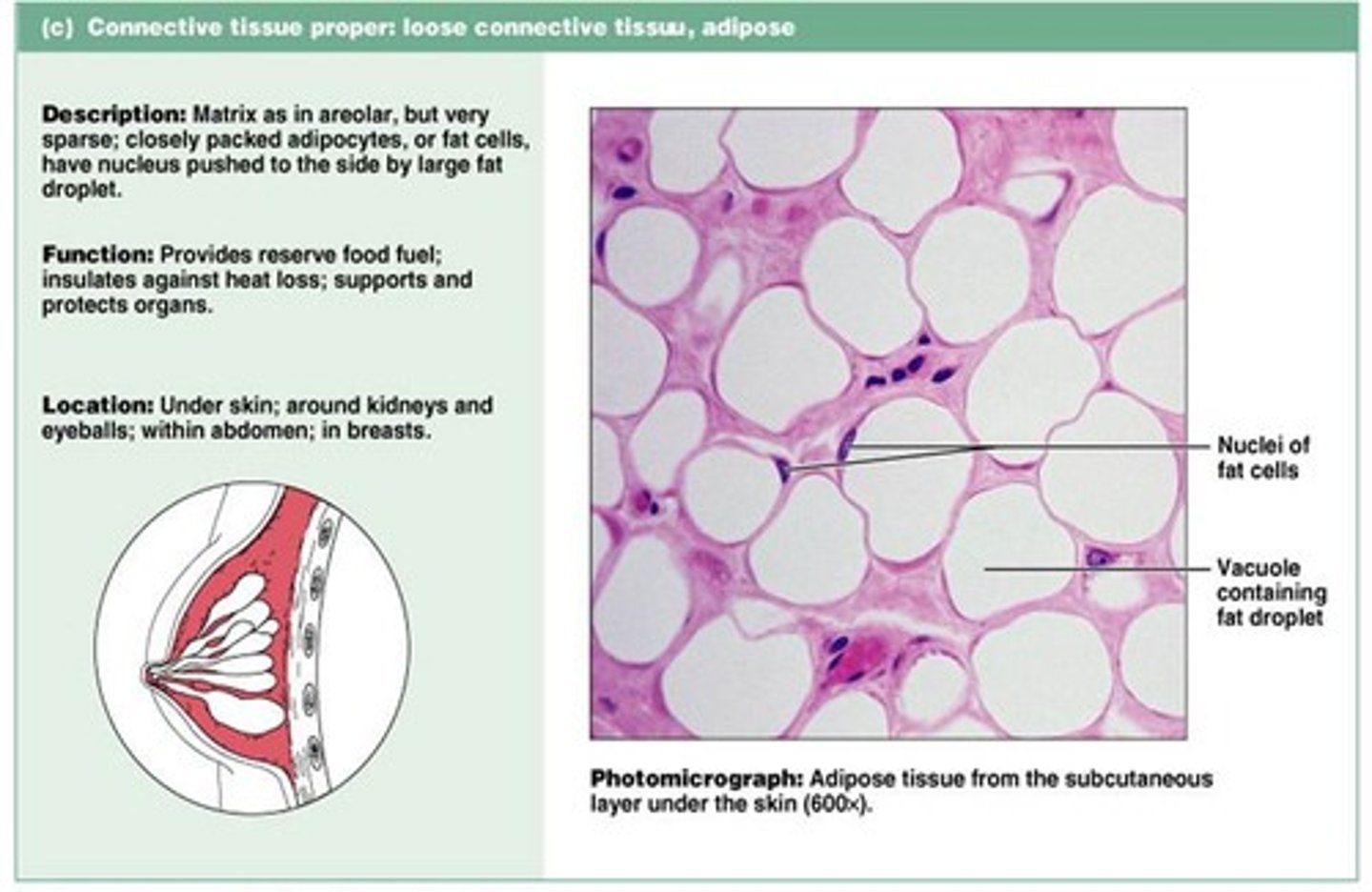
adipose connective tissue
Contains cells called adipocytes, which are full of fat.
This is a very cellular connective tissue.
It is found in the subcutaneous tissue (the fat under the skin), in the breast, around the kidneys, behind the eyes.
It functions in energy storage, insulation, absorbing shock and to cushion.
Three types of dense connective tissues
1. dense regular connective tissue
2. dense irregular connective tissue
3. elastic connective tissue
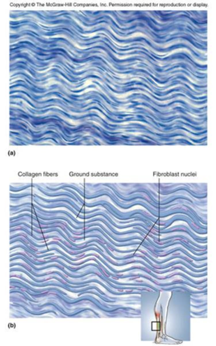
dense regular connective tissue
Made up primarily of collagen fibers that are densely packed and run parallel to each other.
Attaches muscles to bones or to muscles; attaches bones to bones; withstands great tensile stress when pulling force is applied in one direction
This is found where you need strength in 1 plane – ex. Tendons, ligaments
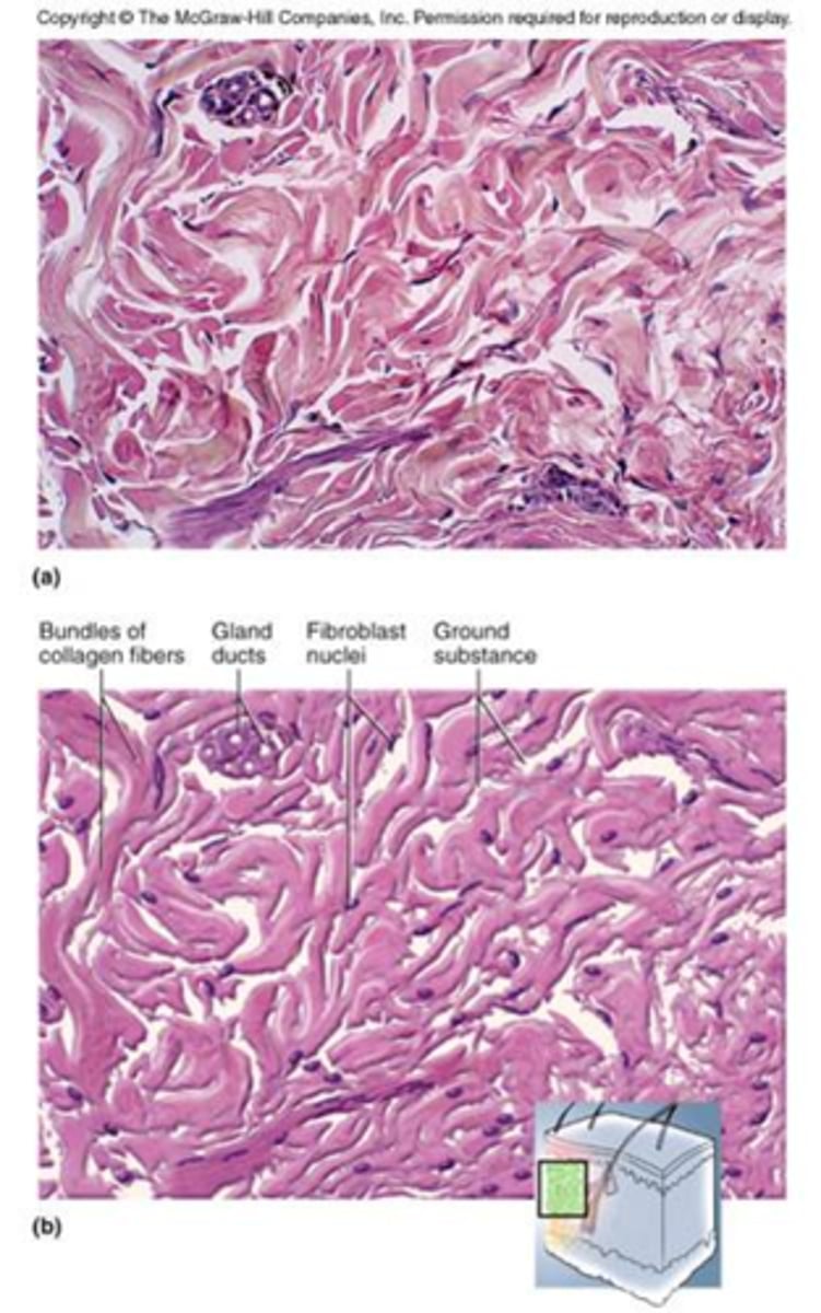
dense irregular connective tissue
Made up of collagen fibers that are densely packed but randomly arranged.
This can withstand stress in many directions.
Found in the dermis of the skin and in capsules around organs.
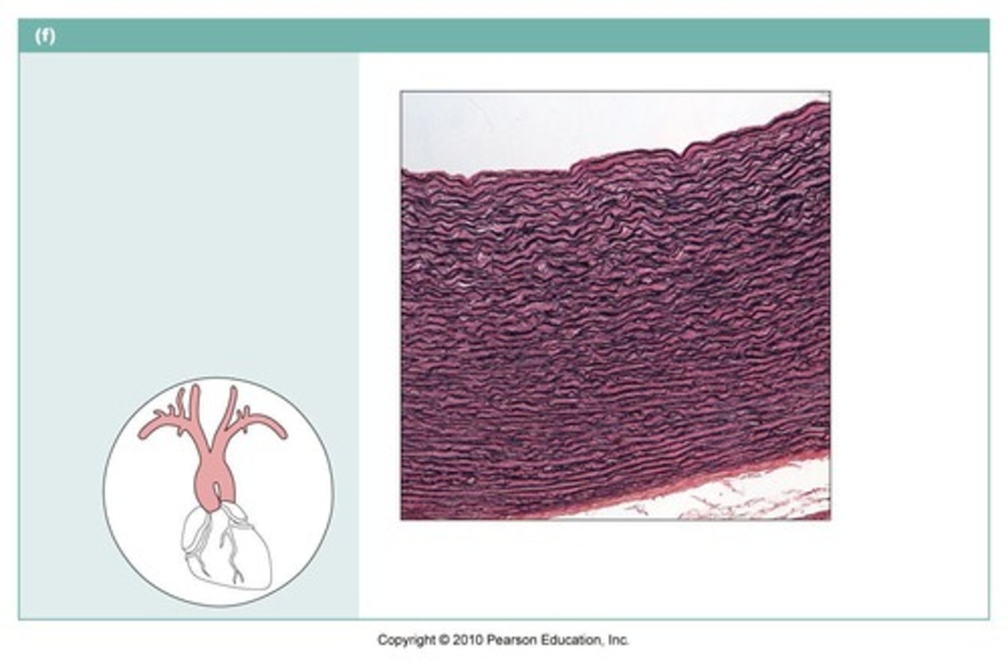
elastic connective tissue
Made up primarily of elastic fibers plus some collagen fibers.
It allows for strength and flexibility.
Found in the aorta and ligaments between the vertebrae
Three types of cartilage tissues
hyaline, elastic, fibrocartilage
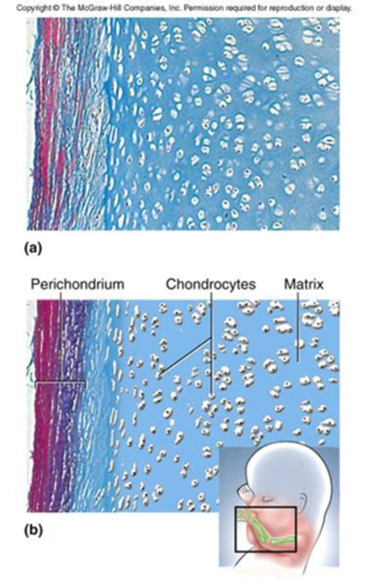
Characteristics of cartilage tissues
Cartilage has a firm matrix and it is tough and flexible.
It does not have nerves or blood vessels.
The cells are chondrocytes and they sit in cavities called lacunae.
It is surrounded by a membrane called the perichondrium (blood vessels are in the perichondrium).
hyaline cartilage
Made up mainly of collagen fibers.
Grossly, it appears bluish white.
Found at the end of long bones, tip of the nose, larynx, trachea, bronchi and in the embryonic skeleton.
elastic cartilage
Has collagen and elastic fibers.
It is found where you need strength and stretch.
Found in external ear and epiglottis.

Fibrocartilage
The strongest cartilage - it resists compression and absorbs stress.
Found in the pubic symphysis, the meniscus of the knee, and intervertebral discs.
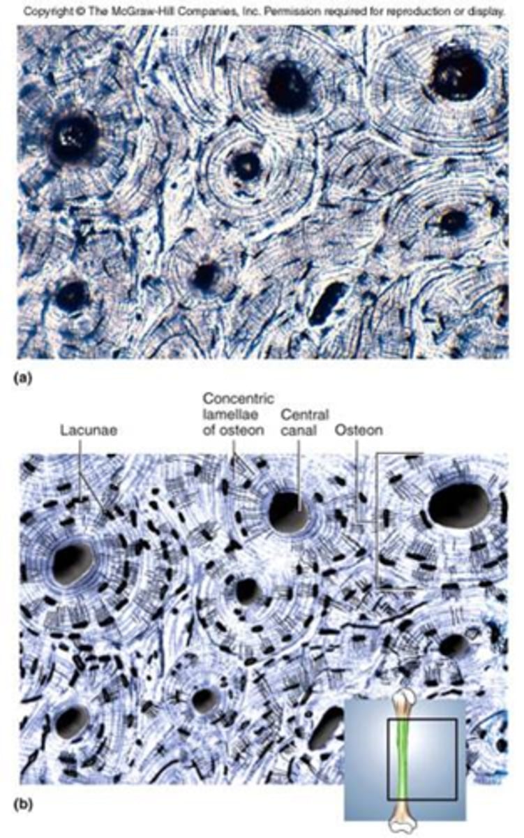
Bone (osseous tissue)
A hard matrix that consists of collagen fibers and Ca++.
It functions to support and protect.
The cells are osteoblasts (they make the matrix) and osteocytes (which sit in lacunae and maintain the matrix in a healthy state).
It is very well vascularized.
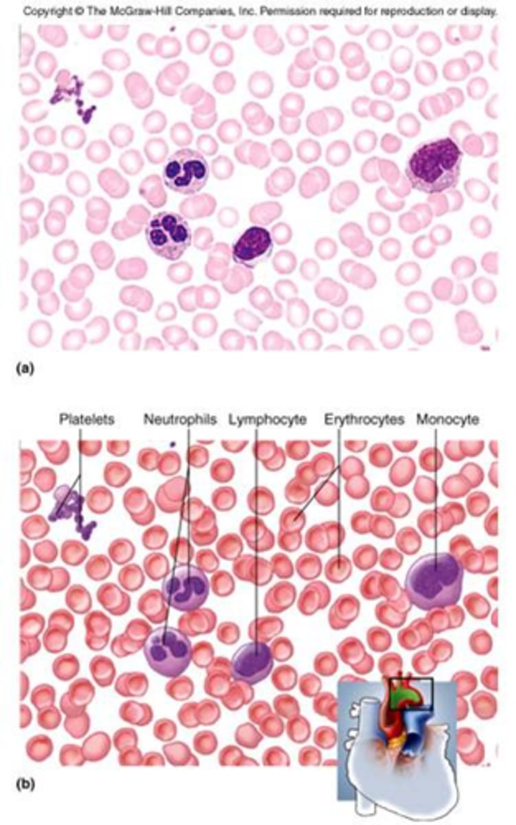
blood (vascular tissue)
The matrix is the plasma of the blood and it contains fibers which are the clotting proteins (you don't see the fibers in the blood that are not clotted because they are dissolved in the plasma).
It also contains cells including
- erythrocytes (red blood cells)
- leukocytes (white blood cells)
- platelets.
The cells are called the formed elements.
It functions in transport.
Three types of muscle tissues
skeletal muscle, cardiac muscle, smooth muscle
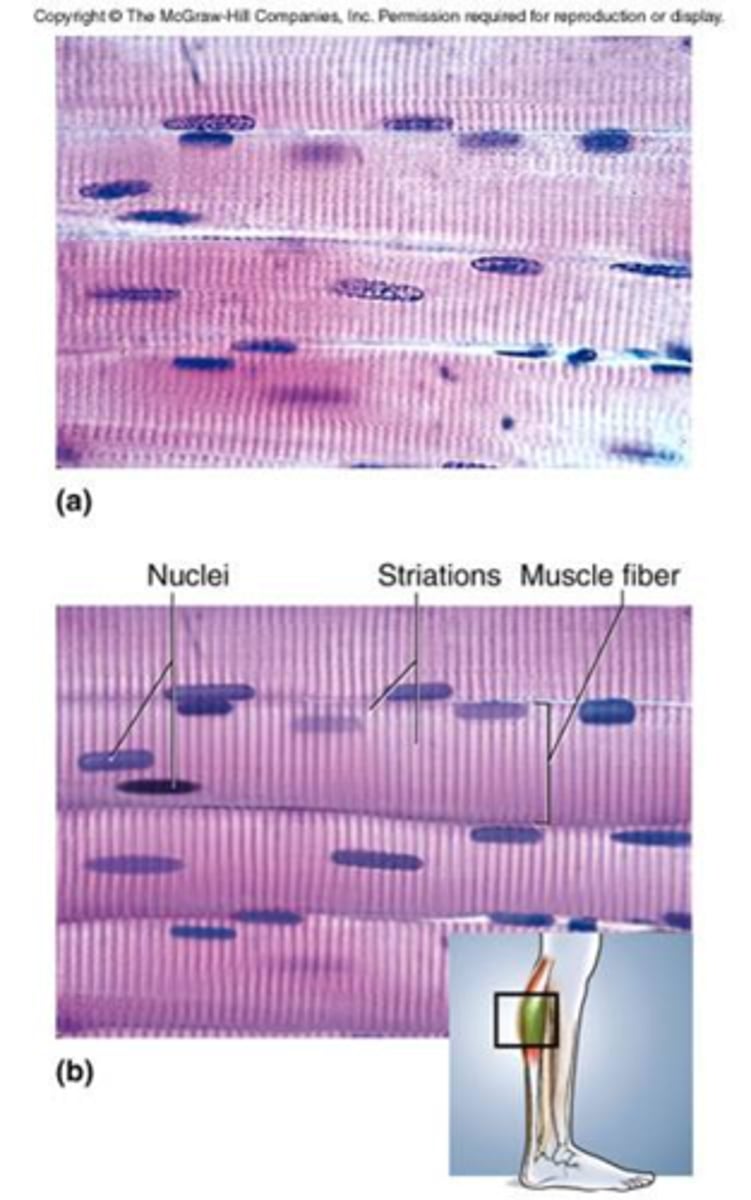
skeletal muscle tissue
This is also called voluntary muscle because it is under voluntary control.
It is also called striated muscle because it has a striated (striped) appearance due to the presence of actin and myosin filaments.
It is attached by tendons to bones and that is why it is called skeletal muscle.
The muscle cells (fibers) are long and cylindrical and have many nuclei.
Examples are gluteus maximus, biceps brachii, deltoid, etc.
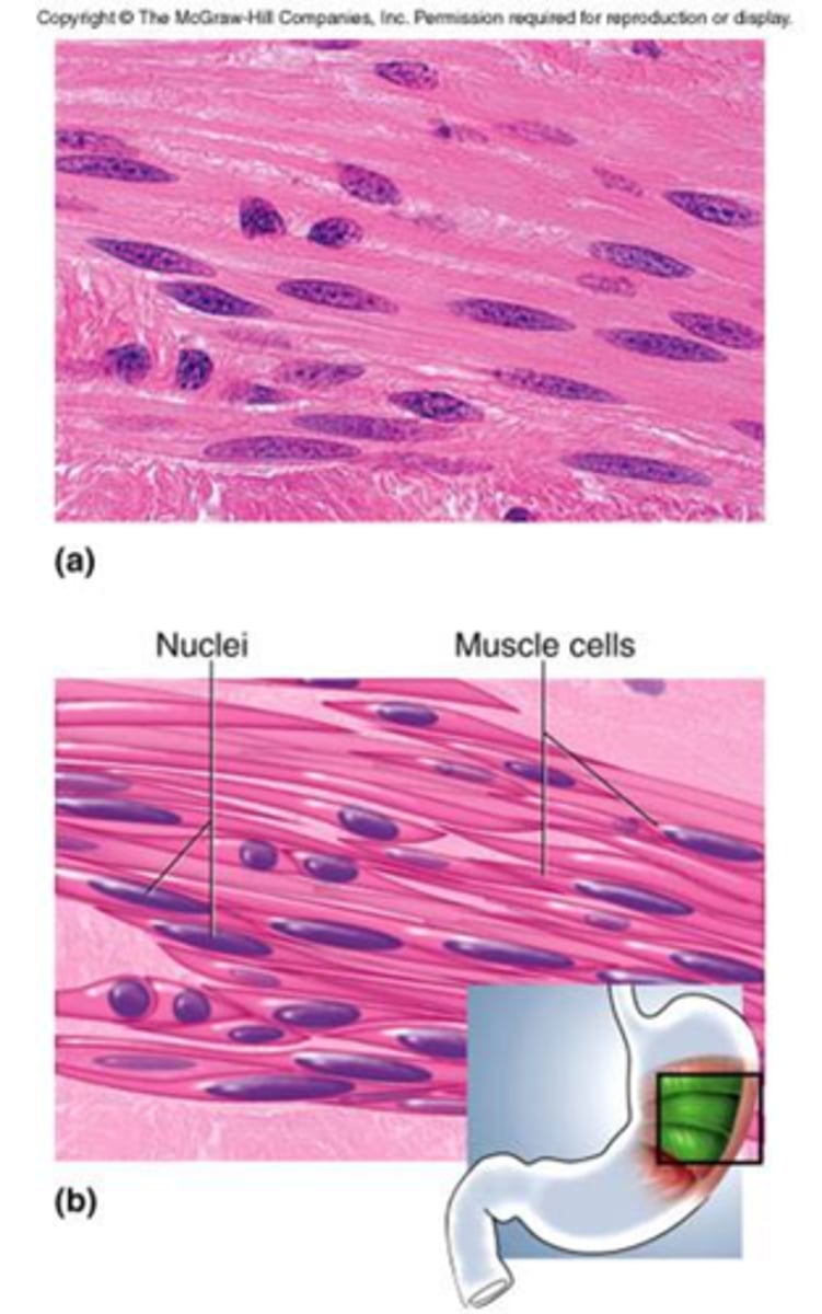
smooth muscle tissue
This is also called involuntary muscle because you do not have voluntary control over it.
It does NOT have striations.
This muscle type is found in the walls of the digestive, respiratory and urinary tracts, blood vessels, the uterus, and other organs and it allows substances to move through those organs.
It contracts more slowly than skeletal muscle but it remains contracted longer than skeletal muscle does.
The muscle cells are long and tapered with a single nucleus (fusiform).
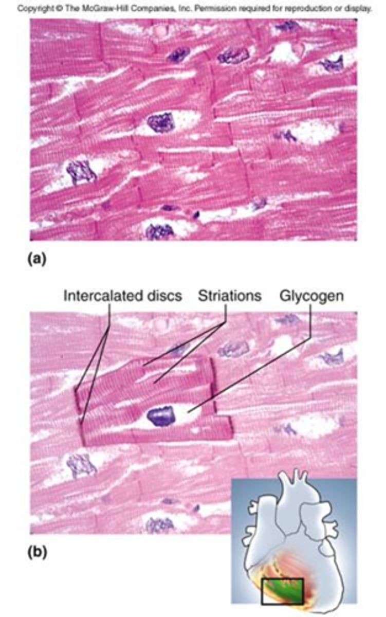
cardiac muscle tissue
This muscle is found only in the walls of the heart.
It has striations but it is involuntary.
The cells (fibers) are branched and join each other at intercalated disks, which are areas where the plasma membranes are connected to each other by gap junctions.
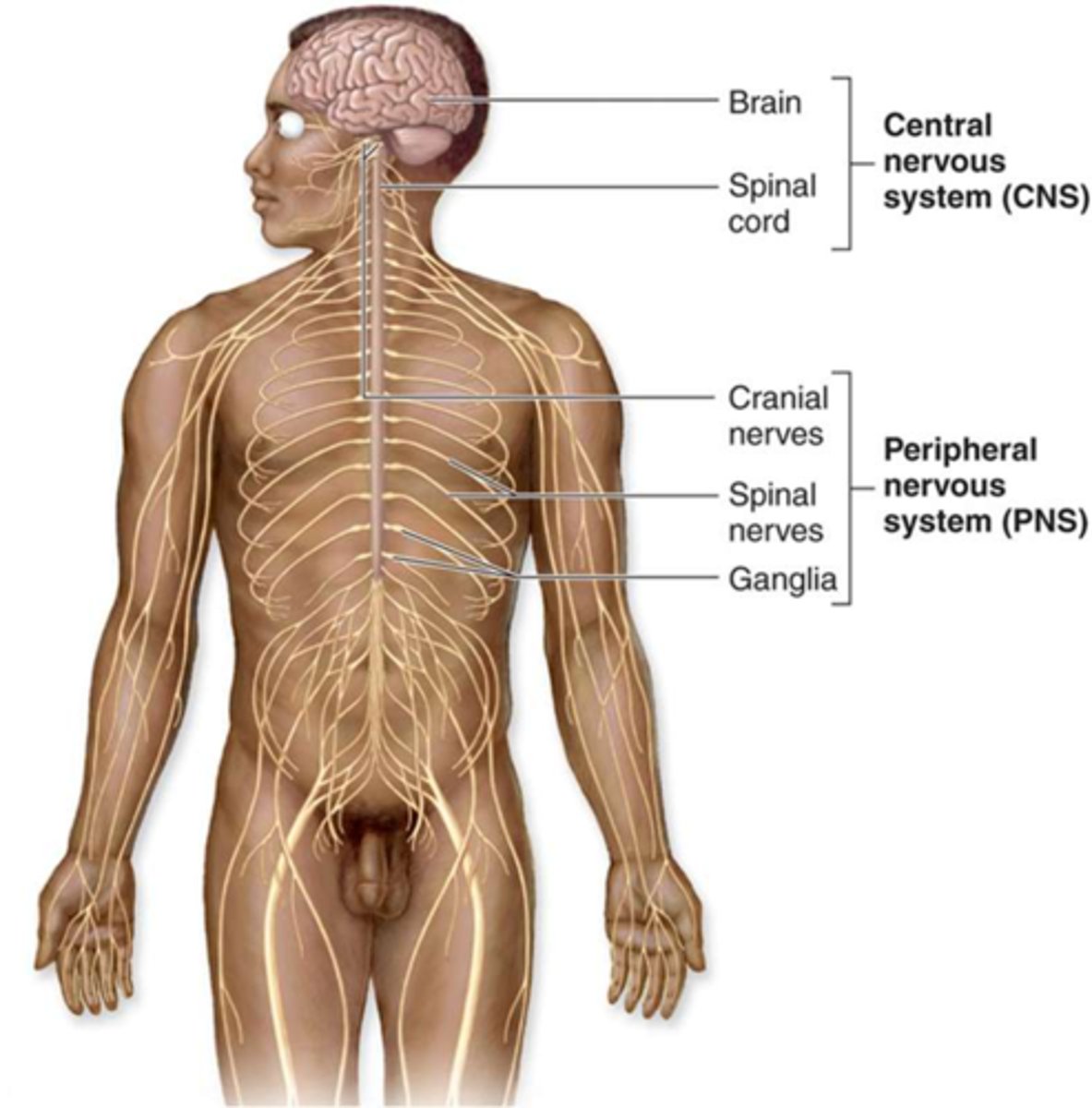
location of nervous tissue
brain, spinal cord, nerves