Medulla Oblongata
1/10
There's no tags or description
Looks like no tags are added yet.
Name | Mastery | Learn | Test | Matching | Spaced |
|---|
No study sessions yet.
11 Terms
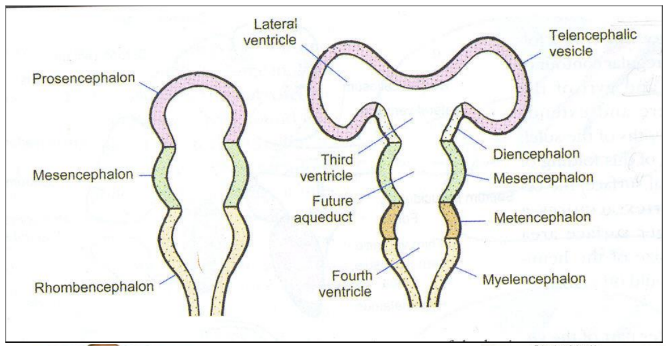
What part of the brain does medulla oblongata come from?
Mylencephalon and the fourth ventricle
Features of the medulla oblongata
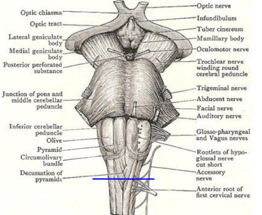
Medulla provides attachment to last four cranial nerves
The junction between the medulla and spinal cord is marked by an imaginary line which passes just above the attachments of first pair of cervical spinal nerves
Ponto-medullary junction is marked by the attachment of 6th, 7th and 8th cranial nerves from medial to lateral side
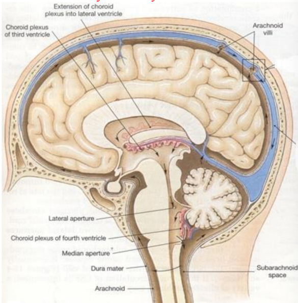
The lower half is closed and contains the central canal below and above opens into the fourth ventricle
Relations of medulla oblongata
Ventrally rests on the basilar part of occipital bone, but separated from the bone by the meninges and basilar venous plexus
Dorsally separated from the cerebellum by the cavity of the fourth ventricle
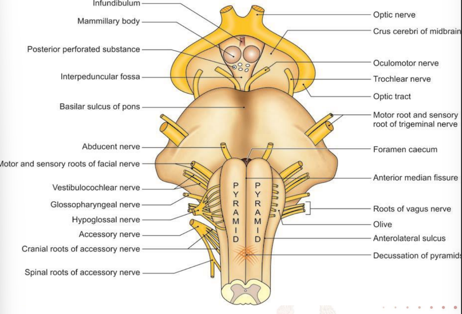
External features
Anterior median fissure
foramen caecum
decussation of pyramidal fibres
anterior external arcuate fibres
Posterior median sulcus
Anterolateral sulcus
gives attachment to the hypoglossal rootlets
Posterolateral sulcus
gives attachment to vagus, glossopharyngeal and accessory nerve
Anterior area
Pyramid- produces by corticospinal, corticopontine and corticobulbar (end in motor nuclei of cranial nerves) tracts
Arcuate nuclei- cover ventral surface of pyramid
Lateral area
Olive- produced by underlying inferior olivary nucleus
Occupied by ventral and dorsal spinocerebellar, lateral spinothalamic, spino-olivary and olivo-spinal
Posterior area
fasciculus gracilis and gracile tubercle
fasciculus cuneatus and cuneate tubercle
tuberculum cinereum (produced by underlying spinal nucleus and tract of trigeminal
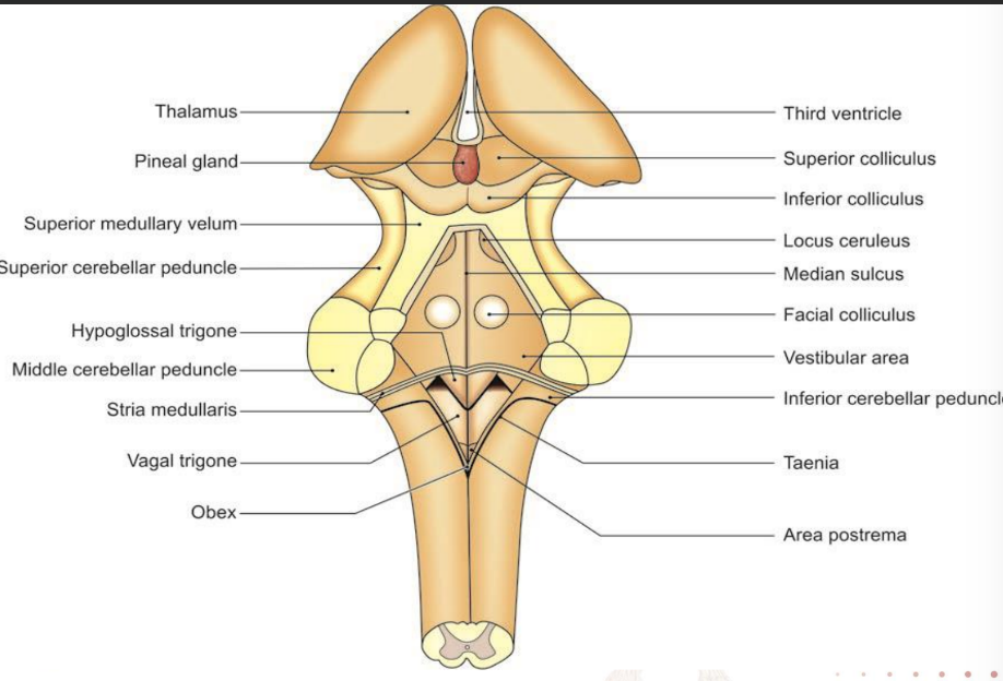
Internal structure of the medulla oblongata
At the level of the pyramidal decussation (motor)
At the level of the sensory decussation
At the level of inferior olivary nucleus
Changes that take place
Appearance cranial nerve nuclei
Appearance of ventricles of the brain
Grey matter loses shape
Appearance of reticular formation
Transverse -section at the level of the pyramidal decussation
Nucleus gracilis and cuneatus appear as narrow strips within the fascicules from the posterior aspect of central grey matter
Anterior horn forms the spinal nucleus of accessory lateral (continuous with hypoglossal nuclei) and supraspinal nucleus medially (in line with nucleus ambiguus
Spinal tract of trigeminal nerve conveys pain and temperature from face and forehead (in line with chief sensory nucleus)
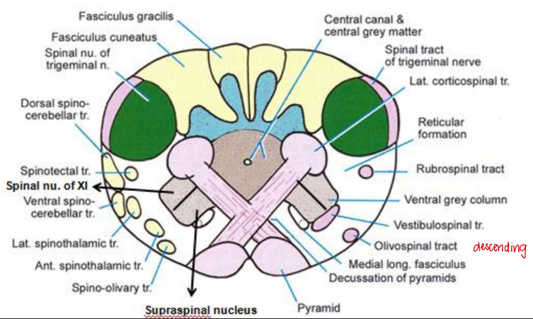
Transverse section of medulla oblongata at level of sensory decussation
Nucleus cuneatus and gracilis become more pronounced and fasciculus fibres terminate here
Internal arcuate fibres from these nuclei courses and contralateral and become medial lemniscus
Accessory cuneate nucleus receives more lateral fibres (convey proprioceptive fibres from upper limb to cerebellum through inferior cerebellar peduncle)
Inferior olivary nucleus begins to appear
Pyramids on either side of the median fissure
Nucleus of tractus solitarius (receives taste fibres from VII, IX and X), hypoglossal nucleus (supplies muscles of tongue)and dorsal nucleus of vagus (supply heart, smooth muscles)
Medial longitudinal bundle
Lateral and anterior spinothalamic tract form spinal lemniscus
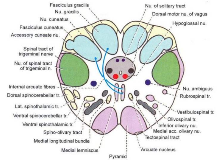
Transverse section at the level of the olive
Inferior olivary nuclear complex present
Consists of:
principal inferior olivary nucleus
Medial accessory olivary nucleus
Dorsal accessory olivary nucleus
Afferents from cerebral cortex, red nucleus, basal nuclei
Efferents from olivo-cerebellar and paraolivocerbellar (climbing fibres)
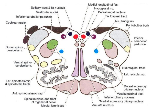
Consists of hypoglossal nucleus, nucleus intercalatus, dorsal nucleus of vagus, nucleus solitarius, vestibular nuclei and nucleus ambiguus
Medial longitudinal fasciculus
Tecto-spinal tract
Medial lemniscus
Pyramidal tract
Arcuate nuclei (displaced pontine nuclei)
Blood supply
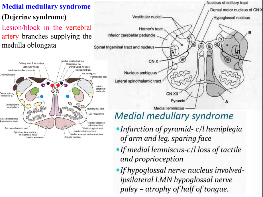
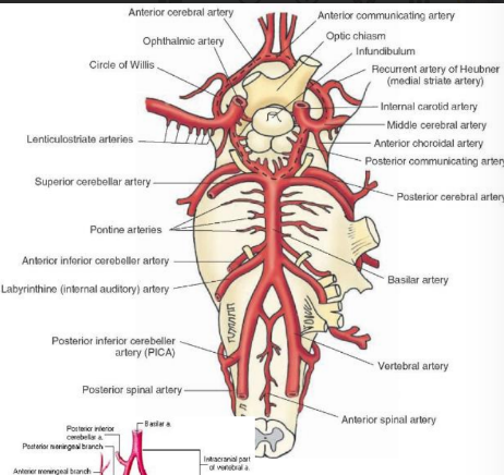
Anterior and posterior spinal arteries from vertebral artery
Posterior inferior cerebellar arteries from vertebral artery
Basilar artery and its branch anterior inferior cerebellar artery
Lateral medullary syndrome
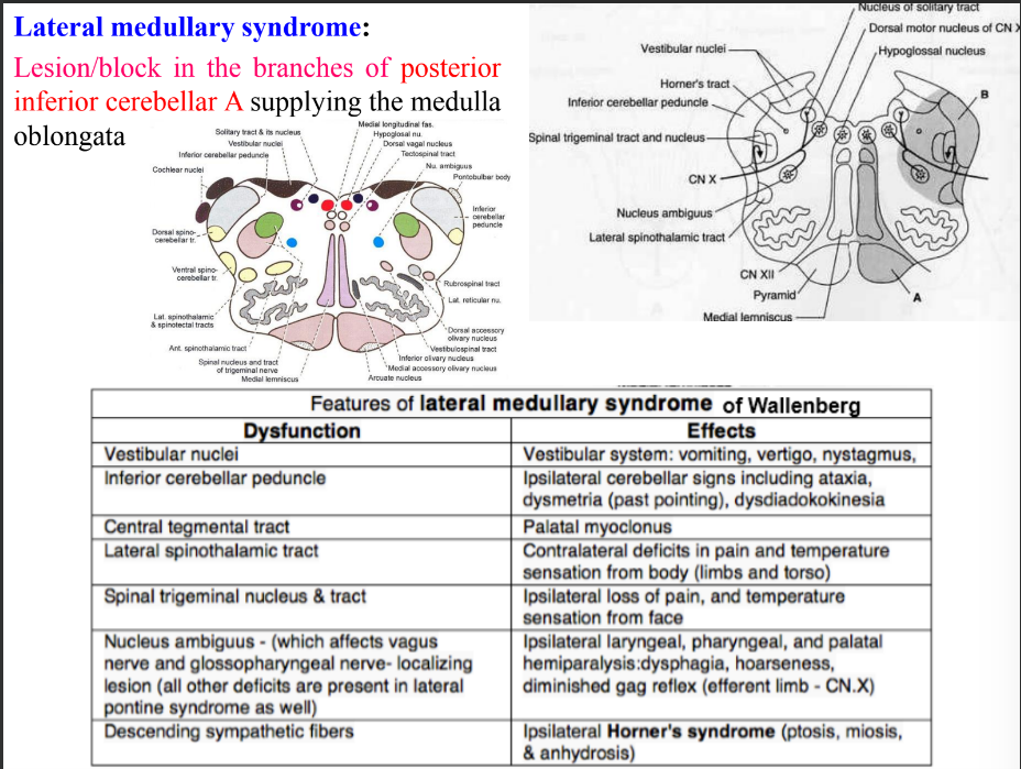
Vertigo – sensation of whirling and loss of balance
Nystagmus – rapid involuntary movements of the eye
Ataxia – loss of full control of bodily movements
Dysmetria – lack of coordination movement due lack of ability to perceive distance
Dysdiadokokinesis – inability to perform rapid alternate movements
Palatal myoclonus – rapid spasm of palatal muscles
Dysphagia – difficulty in swallowing
• Gag reflex or pharyngeal reflex
Miosis – excessive constriction of pupil
Anhydrosis – failure of sweat glands to secrete
Medial medullary syndrome
