Lesson 3: Cytoskeleton
1/42
There's no tags or description
Looks like no tags are added yet.
Name | Mastery | Learn | Test | Matching | Spaced | Call with Kai |
|---|
No study sessions yet.
43 Terms
Cytoskelton definition
Complex network of protein filaments that extends throughout
the cytoplasm.
Characteristics
• Highly dynamic structure that rearranges quickly as the cell changes
its shape
-divides or responds to external stimuli.
• Exclusively found in eukaryotic cells.
Main functions
• Maintenance of cell shape.
• Regulation of the position and displacement of organelles.
• Cell movement (leukocytes, pseudopodia) → control processes
Components: Filaments
• Microtubules (25 nm Ø) - miotic spindle
• Microfilaments or actin (micro)filaments( 7 nm Ø) - contractile ring
• Intermediate filaments (10 nm Ø) - many types
Polymerization of proteins
Filaments are formed by hundreds of molecules that will generate lineal, unbranched polymers with enough length for their functions

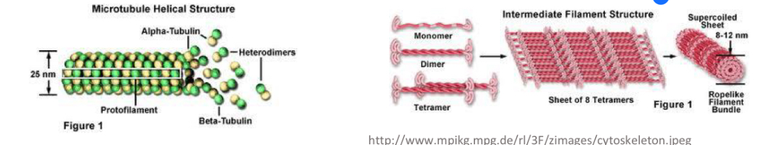
Microtubules
• Hollow cylinders of protein (clear internal space) with a diameter of 25nm and a wall thickness of 5nm.
• Variable length: from tenths of microns to several microns.
• Unbranched.
• Straight, or with a large curvature angle. → tubular shape
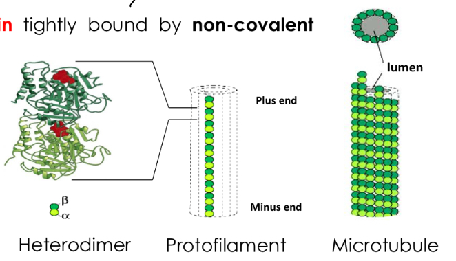
Molecular organization
• Microtubules are made of tubulin.
• Tubulin (heterodimer) = α-tubulin + β-tubulin tightly bound by non-covalent bindings.
• Tubulin heterodimers linked with α and β subunits form the protofilaments
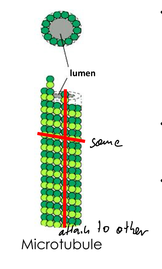
• made of 13 parallel protofilaments with lumen
Structure
-changing alpha and beta subunit
-same row same subunits
-increase the stability in the center of microtubules
-increase dynamism in the ends
Polarized structure
In one end of the microtubule there will be α
subunits exposed, whereas on the other end β subunits will be exposed
Types of Microtubules
• Labile Microtubules:
Originated from the centrosome
-They form parallel groups, forming bundles
- centrosome
- Mitotic spindle.
Stable microtubules
permanent structures.
-Associated forming complex structures:
- Centrioles
- Axoneme of cilia and flagella.
Labile microtubules, dynamic behaviour
• The two ends of the microtubule are constantly incorporating or losing subunits.
• The speed or rate at which subunits are incorporated or lost is dependent on the concentration of free subunits.
• There is a concentration where the gain and loss of subunits occurs at the same rate: Critical concentration.
o If the concentration of free subunits is higher than critical concentration, the microtubule will polymerize.
o If the concentration of free subunits is lower than critical concentration, the microtubule will depolymerize (loses subunits).
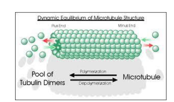
Dynamic instability
Microtubules ends polymerize/depolymerize at different rates:
- Plus (+) end → fast growth or dissociation
- Minus (-) end → slow growth (attached to centrosome)
• MAP proteins control process of growing and shrinking

Functions of Labile Microtubules
• Control of cell SHAPE.
• Control of the POSITION and DISPLACEMENT of intracellular structures.
• Formation of the MITOTIC SPINDLE.

Control of shape
-Form a complex network of tubules
• Other filaments use this network of microtubules to localize and perform their functions
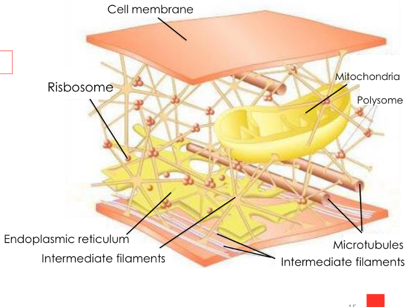
Control of position and displacement of intracellular structures
• Start in the centrosome and extend throughout the cells.
• Determine the position of other organelles in the cell:
- Cisternae and tubules from the endoplasmic reticulum are aligned with microtubules and move towards the periphery of the cell.
- Golgi complex is located near the centrosome.
• Microtubules create a system of guides or "highways" within the cell, along which organelles, vesicles and other cellular components can move
Formation of the miotic Spindle
During cell division, microtubules from the mitotic spindle are responsible for separating chromosomes in the two daughter cells.
Types of stable microtubules
-Centrioles
-Cilia and flagella
Centrioles
-Formed by 9 microtubules triplets -A, B, C
-Bridges between A and C of neighbor triplet
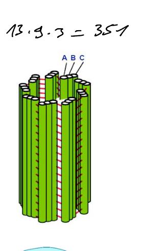
Function of centrioles
Generate miotic spindle
Cilia and flagella
Formed by group of microtubules orientated parallel
Basal body → triplet centriole
Transition body
Axis or atonement → allows movement
Basal body
Located at the base of the cilium/flagellum, under
the plasma membrane.
• centriole structure
• innermost part is attached to the cytoskeleton by protein structures called cilliary roots.
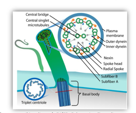
Transition zone
Between the basal body and the axoneme
appears the transition zone, with 9 doublets (9
pairs of microtubules).
• The basal plate, located in the transition zone,
forms a central pair of microtubules.
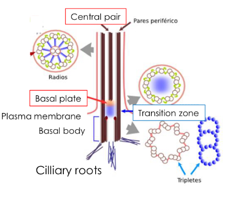
Axis
-9 outer doublets (two microtubules)
-2 central tubules covered by an inner sheath
-inner shealth and binding between A and B keep structure
Actin filaments
2 polymers of globular actin (with Ca2+ helix structure)
-Actin free subunit bound to ATP
During polymerization, ATP is hydrolized toADP.
• More flexible and thinner than microtubules
• Usually branched.
Actin filaments Types
• cortical: the actin filaments are mainly arranged near the membrane.
• cytoplasmic (throughout the cytoplasm)
Dynamic behaviour
Polarity: PLUS end (fast), MINUS end (slow growth)
• Continuous addition and loss of subunits. It is faster than in microtubules.
• ROTARY EXCHANGE: If neither end is protected and the actin filament reaches a stable lengthe addition of subunits at the plus (+) end is equal to the loss at the minus (-) end.
There is a continuous renewal of subunits (treadmilling), which helps maintaining the same filament length.
Actin filaments functions
• Control of the SHAPE of the CELL SURFACE
• CELL MOVEMENTS
• Processes of ENDOCYTOSIS and PHAGOCYTOSIS
• MUSCLE CONTRACTION
• CYTOKINESIS
Control of the shape of the cell surface
-structural function
Form cell cortex
-shape and cell surface movement
-cell cohesion, adherents junctions
They are arranged longitudinally along the axis of the
microvilli.
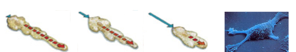
Cell movements
Amoeboid movement, involves changes in the shape of the cell:
- Cells form a pseudopodium in the front to move the cell forward. The cytoplasm follows that movement and retracts at the back of the cell.
When the cytoplasm is located in the place where the pseudopodium was at the beginning, a new pseudopodium is formed.
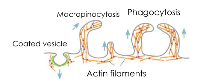
Process of endocytosis and phagocytosis
Actin filaments are involved in coated vesicle formation, pinocytosis and phagocytosis

Muscle contraction
Muscle cell contraction depends on actin and myosin microfilaments (sarcomere):
• Ca2+ release from smooth ER though nervous stimuli
• increase in Ca2+ concentration produces a sliding of the thin filaments (actin) over the thick filaments (myosin).
Use the energy stored in ATP molecules (ATP hydrolysis).
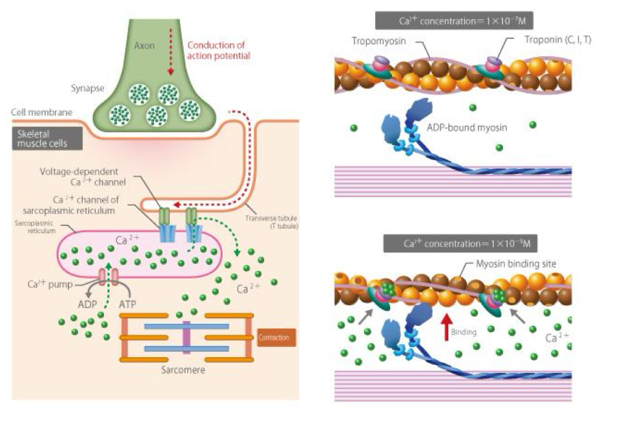
Sarcomere contraction
Happens because actin filaments slide over myosin filaments towers center
Sarcomere structural and function unit of the
myofibrils in the skeletal muscle
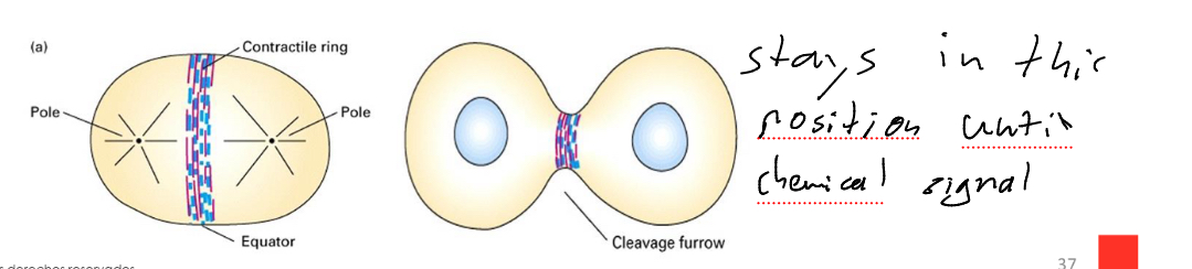
Cytokinesis
In cell division, cytokinesis is the division of the cytoplasm
Characterised by formation of a contractile ring Made of muscle myosin II and actin filaments

Intermediate filaments
-anchored to plasma membrane junctional complexes and also are within the nucleus.
-thicker than actin filaments, but thinner than microtubules
-most stable elements of the cytoskeleton because they are the less soluble
-Not involved in cell movement
-Their function is structural. They confer support and
mechanical stability to the tissues.
Different cell types have intermediate filaments
– Keratin: in epithelial cells and derivatives.
– Desmin: in muscle cells, both smooth and striated.
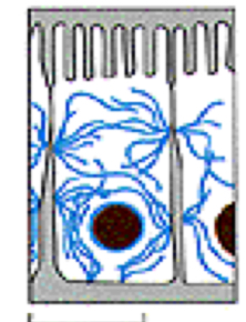
– Neurofilaments: mantain the shape of neurons (e.g. in axons of neurons).
– Vimentin: in cells of mesenchymal origin. They are arranged around the nucleus and hold it in place (the most widely distributed type of intermediate filament).
– Laminin: it is the nuclear filament in the nuclear lamina (inner lining of nuclear envelope).
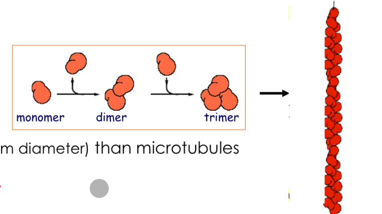
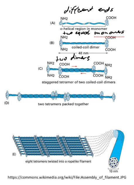
Polymers
A. Monomers: large molecules with folded ends.
B. Dimers: Two monomers parallel in the same direction.
C. Tetramers: 2 dimers in opposite directions
D. Tetramers will group from end to end and form a
protofilament.
E. 8 protofilaments associate and finally form the
intermediate filament.
Functions of intermediate filaments
• RESISTANCE and COHESION between contiguous
cells
• Participates in CELL DIVISION
Résistance and cohesion between contiguous cells
They are a mechanical support for body and cell projections.
– E.g.: in neurons, they are disposed in parallel, in longitudinal direction along the axon.
• Components of structures involved in cell-cell contact (desmosomes) and between cell and matrix (hemidesmosomes).

Participates in cell division
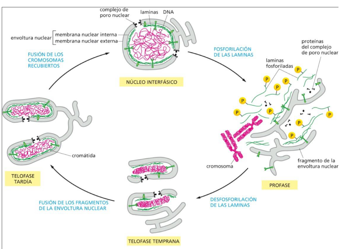
• The nuclear lamina is the inner lining of the nucleus, giving shape and stability to the nucleus of all cells.
• It interacts with chromatin.
• During mitosis and meiosis, it is disorganised
to allow nuclear envelope breakdown and cell division.
Accessory proteins
Hundreds of proteins with different functions:
– Binding of filaments to other filaments or to other organelles.
– Control of tubulin and actin polymerization.
– They produce movements along the microtubules and actin filaments, coupled with hydrolysis of ATP (MOTOR PROTEINS).
-Thanks to the accessory proteins the cytoskeleton is a very organized but flexible structure.
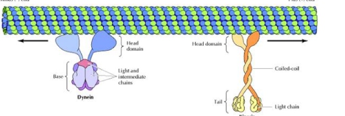
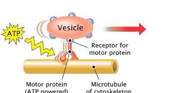
Motor proteins
They bind to a polarized filament, hydrolyze ATP and use the energy obtained to move along the filament.

Types of motor proteins depending on:
-The cargo carried with them (organelles).
-The type of filament bound.
-The direction in which they move.
Types
Dynein and Kinesin
▪ The head or Motor domain (bound to ATP): it binds to the filament and defines
the direction of the movement.
▪ The tail determines the type of intracellular cargo.