NPTE- 2022?
1/248
There's no tags or description
Looks like no tags are added yet.
Name | Mastery | Learn | Test | Matching | Spaced | Call with Kai |
|---|
No analytics yet
Send a link to your students to track their progress
249 Terms
A patient is 12 weeks post talus fracture and has a chief complaint of catching her toe during gait since the removal of the boot cast. Evaluation demonstrates limited ankle dorsiflexion, leg atrophy, and ankle weakness. The physical therapy plan should FIRST emphasize:
Top of Form
1. mobilization of the talocrural joint.
2. stretching the tibialis anterior.
3. strengthening the gastrocnemius.
4. fitting for an ankle-foot orthosis (AFO).
1. MOBILIZATION OF THE TALOCRURAL JOINT. At 12 weeks post fracture the goals of the functional phase should be to restore joint kinematics and attain full range of pain-free motion. Joint mobilization should improve accessory motion, decrease guarding, and lengthen the tissue around a joint. (pp. 1139, 1172-1174)
2. Stretching the tibialis anterior will not help the limited ankle dorsiflexion but may improve plantar flexion. This should not be performed first. (pp. 1172-1174)
3. Before progressing to strengthening the gastrocnemius, emphasis should be on strengthening the dorsiflexors to address the chief complaint (pp. 1172-1174).
4. Catching of the toe, not foot drop, was the chief complaint. Use of an ankle-foot orthosis may be premature, because the patient may improve range of motion with mobilization and strengthening. (p. 1142)
A patient has an irregularly shaped wound at the left medial malleolus. The skin around the wound is darkened. The underlying cause of this wound is MOST likely:
1. lymphedema.
2. venous insufficiency.
3. cellulitis.
4. osteomyelitis.
Rationale
1. Lymphedema presents as swelling over the limb. The most common integumentary complication is cellulitis, which does not present as a single defined ulcer. (pp. 679, 700-701)
2. VENOUS INSUFFICIENCY. The classic presentation of a venous ulcer involves the medial leg and is irregular in shape, with hyperpigmented periwound skin (p. 642).
3. Cellulitis is a painful infection of the soft tissue that is characterized by expanding local erythema, palpable lymph nodes, fever, and chills. Most cases are caused by cuts, abrasions, insect bites, and local burns. (pp. 339-340)
4. Osteomyelitis is an infection of the bone. Clinical characteristics include pain, fever, edema, erythema, and tenderness but not a wound as described in the stem (p. 1236).
A 6-year-old patient with juvenile rheumatoid arthritis involving the cervical spine, bilateral hips, knees, and ankles is referred to the physical therapy department. The patient has developed contractures of all involved joints and continues to complain of morning stiffness. A gait deviation that the physical therapist is likely to observe is:
1. increased cadence.
2. increased plantar flexion range at toe-off (preswing).
3. decreased hip extension at terminal stance.
4. decreased anterior pelvic tilt throughout the gait cycle.
Rationale
1. Children with juvenile rheumatoid arthritis ambulate with a decreased cadence.
2. Children with juvenile rheumatoid arthritis ambulate with decreased plantar flexion at toe off (preswing) and terminal stance.
3. DECREASED HIP EXTENSION AT TERMINAL STANCE. Children with juvenile rheumatoid arthritis ambulate with decreased hip extension at terminal stance and toe off (preswing).
4. Children with juvenile rheumatoid arthritis ambulate with increased anterior pelvic tilt throughout the gait cycle.
A chart review of an adult female patient indicates a hematocrit value of 42% following minor elective surgery. This value is indicative of:
1. anemia. - academic review error
2. inflammation.
3. infection.
4. normal findings.
Rationale
1. Hematocrit is the proportion of the blood that is composed of red blood cells. A low hematocrit is indicative of anemia, but a hematocrit value of 42% is considered normal for an adult female. (p. 268)
2. Inflammation would increase neutrophils and would not influence hematocrit (p. 360).
3. Infection would most influence white blood cell count (p. 360).
4. NORMAL FINDINGS. A hematocrit value of 42% is within normal range for adult females (normal range: 36% to 47%) (p. 268).
A physical therapist uses underwater ultrasound as part of the intervention to treat a patient with an ankle injury. The MOST appropriate mode of application is to immerse the patient's ankle in a:
1. whirlpool filled with degassed water and hold the transducer underwater directly on the skin.
2. metal basin filled with mineral oil and hold the transducer underwater approximately 1 in (2.54 cm) away from the body surface.
3. ceramic basin filled with glycerin and move transducer underwater directly on the skin.
4. plastic basin filled with tap water and move the transducer approximately 0.25 in (0.17 cm) away from the body surface.
Rationale
1. A whirlpool will increase the intensity of ultrasound by reflecting waves. Optimally ultrasound wave reflection should be reduced, not increased.
2. A metal basin will increase the intensity of ultrasound by reflecting waves, which is incorrect.
3. The immersion technique requires holding the ultrasound head away from skin. Holding the transducer directly on the skin is incorrect.
4. Plastic basin filled with tap water and move the transducer approximately 0.25 in (0.17 cm) away from the body surface. This option has the correct basin (with reduced reflection) and application (parallel to and the correct distance from the body surface).
A physical therapist is treating a patient who had a traumatic brain injury 3 weeks ago. The patient is confused and agitated. Physical therapy evaluation found decreased lower extremity coordination and strength. Which of the following would be the MOST appropriate intervention?
1. Participating in biofeedback training for lower extremity muscles with supervision
2. Walking in parallel bars with supervision
3. Participating in an aerobics group exercise class for 30 minutes
4. Performing lower extremity exercises while following a written handout
Rationale
1. With impaired attention, the patient would have difficulty participating in biofeedback training (p. 868).
2. WALKING IN PARALLEL BARS WITH SUPERVISION. Walking in parallel bars permits the patient to use the bars if balance is lost (p. 448). The closed environment is appropriate secondary to the heightened state of activity of the patient (p. 868).
3. In the confused-agitated state, the patient's behavior is bizarre and not purposeful. Group exercise classes would not be appropriate. Gross attention to the environment is very brief. (p. 868)
4. With impaired attention, the patient would have difficulty focusing on the written instructions long enough to complete the task (p. 868).
A patient is referred to physical therapy with a history of ulnar nerve entrapment at the level of the hamate. Which of the following would be the MOST specific exercise to improve this patient's strength deficits?
1. Practice pinching between thumb (1st digit) and the tip of the index finger (2nd digit).
2. Squeeze hand grip with elastic-band resistance.
3. Oppose thumb (1st digit) to the metacarpal phalangeal joint of each finger (2nd through 5th digits). - academic review error
4. Squeeze therapy putty between the sides of the fingers.
Rationale
1. The muscles that are active during this movement are supplied by the median nerve, and impingement occurs in the carpal tunnel, not at the hamate (p. 378).
2. Doing this exercise would not isolate muscles supplied by the ulnar nerve, as only the 4th and 5th digits would be involved. This exercise would be appropriate if the ulnar nerve was entrapped at the level of the cubital tunnel and not in the tunnel of Guyon (at the level of the hamate). (p. 379)
3. This exercise would strengthen muscles supplied by the median nerve at the wrist. Deficits here would be loss of thumb (1st digit) abduction and opposition. (p. 378)
4. SQUEEZE THERAPY PUTTY BETWEEN THE SIDES OF THE FINGERS. This movement isolates the lumbricals and interossei, which are innervated by the ulnar nerve and are affected when entrapment occurs at the tunnel of Guyon (at the level of the hamate) (p. 379).
A 14-year-old baseball player reports shoulder pain of insidious onset. The patient displays apprehension when the shoulder is passively positioned in abduction and full external (lateral) rotation. Which of the following pathologies is MOST likely present in this individual?
1. Adhesive capsulitis
2. Atraumatic instability
3. Acromioclavicular separation
4. Superior labral tear
Rationale
1. The patient is too young for insidious onset of adhesive capsulitis, and full lateral (external) rotation can be achieved in this patient. With adhesive capsulitis, impaired range of motion would be expected. (Dutton, pp. 665-666)
2. ATRAUMATIC INSTABILITY. Symptoms occur with excessive abduction and lateral (external) rotation of the shoulder. Anterior instability should be considered. (Dutton, p. 670)
3. Although the closed packed position for the acromioclavicular joint is at 90° of abduction, apprehension with lateral (external) rotation of the shoulder is not cited in any test for the acromioclavicular joint (Dutton, p. 588).
4. Labral tears are commonly associated with traumatic injury with sudden onset (Magee, p. 318).
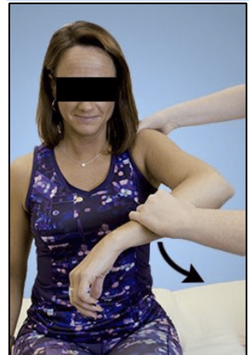
Which of the following is the MOST appropriate technique to improve the flexibility of the hip flexors?
1. Active hip extension to end range, followed by isometric hip flexion
2. Resisted hip extension using cuff weights, followed by active hip flexion
3. Placing the patient in prone with pillows positioned under the abdomen
4. Gentle, sustained passive hip extension
Rationale
1. This exercise requires the addition of active relaxation of the hip flexors and active or passive movement into hip extension to be effective (pp. 94-95).
2. This exercise is a strengthening exercise for the hip extensors; active movement of tight muscles does not activate a relaxation response; an isometric contraction is required (p. 752).
3. Tight muscles need to be taken to their most lengthened position before maintaining the position; lying prone on pillows is not the most lengthened position of the hip flexors (p. 109).
4. GENTLE, SUSTAINED PASSIVE HIP FLEXION. Gentle, sustained passive hip extension is an appropriate method of stretching tight tissues (p. 88).
A patient had a ruptured right middle cerebral artery aneurysm that was repaired. Which of the following functional limitations would the patient MOST likely exhibit?
1. Horizontal nystagmus
2. Ataxic gait
3. Apraxia
4. Rigidity
Rationale
1. Horizontal nystagmus is a symptom of a cerebellar problem, such as a lesion of the anterior inferior cerebellar artery.
2. An ataxic gait is a symptom of a cerebellar problem, such as a lesion of the basilar artery.
3. Apraxia is a clinical symptom of a middle cerebral artery lesion.
4. Rigidity is not caused by a lesion of middle cerebral artery.
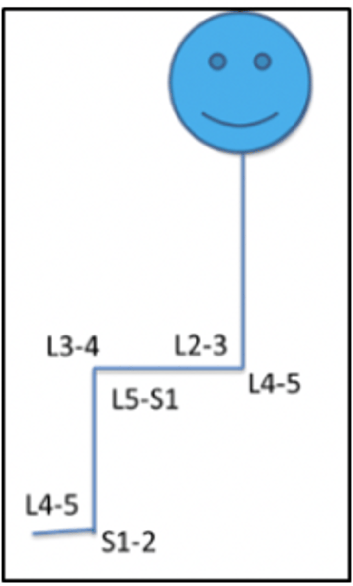
A patient experiences abnormal sensation on the lateral edge of the left foot. Muscle testing reveals weakness of the left hip abductors. Which combination of nerve root levels is MOST likely affected?
1. L2-L3
2. L3-L4
3. L5-S1
4. S2-S3
Rationale
1. L2-L3 sensation is on the lateral thigh (Magee, p. 585).
2. L3-L4 sensation is on the anteromedial thigh and leg (Magee, p. 585).
3. L5-S1 sensation is on the lateral foot; muscles controlling hip abduction are innervated at L4-S1 (Magee, p. 585; Drake, p. 575).
4. S2-S3 sensation is on the plantar foot (Magee, p. 585).
A physical therapist is examining a 50-year-old patient who sustained a right Colles fracture following a fall 6 weeks ago. The patient has a sedentary lifestyle and has rheumatoid arthritis that has been treated with steroids. Which of the following factors will have the GREATEST impact on the patient's fracture healing?
1. Patient's age
2. Rheumatoid arthritis
3. Steroid usage
4. Sedentary lifestyle
3. STEROID USAGE. steroid use - Although age, diagnosis, and sedentary lifestyle may have some impact, the long-term steroid usage will impact healing time to the greatest degree.
A physical therapist is evaluating a patient who reports shoulder pain during overhead activities. During active shoulder abduction on the affected side, the patient demonstrates diminished scapular upward rotation. Weakness of which of the following muscles is MOST likely to contribute to this dysfunction?
1. Upper trapezius
2. Posterior deltoid
3. Rhomboids
4. Teres major
Rationale
1. UPPER TRAP. The upper trapezius elevates the shoulder alone but, coupled with the lower trapezius and serratus anterior, produces upward rotation of the scapula via force coupling (p. 64).
2. The posterior deltoid extends, abducts, and laterally (externally) rotates the shoulder (p. 81).
3. The rhomboids retract, elevate, and downwardly rotate the scapula (p. 65).
4. The teres major extends, adducts, and medially (internally) rotates the shoulder (p. 77).
A patient with a left tibial fracture is restricted to 25% weight-bearing. The patient is currently walking with a single axillary crutch on the left side. Which of the following is the MOST appropriate action for the physical therapist?
1. Have the patient use a walker instead of a crutch.
2. Have the patient use 2 axillary crutches.
3. Switch the crutch to the patient's right side.
4. Prescribe a quad cane to use on the left side.
Rationale
1. A walker would accommodate the weight-bearing restrictions but would be more restrictive than bilateral axillary crutches (pp. 475, 479).
2. HAVE THE PATIENT USE 2 AXILLARY CRUTCHES. Physical therapists should chose the least restrictive device that the patient can safely use. Given the patient is familiar with use of crutches, having the patient use crutches bilaterally would be most appropriate. (pp. 429, 696) Crutches are used to improve balance and to relieve weight-bearing fully or partially on a lower extremity. They are typically used bilaterally. (p. 472)
3. Bilateral axillary crutches are needed to unload the left lower extremity sufficiently. Single devices are not intended for use with restricted weight-bearing gait. (p. 472)
4. Canes are not intended for use with restricted weight-bearing gaits (p. 464).
A patient in a persistent vegetative state in a nursing home has developed a Stage 2 ischial pressure injury. The pressure injury has not improved after 4 weeks of standard wound care treatment. The physical therapist should recommend a consultation with:
1. an orthotist to investigate lower extremity bracing.
2. a nutritionist to investigate level of protein.
3. a respiratory therapist to administer oxygen therapy.
4. a surgeon to perform a skin flap.
1. Since the pressure injury is not a result of contractures that would warrant braces, this would not be the most effective consultation.
2. A NUTRITIONIST TO INVESTIGATE LEVEL OF PROTEIN. Increased protein levels are linked to improved wound healing in patients with pressure injuries. The international guidelines for prevention and treatment of pressure injuries includes referral of all individuals with a pressure injury to a dietician (nutritionist) for early assessment and intervention for nutritional problems.
3. Oxygen is imperative for wound healing in both preventing infection and meeting the metabolic demands of the tissues. In this patient, who is in a persistive vegetative state, inadequate nutrition should be a primary concern and referral to a dietician (nutritionist) is a standard of care.
4. A skin flap would not be indicated for a wound that is not improving. The underlying issues preventing the wound from healing would most likely compromise the integrity of the skin flap.
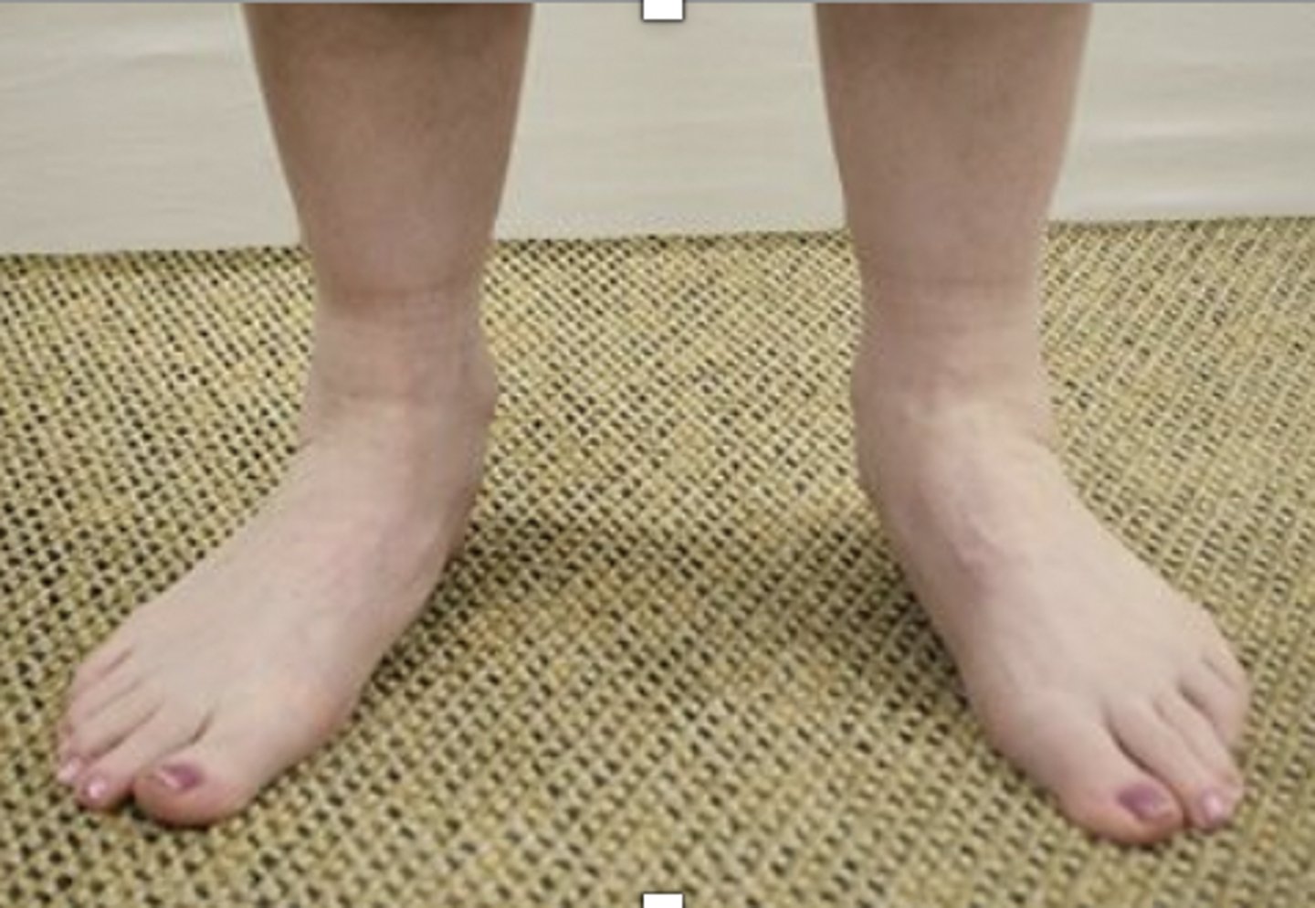
The patient whose feet are shown in the photograph is a waitress who reports 5/10 pain at the anterior calcaneus on the plantar aspect of the left foot. Pain is worse early in the morning and during weight-bearing activities throughout the day. Which of the following associated findings would result in the GREATEST delay in recovery?
1. Fibularis (peroneus) brevis strength of Fair plus (3+/5) and 0° to 10° ankle dorsiflexion range of motion
2. Tibialis posterior strength of Good (4/5) and 0° to 10° ankle dorsiflexion range of motion
3. Body mass index of 36 kg/m2 and 0° to 35° ankle plantar flexion range of motion
4. Body mass index of 36 kg/m2 and 0° to 25° hallux extension range of motion
Rationale
1. Factors that may prolong recovery are a history of an additional lower extremity pathological condition, presence of other medical conditions, or severe obesity. The fibularis (peroneus) brevis is not a medial longitudinal arch support, and the level of dorsiflexion is functional. Therefore, this option should not result in prolonged recovery of the fasciitis. (Magee, pp. 903-904, 915)
2. The tibialis posterior can act as a support for the medial longitudinal arch of the foot and thereby reduce the strain on the plantar fascia in the pronated position. However, the level of tibialis posterior weakness is small, and the degree of talocrural dorsiflexion is functional. Therefore, these factors would not alter the rate of recovery to a substantial degree. (Magee, pp. 903-904, 915)
3. This option includes a body mass index (BMI) in the morbid obesity classification. However, the limitation of plantar flexion is minimal, compared to the discharge criteria of 40°. In addition, limited plantar flexion should not strain or elongate the plantar fascia. Therefore, this option only has one factor that would prolong recovery. (Kisner, p. 868)
4. BODY MASS INDEX 36 KG/M^2 AND 0˚ TO 25˚ HALLUX EXTENSION ROM. The description of the patient is consistent with a diagnosis of plantar fasciitis (Magee, p. 947). Interventions for the diagnosis of plantar fasciitis should focus on the goals of midfoot stability, functional foot and ankle range of motion, Normal (5/5) foot and ankle strength, minimal pain, and return to functional status for activities of daily living and vocational activities. Extra body weight places increased loads on the plantar fascia. Obesity and even a body mass index greater than 25 kg/m2 would be a contributing factor. In addition, the range of motion for hallux extension is more than 50% limited from the normal range of 70°. Therefore, the factors in this option should result in the greatest delay in recovery. (Magee, p. 915)
Underweight = <18.5
Normal weight = 18.5-24.9
Overweight = 25-29.9
Obesity = BMI of 30 or greater
To assess an anterior tibiofibular ligament injury, which of the following tests is MOST appropriate?
1. Talar tilt with the ankle in neutral dorsiflexion
2. Anterior drawer at the ankle with the ankle in neutral dorsiflexion
3. Compression of the shafts of the tibia and fibula at mid calf
4. Squeezing the calf with the ankle in neutral dorsiflexion (Thompson Test)
1. The talar tilt test with the ankle in neutral dorsiflexion is used for evaluating the integrity of the calcaneofibular ligament.
2. The anterior drawer test with the ankle in neutral dorsiflexion is used for evaluating the integrity of the anterior talofibular ligament.
3. Compression of the shafts of the tibia and fibula at mid calf is used to test for syndesmosis ligament injury, including injury to the anterior tibiofibular ligament.
4. Squeezing the calf with the ankle in neutral dorsiflexion is used to test the integrity of the Achilles tendon.
Paraffin would be MOST beneficial for a patient with which of the following conditions?
1. Edematous wrist 1 week following carpal tunnel surgery
2. Swollen elbow resulting from rheumatoid arthritis exacerbation
3. Aching fingers resulting from chronic osteoarthritis
4. Painful hand resulting from early-stage complex regional pain syndrome
Rationale
1. Applying heat may increase the edema due to vasodilation and increased metabolic rate, leading to an increase in inflammation (p. 152). Paraffin as a thermotherapy agent would also be difficult to remove from the healing site of the incision (p. 153).
2. Paraffin wax is used for thermotherapy (p. 157). Cryotherapy is usually recommended for chronic inflammatory conditions such as rheumatoid arthritis (p. 133).
3. As long as no active swelling is noted, paraffin will help increase motion and decrease pain associated with chronic osteoarthritis in the fingers (p. 150).
4. Thermotherapy, aside from the application of neutral warmth, may aggravate the pain associated with complex regional pain syndrome (p. 7).
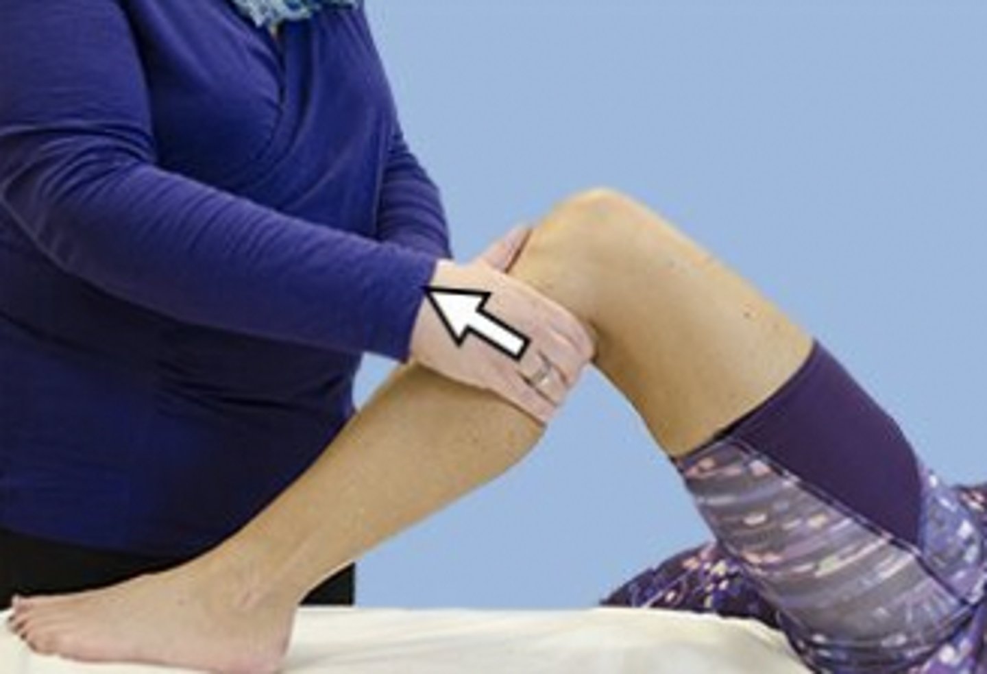
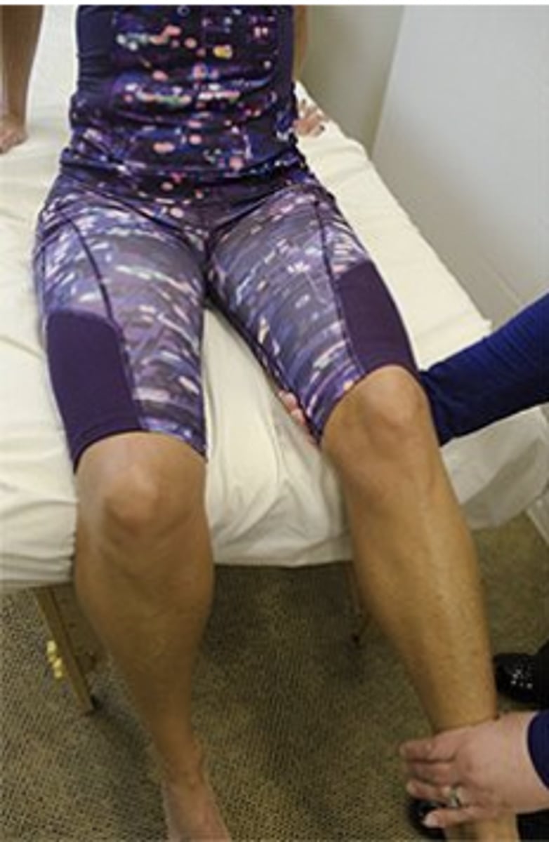
A patient has a positive result on the test shown in the photograph. During the subacute phase of treatment, the MOST appropriate intervention for the patient is independent performance of which of the following exercises?
1. Lower extremity partial squats
2. Open kinetic chain knee extension
3. Straight leg raises
4. Plyometric exercises
Rationale
1. The photograph shows the anterior drawer test for the knee, which is used to assess the integrity of the anterior cruciate ligament. Closed kinetic chain extension exercises will put less stress on the anterior cruciate ligament and are appropriate for the subacute phase (week 4) of treatment.
2. Open kinetic chain knee extension, especially the last 25°, will put increased tension on the anterior cruciate ligament.
3. Although straight leg raises put no stress on the anterior cruciate ligament, this would be an exercise for the acute (not subacute) phase of treatment.
4. Plyometric exercises are an important part of the functional phase (week 10) of rehabilitation after an anterior cruciate ligament tear.
A patient with chronic low back pain had a baseline Oswestry Disability Questionnaire score of 60 points. Three weeks later, the score was 8 points. With regard to the patient's current self-reported level of disability, which of the following courses of action should the physical therapist pursue NEXT?
1. Continue physical therapy until the patient returns to a score of 60.
2. Instruct the patient in a functional conditioning program to prepare for discharge.
3. Immediately refer the patient to the emergency department.
4. Have the patient return to the physician within the next few days.
Rationale
Lower score = better
1. A change in the Oswestry score from 8 to 60 would be consistent with worsening of the patient's condition.
2. An Oswestry score of 8 is favorable, and a functional conditioning program is appropriate.
3. An Oswestry score of 8 does not indicate the need for an emergency department visit.
4. An Oswestry score change of 60 to 8 does not require a (medical) physician visit; it signifies improvement.
The resting heart rate of a 32-year-old runner is measured at 46 bpm. Which of the following explanations for this heart rate is MOST likely?
1. The individual has a hypotensive disorder.
2. The rate is secondary to an increased stroke volume.
3. The individual has an atrioventricular block.
4. Endurance training has stimulated the sympathetic nervous system.
Rationale
1. It is more likely that the bradycardia is a training effect associated with a greater stroke volume (pp. 144-146).
2. Cardiac output is the product of stroke volume multiplied by heart rate. A training effect is an increase in stroke volume. There is a resultant decrease in heart rate to maintain the same cardiac output at rest. (pp. 144-146)
CO = SV x HR
3. It is more likely that the bradycardia is a training effect associated with a greater stroke volume (pp. 144-146).
4. Exercise training increases parasympathetic activity and causes a small decrease in sympathetic discharge. Training also decreases the intrinsic firing rate of the sinoatrial node. These training adaptations explain the resting bradycardia in individuals who train aerobically. (p. 140)
*A patient reports fatigue, proximal upper extremity weakness, and double vision that increases in intensity as the day progresses. The patient demonstrates bilateral ptosis of the eyelids, difficulty chewing, dysphagia, and inability to raise the eyebrows. Which of the following conditions is MOST likely present?
1. Bell palsy
2. Myasthenia gravis
3. Trigeminal neuralgia
4. Amyotrophic lateral sclerosis
Rationale
1. Bell palsy would not result in dysphagia or difficulty chewing, although there may be residual food between the teeth and the cheek due to weakness of the buccinator (Lundy-Ekman, p. 349).
2. The loss of function described in the scenario involves multiple cranial nerves. The fact that it increases as the day progresses implies fatigue that is typical of myasthenia gravis. (Goodman, pp. 1696-1698)
Myasthenia Gravis
Etiology: autoimmune disorder resulting in neuromuscular junction disorder – defect in transmission of nerve impulses; antibodies block/destroy receptors that are needed for acetylcholine uptake & this prevents muscle contraction; enlarged thymus
S&S: extreme fatiguability & skeletal muscle weakness that can fluctuate (periods of remissions and exacerbations); ocular muscles typically affected first, half of pts experience ptosis & diplopia; dysphagia, dysarthria, and CN weakness are also common, like affecting facial expression; other neurological findings are normal (reflexes, sensation, etc)
Triggers: activity, heat, stress, illness, certain meds, menstruation, pregnancy
Tx: MG “crisis” is a medical emergency (involves exacerbation of respiratory muscles & requires a ventilator; Meds to inhibit acetylcholinesterase (the enzyme that breaks down Ach) to allow Ach to buildup at neuromuscular junction (will diminish symptoms of weakness and fatiguability; corticosteroids to suppress immune system; PT focus on obtaining respiratory baseline & pulmonary intervention (breathing techniques), energy conservation techniques, strengthening using isometric contractions, endurance; caution to avoid overexertion
3. Trigeminal neuralgia is a dysfunction of the trigeminal nerve (CN V) that produces sharp, severe, stabbing pain in the distribution of one or more branches of the trigeminal nerve (CN V). It does not cause ptosis, dysphagia, and fatigue, which are described in the stem. (Lundy-Ekman, p. 347).
4. Amyotrophic lateral sclerosis symptoms more typically include tripping, stumbling, and falling; loss of muscle control and strength in hands and arms; difficulty speaking, swallowing, and/or breathing; chronic fatigue; and muscle twitching and/or cramping. Amyotrophic lateral sclerosis is characterized by both upper and lower motor neuron damage. Symptoms of upper motor neuron damage include stiffness (spasticity), muscle twitching (fasciculations), and muscle shaking (clonus). Symptoms of lower motor neuron damage include muscle weakness and muscle shrinking (atrophy). (Lundy-Ekman, pp. 224-225)
When is the BEST time to determine a patient's baseline respiratory pattern?
1. While the patient is unaware of the observation
2. While the patient is providing a medical history
3. After measuring the patient's heart rate
4. After measuring the patient's blood pressure
Rationale
1. The patient will not alter the respiratory pattern if the patient is unaware of the observation.
2. The patient will be speaking, which will affect the baseline respiratory pattern.
3. The order of measuring heart rate and respiratory pattern is not critical to obtaining an accurate measurement.
4. The order of measuring blood pressure and respiratory pattern is not critical to obtaining an accurate measurement.
*A physical therapist is evaluating a 55-year-old male patient with low back pain. The therapist asks the patient if he has noticed any changes in bowel or bladder function. The patient reports he is having difficulty initiating urination. This symptom is MOST often a result of which of the following conditions?
1. Bladder cancer
2. Stress incontinence
3. Prostate enlargement
4. Renal failure
Rationale
1. Difficulty initiating a urine stream is not consistent with bladder cancer (p. 982).
2. Stress incontinence is characterized by loss of urine with coughing or sneezing (activities that increase intraabdominal pressure) (pp. 985-986).
3. The most common presentation for enlargement of the prostate is difficulty initiating the urine stream (pp. 998-999).
4. Difficulty initiating a urine stream is not consistent with renal failure (pp. 969-970).
A nonathletic male patient reports occasional brief palpitations that occur in the absence of pain, dizziness, or light-headedness. The patient has no personal or familial history of heart disease and is otherwise healthy. Which of the following factors is the MOST likely source of the palpitations?
1. Gender
2. Sedentary activity level
3. Excess caffeine intake
4. Cardiac abnormality
Rationale
1. Palpitations can occur as a result of hormonal changes (i.e., during menopause or with ovulation). Since this is a healthy male patient, hormonal changes associated with gender can be ruled out.
2. Generally, a low-activity-level/nonathletic lifestyle does not cause or increase the likelihood of palpitations. Exercise can both induce and reduce the frequency and onset of palpitations.
3. Palpitations can occur due to diet, particularly with excessive intake of caffeine. Typically, caffeine intake precipitates the palpitations and causes brief palpitations of gradual onset and without any associated pain, dizziness, or light-headedness.
4. Typically, palpitations of cardiac origin are associated with dyspnea, fainting, or severe light-headedness or dizziness. This patient does not have any of these additional symptoms.
*A patient admitted to the hospital with a diagnosis of exacerbation of heart failure is preparing for discharge to home. Which of the following clinical characteristics would be MOST important to monitor as part of the home program?
1. Blood pressure and fatigue level
2. Heart rate and cough productivity
3. Presence of cyanosis and diaphoresis
4. Presence of shortness of breath and dependent edema
Rationale
1. An increase in fatigue is a symptom of heart failure exacerbation; blood pressure changes are not.
2. A productive cough is a symptom of heart failure; heart rate changes are not.
3. Cyanosis is a symptom of heart failure; diaphoresis (sweating) is not.
4. Shortness of breath and dependent edema are symptoms of heart failure exacerbation.
HF Exacerbation: fatigue, productive cough, cyanosis, SOB, dependent edema, wheezing
NOT HF: BP changes, HR changes, diaphoresis
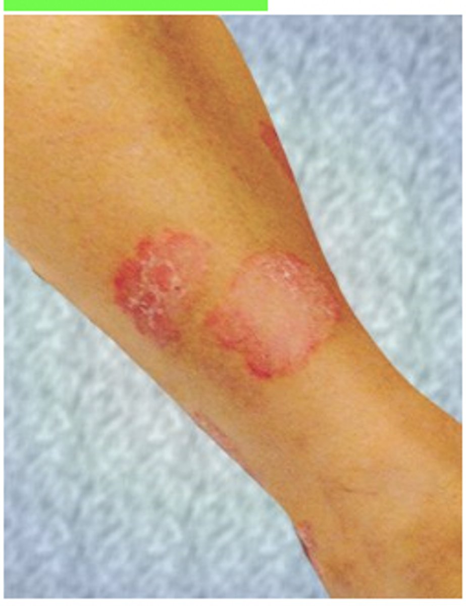
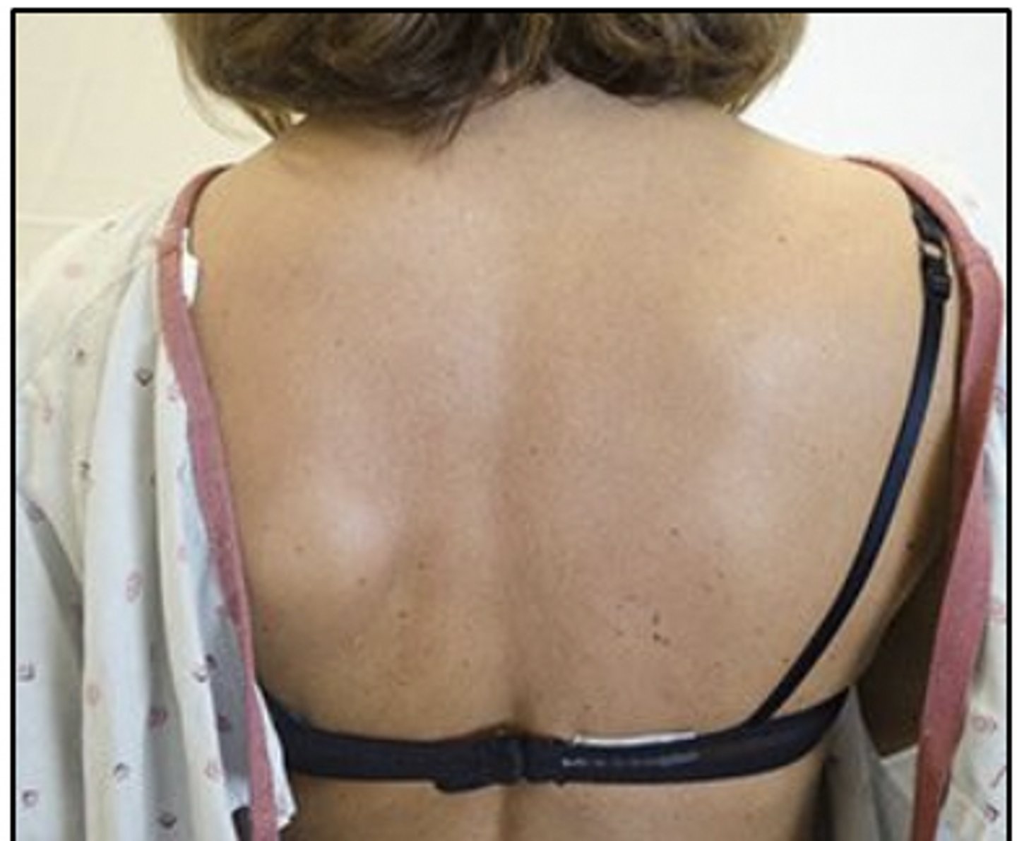
A patient's leg has the skin changes shown in the photograph. Further examination reveals the presence of similar lesions on the opposite extremity, elbows, knees, and scalp. The patient MOST likely has which of the following conditions?
1. Melanoma
2. Lyme disease
3. Scleroderma
4. Psoriasis
Rationale
1. Melanoma typically is characterized by a colored, irregularly shaped lesion that can be mottled with light brown to black colors (p. 435).
2. The rashes associated with Lyme disease typically start as a red spot that expands with clearing of redness in the central area (p. 359).
3. Skin changes associated with scleroderma mainly include Raynaud phenomenon and tightening of the skin. Appearance of a rash is not typical of this disease. (pp. 445-447)
4. The image shows well-defined, dry, erythematous keratinous plaques, which are typical of psoriasis. These plaques are most commonly found in the scalp, extensor surfaces of extremities, and, in severe cases, the trunk. Identifying these plaques is important for the physical therapist in making decisions regarding referral for further medical attention. (pp. 440-441)
*Which of the following factors is MOST important when considering footwear for a patient with diabetes?
1. Leather soles and heels
2. Selection of a shoe without laces
3. Snug fit around the heel
4. Non-leather material uppers
Rationale
1. For all patients who have diabetes, footwear should have a soft lining for protection from and prevention of excessive friction and pressure (Nather, p. 528). Shoes with leather soles are made of firmer material and are typically not soft and cushioned.
2. Shoes should be fastened with adjustable laces, straps, or Velcro high on the foot to keep the foot secure and reduce frictional force (Nather, p. 528).
3. For all patients who have diabetes, footwear should offer a supportive structure for stability and offer protection from and prevention of excessive friction (Nather, p. 528). A snug fit around the heel can provide this stability.
4. Shoes for the insensitive foot should be soft leather that will conform to abnormalities on the dorsal surface (Lusardi, p. 175).
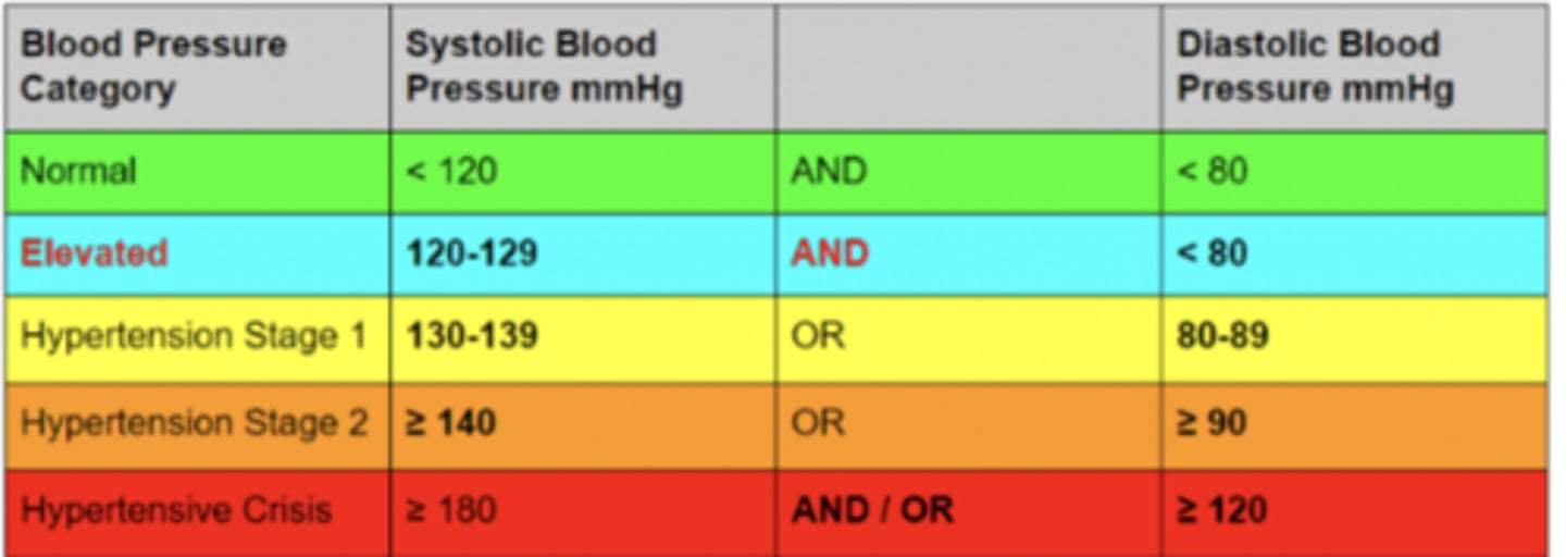
Which of the following findings is CONSISTENT with low risk for development of metabolic syndrome?
1. Triglyceride level of 135 mg/dL (1.5 mmol/L)
2. Blood pressure reading of 135/85 mm Hg
3. Fasting blood glucose level of 126 mg/dL (7.0 mmol/L)
4. Waist measurement of 41 in (104.1 cm)
Rationale
1. Triglyceride level below 150 mg/dL is normal.
2. Blood pressure reading equal to or greater than 130/85 mm Hg is a risk factor for the development of metabolic syndrome.
3. Fasting glucose of 100 mg/dL (5.5 mmol/L) or more is a risk factor for the development of metabolic syndrome.
4. Waist measurement greater than 35 inches (89 cm) for women or 40 inches (102 cm) for men is a risk factor for the development of metabolic syndrome.
Metabolic Syndrome: a cluster of biochemical and physiological abnormalities associated with the development of cardiovascular disease and type 2 diabetes.
Risk Factors for developing Metabolic Syndrome:
· BP ≥ 135/85mmHg
· blood sugar/fasting glucose ≥ 100mg/dL
· waist measurement >35in F or >40in M
· triglycerides > 150mg/dL
· HDLs (good cholesterol) <50mg/dL F or <40 mg/dL M
· If 3 risk factors are present, suspect metabolic syndrome
A patient who has meralgia paresthetica has been referred to physical therapy. Which of the following clinical features is MOST likely to be assessed by the physical therapist during the examination?
1. Strength of the adductor longus
2. Strength of the quadriceps femoris
3. Sensation of the superior medial aspect of the thigh
4. Sensation of the lateral aspect of the thigh
Rationale
1. The adductor longus is innervated by the obturator nerve, and strength testing would assess the motor integrity of this nerve. Meralgia paresthetica does not involve the obturator nerve.
2. The quadriceps femoris is innervated by the femoral nerve, and strength testing would assess the motor integrity of this nerve. Meralgia paresthetica does not involve the femoral nerve.
3. Meralgia paresthetica is an entrapment or injury to the lateral femoral cutaneous nerve, a purely sensory nerve. Injury affects sensation to the lateral thigh. Sensory testing of the superior medial aspect of the thigh would be an assessment of the ilioinguinal nerve.
4. Meralgia paresthetica is an entrapment or injury to the lateral femoral cutaneous nerve, a purely sensory nerve. Injury affects sensation to the lateral thigh. Sensory testing of this region is the most appropriate assessment.
An exercise session that includes 25 minutes of continuous practice and 5 minutes of rest BEST represents which of the following types of practice?
1. Massed
2. Distributed
3. Blocked
4. Random
1. Massed practice refers to a sequence of practice and rest times in which rest time is much less than the practice time (p. 34).
2. Distributed practice refers to practice intervals in which the practice time is equal to or less than the rest time (p. 34).
3. Blocked practice refers to a practice sequence organized around one task performed repeatedly, uninterrupted by practice of any other tasks. Therefore, this type of practice is not related to the amount of time spent on performing a task. (p. 35)
4. Random practice refers to a practice sequence in which several various tasks are ordered randomly across trials (p. 35).
· Massed: Practice time > rest time
· Distributed: Practice time = rest time
· Blocked: Practice of one task repeatedly (111) (222) (333)
· Serial: Predictable, repeated order of multiple tasks (123123123)
· Random: Tasks practiced in random order (123321312)
· Parts-to-Whole: Tasks broken into component parts for separate then integrated practice
· Mental: Motor task is envisioned without overt physical practice
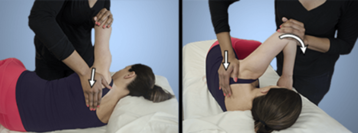
A patient who has diabetes mellitus reports a progressive loss of shoulder mobility. A physical therapist performs the test shown in photographs A and B. Which of the following conditions is MOST likely being assessed?
1. Functional horizontal adduction
2. Posterior capsule tightness
3. Acromioclavicular joint tightness
4. Scapular dyskinesia
Rationale
1. Functional horizontal adduction is performed in sitting or standing position, and strength testing for horizontal adduction is performed with the patient in supine position (Hislop, p. 128). Functional activities require accompanying movements at the scapula. The procedure shown in the photographs prevents scapular movement.
2. The photograph shows the technique to test for posterior capsular tightness. Retracting the scapula as illustrated removes the confounding compensatory effect of scapular protraction and allows isolation of the glenohumeral joint. The posterior capsule has been implicated as the source of restriction in this test (Magee, p. 285). In addition, patients who have diabetes are likely to experience shoulder disorders/mobility limitations (Goodman, p. 429).
3. Horizontal adduction does indeed test for pathological conditions of the acromioclavicular joint. However, the test is performed in sitting or standing position, and the scapula is not retracted, because one would need to be able to adduct the humerus to end range to elicit acromioclavicular joint symptoms. Retracting the scapula will limit horizontal adduction. (Magee, pp. 285, 330)
4. Assessment of scapular dyskinesia would require assessment of scapular mobility. The procedure shown in the photographs prohibits movement of the scapula. (Magee, p. 260).
A patient who has chronic obstructive pulmonary disease becomes short of breath when walking 5 feet (1.5 m) with a rolling walker. Which of the following techniques would be MOST appropriate in order to increase the distance the patient is able to walk without becoming short of breath?
1. Incentive spirometry
2. Pacing
3. Diaphragmatic breathing
4. Segmental breathing
Rationale
1. Incentive spirometry is used to improve inspiratory volumes and chest expansion. It should be used with caution in patients who have chronic obstructive pulmonary disease. It does not improve exercise tolerance. (Reid, p. 261)
2. Using pacing, the patient would learn to work within his or her exercise tolerance. This may mean walking slower or walking with breaks and would allow greater total walking distance without shortness of breath. (O'Sullivan, p. 513)
3. Diaphragmatic breathing is used to decrease the work of breathing and improve diaphragmatic movement, not to improve exercise tolerance (Frownfelter, pp. 357-358; Hillegass, pp. 550-551).
4. Segmental breathing is used for patients with chest hypomobility to augment localized lung expansion (Frownfelter, pp. 362-364). Chest hypomobility is not usually an issue in patients who have chronic obstructive pulmonary disease.
Which of the following procedures is MOST appropriate for measuring a wound that has well-defined margins?
1. Clean the skin around the wound, place a nonsterile ruler on the wound to obtain measurements, and then clean the ruler for future use.
2. Clean the skin around the wound, place a nonsterile ruler on the wound to obtain measurements, and then discard the ruler after use.
3. Place a nonsterile ruler close to the wound to obtain measurements and then clean the ruler for future use.
4. Place a nonsterile ruler close to the wound to obtain measurements and then discard the ruler after use.
Rationale
4.
Despite cleaning the area, the ruler should not make contact with the wound, and the ruler should be discarded after a single use.
A patient with weakness of the muscle group being tested in the photograph would have the MOST difficulty with which of the following activities?
1. Stepping up on a curb
2. Walking on a level surface
3. Sitting up from a reclining position
4. Bringing the trunk forward in sitting position
The quadriceps group is being tested in the photograph (Hislop, p. 248). This option is correct because maximum torque of the knee extensors reaches a peak at about 60° of knee flexion and decreases with further extension of the knee. Stepping up on a curb requires a greater workload for the quadriceps muscle group, compared to the other activities listed. (Houglum, p. 460).
Walking on a level surface requires less quadriceps work than stepping up on a curb because maximum torque of the knee extensors reaches a peak at about 60° of knee flexion and decreases with further extension of the knee (Houglum, p. 460). The quadriceps group is being tested in the photograph (Hislop, p. 248).
With the femur fixed, the hip flexors will flex the trunk forward. Although the rectus femoris is a hip flexor and knee extensor, weakness of the rectus femoris can be compensated for in this action by other hip flexors such as the iliopsoas. (Loudon, p. 270)
A patient has been prescribed warfarin (Coumadin) following total hip arthroplasty. Which of the following over-the-counter medications listed in the patient's medical history at the first postoperative visit would be of GREATEST concern to a physical therapist?
1. Diphenhydramine (Benadryl)
2. Cetirizine (Zyrtec)
3. Omeprazole (Prilosec)
4. Acetylsalicylic acid (Aspirin) - beta blocker
Rationale
1. Benadryl is a histamine antagonist and is not listed as a drug that has anticoagulant effects (p. 278).
2. Cetirizine is a histamine antagonist and is not listed as a drug that has anticoagulant effects (p. 278).
3. Omeprazole is a proton pump inhibitor. It is not described to interact with warfarin or have anticoagulant effects. (pp. 1085-1089)
4. Acetylsalicylic acid (Aspirin) and warfarin are both anticoagulants. The most serious interactions with warfarin are those that increase anticoagulant effects and the risk of bleeding (p. 610). Patients who are taking warfarin should be instructed not to take acetylsalicylic acid (Aspirin) simultaneously without discussing the combination with a physician. Taking both could cause excessive anticoagulation, which could be harmful.
The condition shown for the patient's left hand in the photograph is MOST likely caused by entrapment of which of the following nerves?
1. Anterior interosseous nerve
2. Radial nerve
3. Posterior interosseous nerve
4. Ulnar nerve
Rationale
1. The image shows an anterior interosseous syndrome (Kiloh-Nevin syndrome) in the patient's left hand. The patient is unable to flex the distal phalanx of the thumb and index fingers (1st and 2nd digits) because the anterior interosseous nerve, which supplies the flexor pollicis longus and the radial half of the flexor digitorum profundus, is entrapped. (p. 411)
2. With entrapment of the radial nerve, all extensor muscles of the forearm would be affected (p. 416). The image shows a deficit in the pinch of the thumb and index finger (1st and 2nd digits).
3. Entrapment of the posterior interosseous nerve results in functional wrist drop (p. 416). The image shows a deficit in the pinch of the thumb and index finger (1st and 2nd digits).
4. When the ulnar nerve is affected, the patient cannot fully adduct the little finger (5th digit) and hold the finger abducted and extended (p. 415). The image shows a deficit in the pinch of the thumb and index finger (1st and 2nd digits).

A patient reports upper extremity numbness and tingling that extends from the neck to the thumb and index finger (1st and 2nd digits). Which of the following shoulder positions would MOST likely exacerbate the patient's symptoms?
1. Lateral (external) rotation with abduction
2. Medial (internal) rotation with abduction
3. Lateral (external) rotation with adduction
4. Medial (internal) rotation with adduction
Rationale
1. Numbness and tingling over the thumb and index finger (1st and 2nd digits) involves the median nerve. Shoulder lateral (external) rotation with abduction is used to test the median nerve (upper limb tension test [ULTT 2a]). Shoulder lateral (external) rotation is added to 90° of shoulder abduction combined with shoulder girdle depression to place tension on the median nerve.
2. Shoulder medial (internal) rotation is used when testing for radial nerve involvement (upper limb tension test [ULTT 2b]).
3. Adduction of the shoulder would reduce tension on the median nerve; abduction increases tension on the nerve.
4.. Medial (internal) rotation with adduction is not used to test upper extremity neural tension because it does not create adequate neural tension.
A physical therapist is testing the strength of a patient's latissimus dorsi with the patient in prone position and arms at the side with palms facing the ceiling. Following instruction in the desired motion, the patient lifts the arm off the table after first turning the palm downward. The therapist should ask the patient to do which of the following actions NEXT?
1. Repeat the motion with the palm upward.
2. Extend the arm while seated without back support.
3. Repeat the motion while the therapist adds resistance.
4. Extend the arm while in sidelying position with the upper arm supported.
Rationale
1. Extending the shoulder with lateral (external) rotation allows the long head of the triceps to substitute for the latissimus dorsi. The therapist must ensure that the patient understands the desired motion before determining whether to add resistance or change the patient's position. (pp. 58-60)
2. Sitting is not a position used to test latissimus dorsi strength. Sitting would decrease the effect of gravity. (pp. 57-58)
3. The patient must be able to perform the proper motion against gravity before resistance can be added (p. 59).
4. The therapist may have chosen to start testing in the gravity-eliminated position (sidelying) if less than Fair (3/5) strength was suspected, but, having chosen to start in the against-gravity position, the therapist should first determine whether the patient truly cannot perform the motion against gravity before changing position (p. 60).
Lat Action: The latissimus dorsi is responsible for extension, adduction, horizontal abduction, flexion from an extended position, and (medial) internal rotation of the shoulder joint. It also has a synergistic role in extension and lateral flexion of the lumbar spine.
Which of the following locations of pain is MOST consistent with bladder infection?
1. Groin
2. Sacral area
3. Lower buttocks
4. Suprapubic area
1. Groin pain is associated with upper urinary tract problems, such as kidney or ureter infection. (ureter referral)
2. Sacral pain is associated with colon cancer and colitis.
3. Lower back, not lower buttocks, is associated with bladder infection. (kidney referral)
4. Pain generated by the bladder typically manifests in the suprapubic area and the lower back.
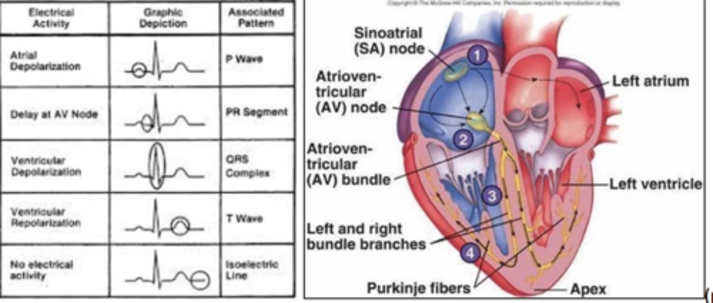
A patient's electrocardiogram shows a junctional rhythm. The patient's heart rate is 60 bpm and regular. Which of the following waves will MOST likely be absent from the rhythm strip?
1. P
2. R
3. S
4. T
Rationale
1. Junctional rhythm originates from the atrioventricular junction instead of the sinoatrial node, which normally causes the P wave. Therefore, the P wave will be missing. R, S, T waves come from the ventricles after stimulation from the atrioventricular junction and will be unaffected.
2. R waves come from the ventricles after stimulation from the atrioventricular junction and will be unaffected.
3. S waves come from the ventricles after stimulation from the atrioventricular junction and will be unaffected.
4. T waves come from the ventricles after stimulation from the atrioventricular junction and will be unaffected.
The asymmetrical position in the photograph is MOST likely due to a lesion in which of the following nerves?
1. Long thoracic
2. Spinal accessory
3. Axillary
4. Dorsal scapular
Rationale
1. The long thoracic nerve innervates the serratus anterior. Weakness of the serratus anterior results in winging of the scapula, which is the pathological position shown in the photograph. (Magee, p. 281; Drake, pp. 726-727, 744)
2. The spinal accessory nerve innervates the sternocleidomastoid and trapezius. The trapezius adducts and upwardly rotates the scapula (Drake, pp. 895, 1024). The sternocleidomastoid flexes the head to the side and rotates the head to the contralateral side (Magee, p. 174). These muscles are not involved in the asymmetrical position shown in the photograph.
3. The axillary nerve innervates the deltoid and teres minor, which are not involved in the asymmetrical position shown in the photograph (Magee, pp. 177, 287).
4. The dorsal scapular nerve innervates the rhomboids, which elevate, retract, and downwardly rotate the scapula and are not involved in the asymmetrical position shown in the photograph (Drake, pp. 715-716, 744; Magee, p. 287).
A patient has a left thoracolumbar scoliosis. Pelvic landmarks are symmetrical. Which of the following muscles will MOST likely be tight?
1. Right hip abductors
2. Left latissimus dorsi
3. Right quadratus lumborum
4. Left iliocostalis lumborum
Rationale
1. The right hip abductors will be normal length since the pelvis is level.
2. The left latissimus dorsi will be normal or lengthened dependent on the severity of the curve.
3. With a left thoracolumbar scoliosis, the C curve is concave on the right, resulting in shortened trunk musculature on the right, i.e., quadratus lumborum.
4. The left iliocostalis lumborum will be of normal length or lengthened, depending on the severity of the curve.
A patient who has hypothyroidism is MOST likely to exhibit which of the following signs or symptoms?
1. Ptosis
2. Muscle ache
3. Dysphagia
4. Tachycardia
Rationale
1. Ptosis is not a common symptom of hypothyroidism.
2. Muscle ache (myalgia) is a common musculoskeletal symptom of hypothyroidism.
3. Dysphagia is not a common symptom of hypothyroidism.
4. Bradycardia, not tachycardia, is a common symptom of hypothyroidism.
Common signs of hypothyroidism: fatigue, muscle ache, weakness, bradycardia, weight gain, constipation, delayed puberty, retarded growth/development
A patient who sustained a traumatic brain injury and is unable to follow commands has been referred for physical therapy evaluation. When the physical therapist arrives at bedside, the patient is agitated. Which of the following actions should the therapist take INITIALLY?
1. Carefully observe the patient's spontaneous behavior.
2. Postpone the assessment until the patient has become calm.
3. Apply soft restraints to calm the patient before assessment.
4. Proceed with the assessment regardless of the patient's agitated state.
Rationale
1. Confused-agitated is a state common in patients following traumatic brain injury. Observing the patient without touching the patient will reveal information that is important for the evaluation.
2. The agitated state may exist for some time, and the assessment should not be postponed.
3. The patient needs to feel safe. The therapist should model calm behavior. Restraining the patient would produce more fear and agitation.
4. Formal measurements of range of motion and strength are difficult, and the patient is unable to cooperate. The therapist should use only observation and estimate functional abilities.
A patient with severe arthritis of the hips and knees is able to partially stand but cannot clear the armrest of the wheelchair adequately during stand-pivot transfers. Which of the following strategies is BEST to facilitate the transfer?
1. Design a therapy program for increasing strength of the lower extremities.
2. Design a therapy program for improving active range of motion of the lower extremities.
3. Recommend that the family acquire a wheelchair with removable armrests.
4. Recommend that the family acquire a mechanical lift for transfers.
Rationale
1. A strengthening program should be encouraged, but a wheelchair with removable armrests would allow the patient to transfer even during periods of exacerbation of the severe arthritis.
2. A range of motion program should be encouraged, but a wheelchair with removable armrests would allow the patient to transfer even during periods of exacerbation of the severe arthritis.
3. The patient is able to partially stand. Removable armrests are recommended for patients who will perform a lateral swinging or squat-pivot transfer.
4. The patient is able to partially stand. The least restrictive device should be used to encourage independence.
Which of the following activities should be the PRIMARY emphasis of a physical therapy treatment program for a child who has athetoid cerebral palsy?
1. Facilitating cocontraction patterns and encouraging control in voluntary movement gradation
2. Increasing muscle strength using progressive resistive exercises
3. Facilitating use of primitive reflexes to perform gross motor tasks
4. Preventing development of contractures and ensuring full voluntary range of motion
Rationale
1. Athetoid cerebral palsy is characterized by involuntary movements that are slow and writhing. In therapy, the emphasis should be on facilitating cocontraction and encouraging control in voluntary movement.
2. Although strength training is indicated in children with cerebral palsy, a child with athetoid cerebral palsy lacks the control to consistently produce a maximal effort in a controlled movement. Therefore, the focus must first be on gaining control, then on traditional strength training.
3. A goal of therapy would not be to reinforce primitive reflexes. The primary need for this child is to gain controlled movement.
A patient has acute rheumatoid arthritis involving the wrist joints. Which of the following interventions is MOST appropriate?
1. Resistive exercises to end range
2. Functional fine motor tasks
3. Splints with wrists in neutral position
4. Passive stretching exercises
Rationale
1. Strengthening can be difficult especially with pain of the acute phase.
2. Active exercise (needed for functional fine motor tasks) has questionable benefit in the acute phase.
3. Splints can be applied to rest the involved joints, prevent excessive movement, and reduce mechanical stresses, all of which are desired outcomes in the acute phase of rheumatoid arthritis.
4. Passive stretching exercises are important as a part of a rehabilitation effort; however, given the acute nature of the problem, rest and protection are paramount, making this option inappropriate.
A patient who has a history of heart disease is being treated for left glenohumeral dysfunction. The patient reports left upper quadrant pressure that continues after joint mobilization has ceased. Which of the following actions is MOST appropriate for the physical therapist?
1. Assess the patient's cervical spine nerve root integrity.
2. Have the patient perform relaxation exercises and inquire about cardiac symptoms.
3. Stop the treatment and monitor the patient's vital signs.
4. Resume joint mobilization at a lower intensity and reassess the patient's status.
Rationale
1. The cervical spine could be of concern, but neurologic symptoms typically include tingling, numbness, weakness, or burning pain, not "pressure" (p. 701). Left upper quadrant pain is a red flag (warning sign), especially with the patient's history of heart disease.
2. The patient is already having a potential cardiac symptom (p. 255). Relaxation may decrease sympathetic tone and decrease anginal symptoms, but first vital signs should be assessed.
3. Vague left upper quadrant pressure pain can be an anginal equivalent and indicate myocardial infarction. Given this possibility, one should stop and assess vital signs, especially in a patient with a past medical history of heart disease. (pp. 701-702)
4. The symptoms are present at rest after joint mobilization; thus, joint mobilization should not be resumed. Left quadrant pain is a red flag (warning sign), especially with the patient's history of heart disease; therefore, it is appropriate to stop treatment and seek medical attention. (p. 701)
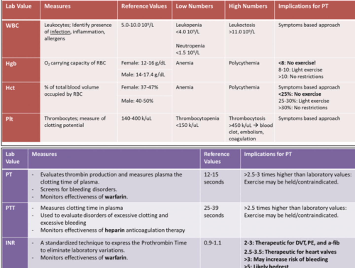
Which of the following laboratory values should a physical therapist monitor when treating a patient who is taking warfarin (Coumadin)?
1. Hemoglobin
2. Red blood cell count
3. International normalized ratio
4. Erythrocyte sedimentation rate
1. Hemoglobin would not be changed by anticoagulant medication. Hemoglobin values measure the oxygen-carrying capacity of the red blood cells. (p. 1713)
2. Red blood cell counts are not changed by anticoagulants. The red blood cell count is a method used to assess the oxygen-carrying capacity of the blood. (p. 1712)
3. Warfarin (Coumadin) is an anticoagulant. The physical therapist must be aware when a patient is taking an anticoagulant so that treatment can be modified if there is an increased risk of hemorrhage. The international normalized ratio (INR) was developed to provide results that would not vary between laboratories. Therapeutic anticoagulation requires an INR of 2 to 3. As the INR increases above these values, the risk of bleeding during activity is increased. (pp. 1712-1713)
4. The erythrocyte sedimentation rate is not expected to be affected by anticoagulant medication. The erythrocyte sedimentation rate is used to identify inflammatory or necrotic processes. (p. 1715)
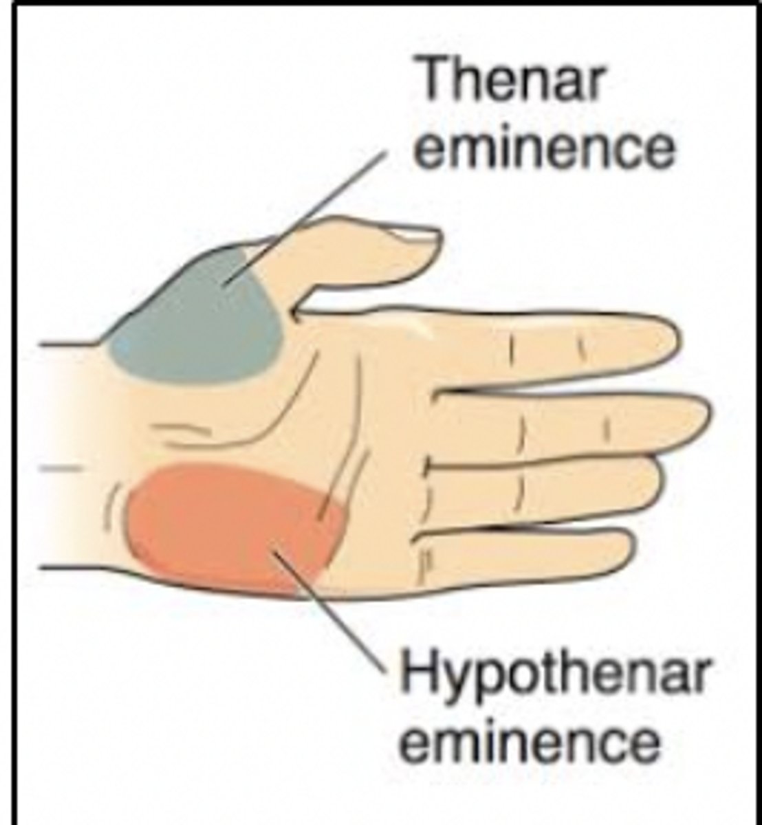
*Initial examination of a patient reveals paresthesia over the hypothenar eminence. The MOST probable cause of this condition is:
1. carpal tunnel syndrome.
2. C8 nerve root involvement.
3. de Quervain tenosynovitis.
4. pronator teres syndrome.
Rationale
1. Carpal tunnel syndrome is a result of entrapment of the median nerve, which innervates the thenar eminence.
2. The C8 nerve root innervates the hypothenar eminence. Injury to the C8 nerve root will cause paresthesia in the hypothenar eminence
3. De Quervain tenosynovitis affects the first dorsal compartment. It would not cause paresthesia in the hypothenar eminence. (Dutton, pp. 847-848)
4. Pronator teres syndrome affects the median nerve. It would cause paresthesia in the thenar eminence.
The rationale for using superficial heat prior to exercise includes all of the following EXCEPT:
1. increasing core temperature.
2. increasing tissue temperature.
3. promoting relaxation.
4. reducing pain.
1.
Rationale
Superficial heat cannot increase core (body) temperature. Superficial heat can increase tissue temperature, decreases the nerve firing rate and muscle spasm, thereby relaxing the muscle, and can increase the pain threshold (reduce pain).
A person with spina bifida uses a knee-ankle-foot orthosis to:
1. provide support for muscle incoordination.
2. facilitate muscular activity.
3. prevent development of muscle contractures.
4. substitute for the lack of muscle activity.
Rationale
1. A person who has spina bifida uses a knee-ankle-foot orthosis when motor function is weak or absent or to address knee instability. It is not commonly used for incoordination of muscles.
2. A knee-ankle-foot orthosis is not able to facilitate muscle activity.
3. A knee-ankle-foot orthosis is not commonly used to prevent muscle contractures. It may be used when contractures are already present, if these contractures prevent upright positioning.
4. A knee-ankle-foot orthosis is commonly used when muscles of the knee are weak and muscles of the ankle are absent. It substitutes for lack of muscle activity.
Spina Bifida: a congenital defect of the spine in which part of the spinal cord and its meninges are exposed through a gap in the backbone. It is a developmental abnormality due ti insufficient closure of the neural tube by the 28th day of gestation. It often causes paralysis of the lower limbs, and sometimes mental handicap.
Shoulder pain during the test shown in the photograph MOST likely indicates which of the following pathologies?
1. Anterior glenohumeral instability
2. Cubital tunnel syndrome
3. Shoulder impingement syndrome
4. Thoracic outlet syndrome
Rationale
1. The photograph shows the Hawkins-Kennedy test. Anterior glenohumeral instability is not tested with the Hawkins-Kennedy test. The Hawkins-Kennedy test is used to test for impingement signs for the diagnosis of subacromial bursitis or rotator cuff pathology. (p. 630)
2. Cubital tunnel syndrome is not tested with the Hawkins-Kennedy test. The elbow flexion test or Tinel sign is used to test for cubital tunnel syndrome. (pp. 738-739)
3. The photograph shows the Hawkins-Kennedy test, which is used to test for impingement syndrome of the shoulder (p. 630).
4. Thoracic outlet syndrome is not tested with the Hawkins-Kennedy test. It is tested with the Roos test. (p. 1300)
Patients with advanced emphysema experience difficulty in breathing during exercise because of:
1. hypocapnia.
2. atrophy of secondary breathing muscles.
3. alveolar dilation.
4. damage to the phrenic nerve.
Rationale
1. Patients with emphysema have normal or slightly elevated partial pressure of arterial carbon dioxide (PaCO2) (p. 88), not hypocapnia (or decreased CO2).
2. Patients who have emphysema tend to breathe with accessory muscles of respiration (p. 87), which may lead to hypertrophy, not atrophy of those muscles.
3. Emphysema is characterized by abnormal and permanent enlargement of the air spaces distal to the terminal nonrespiratory bronchioles accompanied by destructive changes of the alveolar walls (p. 86).
4. There is no involvement of the phrenic nerve in this condition.
Emphysema results from a long history of chronic bronchitis, recurrent alveolar inflammation or from genetic predisposition of a congenital alpha 1-antitrypsin deficiency; results from a non-reversible injury and destruction of elastic protein within the alveolar walls à permanent enlargement of airspaces distal to terminal bronchioles; chronic progressive disease; blebs and bullae
risk factors: chronic bronchitis, cig smoking, lower respiratory infections, genetics
S&S: wheezing, persistent cough, difficulty breathing – especially with expiration, increased RR, barrel chest, rounded shoulders d/t tight pecs, pursed-lip breathing strategy
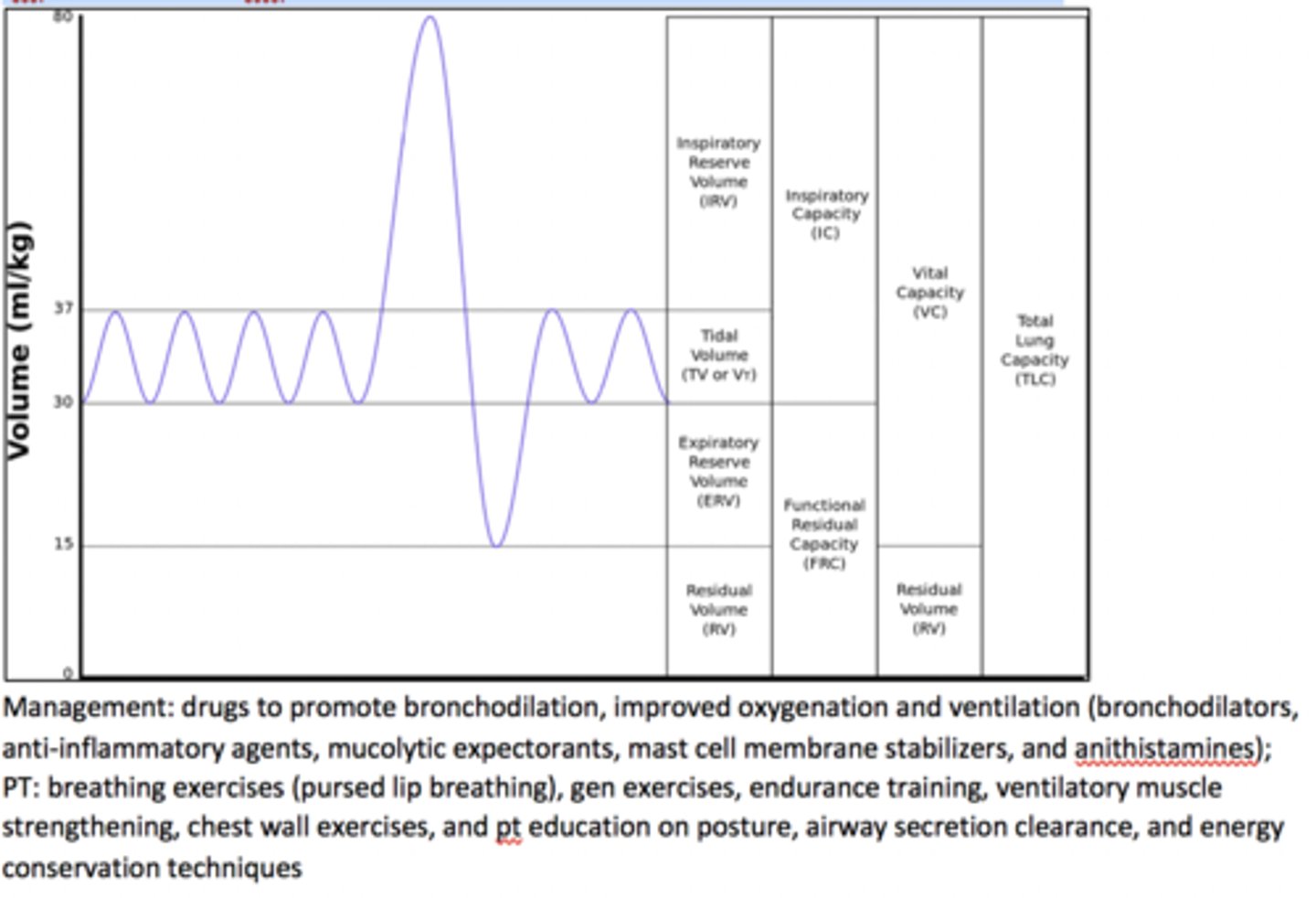
(Pg 431)Pulmonary fxn tests: impaired FEV1, VC, and FVA; increased TLC, RV, and FRC
*A therapist is treating a patient who recently had a myocardial infarction. At the beginning of treatment, blood pressure was 120/80 mm Hg and heart rate was 90 beats/min. Midway through treatment, blood pressure was 130/84 mm Hg and heart rate was 105 beats/min. The BEST action for the therapist to take is to:
1. continue with treatment.
2. increase the intensity of treatment.
3. stop the treatment, and notify the physician.
4. decrease the intensity of the next treatment.
Rationale
1.CONTINUE WITH TX
Systolic blood pressure is expected to rise in direct proportion to the level of exertion performed. A hypertensive response to low-level exercise (over 160/90 mm Hg) in the patient who is at least 3 days post myocardial infarction may be indicative of cardiac ischemia. Heart rate should increase between 12-24 bpm above the resting heart rate. The patient is showing a normal response to exercise and should continue with treatment. There is NO indication to increase activity level or to stop treatment. After a recent myocardial infarction, the patient should avoid activities that cause a significant change in vital signs.
Responses to exercise: HR increases linearly as a function of increasing workload and oxygen uptake (VO2) but plateaus just before maximal oxygen uptake (VO2max); systolic BP should rise with increasing workloads and VO2, but diastolic BP should remain about the same.
See pg~160 of ACSM!!!!
A peak DBP >90 mm Hg or an increase in DBP >10 mm Hg during exercise above the
pretest resting value is considered an abnormal response (17) and may occur with exertional
ischemia (53). A DBP >115 mm Hg is an exagerated response and a relative indication to stop a test
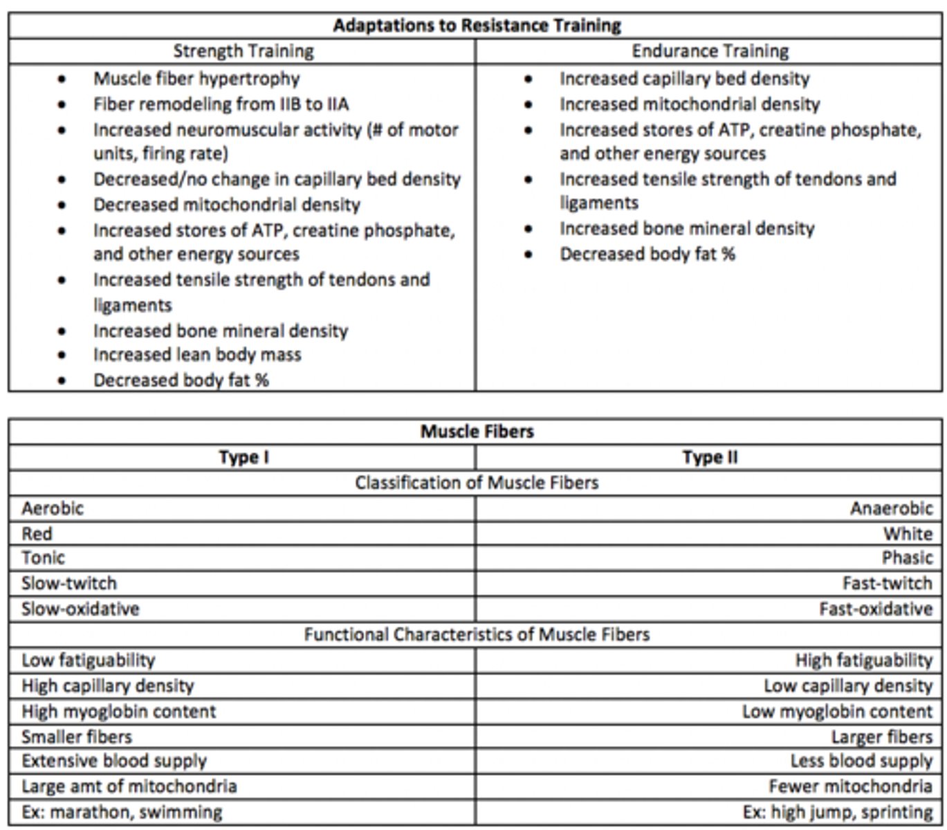
After 1 week of a progressive resistance exercise training program, an individual demonstrates significant strength gains. The MOST likely explanation for the observed strength gains is:
1. an increased ratio of fast- to slow-twitch fibers.
2. improved neuromuscular recruitment.
3. muscle-fiber hyperplasia.
4. muscle hypertrophy.
Rationale
1. Although transformation of type IIB to type IIA fibers occurs in the early weeks of resistance training, transformation from slow-twitch to fast-twitch fibers is unlikely (p. 169).
2. The initial rapid gain in the tension-generating capacity of skeletal muscle is largely attributable to neural responses, including increased recruitment in number of motor units firing and increased rate of synchronization of firing (p. 168).
3. Muscle-fiber hyperplasia is an increase in the number of muscle fibers, and if it occurs, it is in response to heavy resistance training and only accounts for a small percentage of the increase in strength (p. 168).
4. Muscle hypertrophy, or increase in the size of individual muscle fibers, requires an extended period (4-8 weeks) of moderate-intensity to high-intensity resistance training (p. 168). One week is too short of a duration for such change.
*A home health physical therapist is working with a patient who had a myocardial infarction 2 weeks ago. The patient reports interrupted sleep, increased swelling of the feet, and shortness of breath. The patient's heart rate is 120 bpm and respiratory rate, 28 breaths/minute. Auscultation reveals crackles in both lung bases. The therapist should suspect:
1. acute congestive heart failure.
2. pneumonia in bilateral lower lobes.
3. atelectasis.
4. renal failure.
Rationale
1. These signs and symptoms are consistent with congestive heart failure (Hillegass, p. 97).
2. Pneumonia may result in orthopnea (shortness of breath (dyspnea) that occurs when lying flat) and disrupted sleep, but it would not cause lower extremity edema
3. Atelectasis may be associated with crackles (rales) and shortness of breath, but the other symptoms are not consistent with atelectasis (partial or complete collapse of the lung)
4. Renal failure may result in lower extremity edema, shortness of breath, and tachypnea (rapid breathing) but not crackles and tachycardia (Goodman, pp. 396-397).
*A patient with a transtibial amputation is being treated by a physical therapist for gait training with a prosthesis. The patient reports tingling and shooting pain at the end of the residual limb. The pain occurs whether or not the patient is wearing the prosthesis. The pain is MOST likely caused by which of the following?
1. A neuroma
2. Inadequate prosthetic tibial relief
3. Distal soft-tissue adhesions
4. Osteomyelitis
Rationale
1. A neuroma is a collection of axons and fibrous tissue that can cause sharp, shooting, and localized pain (Lusardi, p. 707). Localized hypersensitivity may be an indicator that a neuroma has developed.
2. Inadequate prosthetic tibial relief may result in skin breakdown, which would be seen and is not indicated in the stem (Lusardi, p. 714).
3. Adherent scar tissue near the end of the bone is a particular problem that may lead to skin breakdown, which is not indicated in the stem (Lusardi, p. 819).
4. Clinical manifestation of pain with osteomyelitis may be described as deep, constant, and increasing with weight-bearing when present in the lower extremity. Patients may report local pain and swelling, which is not indicated in the stem. (Goodman, p. 1236)
A physical therapist is obtaining the medical history of a patient with amyotrophic lateral sclerosis. Which of the following is MOST important to ask about in order to determine the prognosis for this patient?
1. Swallowing difficulties
2. Cognitive deficits
3. Bowel and bladder function
4. Neck pain
Rationale
1. Patients with an initial onset of bulbar and respiratory weakness tend to have a more rapid progression to death than patients whose weakness begins in the distal extremities.
2. Cognitive deficits are not associated with amyotrophic lateral sclerosis.
3. Sphincter control problems are not a component of amyotrophic lateral sclerosis.
4. Musculoskeletal pain is not predictive for prognosis in amyotrophic lateral sclerosis.
ALS: a disease affecting UPPER AND LOWER motor neurons of the spinal cord, which causes progressive weakness and atrophy of muscles.
* A physical therapist is examining a patient with low back pain which began 2 months ago while mopping the floor at work. The patient has pain radiating to the buttocks and posterior thigh, has limited lumbar spine range of motion, is unable to perform repeated movements into lumbar flexion, and can only tolerate standing for 5 minutes. Based on this information, which of the following is the MOST appropriate goal for this patient to be met in 2 weeks?
1. Demonstrate normal lumbar spine flexion range of motion.
2. Be able to bend forward 20 times without an increase in leg pain.
3. Return to work with no job modifications.
4. Stand for 10 to 15 minutes without an increase in leg pain.
Rationale
1. Restoring flexion range of motion is not necessarily a functional goal, and the patient would be unlikely to achieve this goal within 2 weeks.
2. Bending forward 20 times without an increase in leg pain is not necessarily a functional goal, and the patient would be unlikely to achieve this goal within 2 weeks.
3. This is a long-term goal, but not one likely to be attained in 2 weeks with a patient who has been symptomatic for 2 months.
4. Improved standing for 10 to 15 minutes within 2 weeks without leg pain is a functional level goal and may reasonably be achieved in 2 weeks.
When compared to maximal oxygen uptake values obtained in a lower extremity exercise test, values obtained in an upper extremity exercise test are typically:
1. 30% to 40% lower.
2. the same.
3. 10% to 20% higher.
4. 30% to 40% higher.
Arm exercise typically results in 30% to 40% lower maximal oxygen uptake than leg exercise.
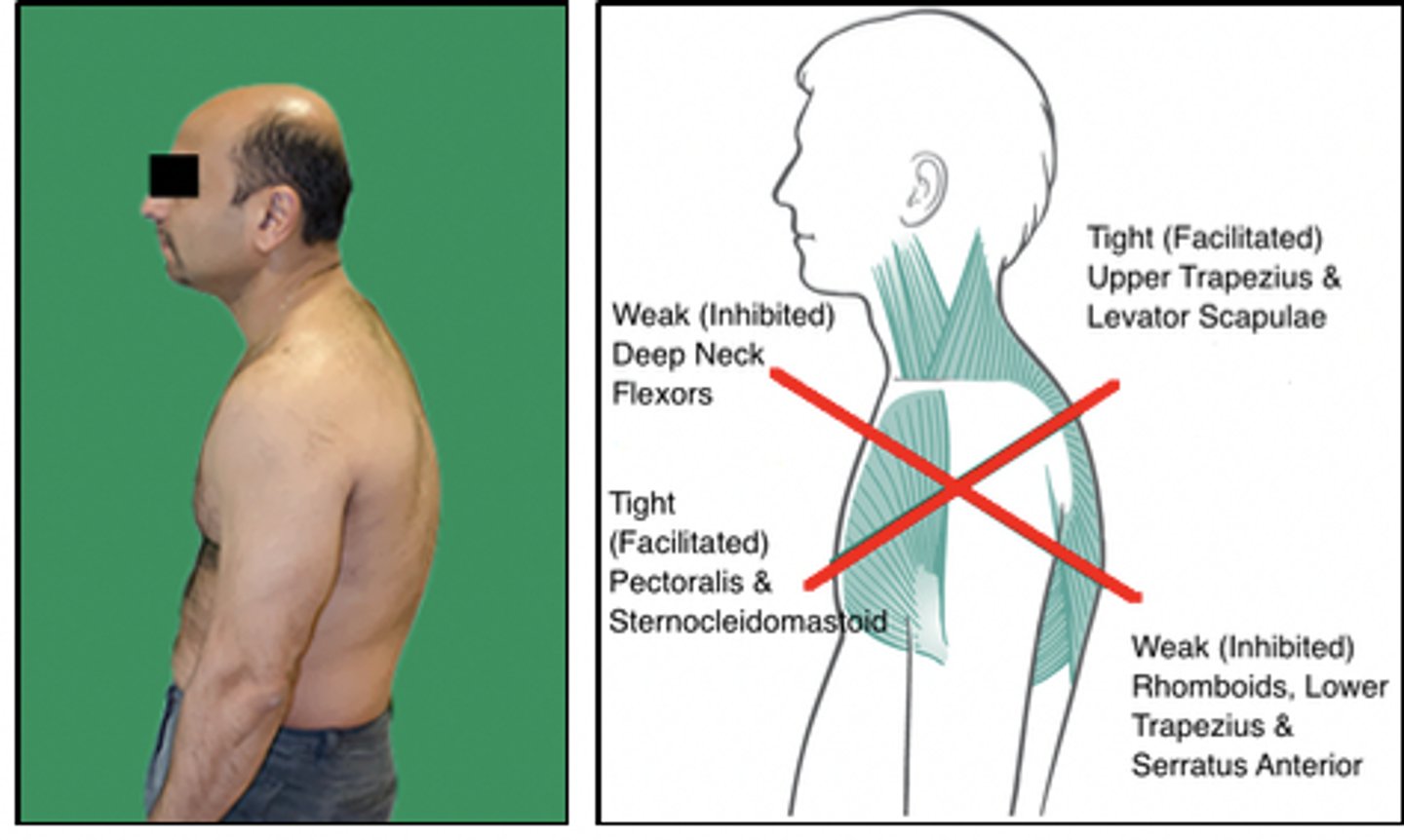
Which of the following muscles is MOST likely to demonstrate postural weakness in the patient shown in the photograph?
1. Long thoracic extensors
2. Pectoralis minor muscles
3. Sternocleidomastoid muscles
4. Suboccipital extensors
Rationale
1. This is correct because the long thoracic extensors demonstrate a stretch weakness in this posture
2. This is incorrect because this patient would use this muscle chronically in its shorter range. Manual muscle testing done in its longer range would have a weaker than normal result. Manual muscle testing in its shorter range would result in a normal or slightly stronger than normal result. Muscle imbalances can occur when a patient strengthens the anterior pectoral muscles and ignores the upper back. This results in tight pectoral muscles and weak rhomboids and trapezius. (Shultz, p. 201)
3. The sternocleidomastoid has increased use during cervical extension (forward head). This muscle would demonstrate a short-strong muscle imbalance. (Hueter-Becker, p. 114)
4. This is incorrect because the suboccipital extensors are chronically in a shorter range. This muscle would demonstrate a short-strong muscle imbalance. (Hueter-Becker, p. 114)
*A physical therapist is examining a 4-year-old child with a history of prematurity and developmental delay. To determine if the child has age-appropriate gross motor skills, the therapist's assessment should include:
1. kicking a rolling ball, catching a small ball, and hopping on one foot.
2. kicking a stationary ball, fast walking, and walking with assistance on stairs.
3. dribbling a basketball, riding a bicycle, and skipping.
4. catching a large ball, riding a tricycle, and running short distances.
Rationale
1. Kicking a rolling ball, catching a small ball, and hopping on one foot are gross motor tasks that are most age-appropriate for a 4-year-old. Gross motor developmental assessment at age 4 years should include functional tasks. (Palisano, p. 62; Tecklin, p. 64)
2. Kicking a stationary ball, fast walking, and walking with assistance on stairs are skills that are appropriate for children age 18 months to 3 years (Palisano, p. 62; Tecklin, p. 64).
3. Dribbling a basketball, riding a bicycle, and skipping are skills that are appropriate for children age 5-6 years (Palisano, p. 63; Tecklin, p. 64).
4. Catching a large ball, riding a tricycle, and running short distances are skills that are appropriate for children age 2-3 years (Palisano, p. 62; Tecklin, p. 64).
A physical therapist is examining a patient by using the test shown in the photograph. Which of the following structures is MOST likely injured?
1. Anterior glenohumeral joint
2. Long head of the biceps brachii
3. Supraspinatus tendon
4. Glenohumeral labrum
Rationale
1. The photograph depicts the apprehension sign test for the presence of anterior glenohumeral instability
2. A test for biceps tendinopathy has the patient producing force into supination with the shoulder in neutral, the elbow bent to 90°, and the forearm starting in pronation (p. 520).
3. The empty can test and the drop arm test are used to check for supraspinatus injuries. In both cases the patient would be sitting upright with the arm raised against gravity. (pp. 522-523)
4. There are several tests used to assess glenohumeral labral tears. The biceps load test has a similar starting position and is used to check for glenoid labrum tear, but the patient's reaction to this position is positive for the apprehension sign, indicating possible anterior glenohumeral instability. (p. 524)
A physical therapist is conducting a graded exercise stress test of an apparently healthy adult using a treadmill. The test should be discontinued if which of the following events occurs?
1. Heart rate continues to increase throughout the test.
2. Borg rating of perceived exertion is reported as 13/20.
3. Diastolic blood pressure reaches 120 mm Hg.
4. Significant redness of the skin and perspiration are observed.
Rationale
1. Monitoring heart rate response to exercise is the purpose of conducting the test, and, therefore, an increasing heart rate is not a reason to stop. Failure of the heart rate to rise with increasing exercise intensity would be a reason to stop. (ASCM, p. 84)
2. A Borg rating of perceived exertion of 13/20 converts to 70% of maximum heart rate and should not be a reason to stop the test (ASCM, p. 83; Kenney, p. 515).
3. A diastolic blood pressure of 120 mm Hg is an indicator for ending the test. A diastolic blood pressure greater than 115 mm Hg is too high to continue testing. (ASCM, p. 84)
4. Significant redness of the skin and perspiration are normal responses to exercise testing. Cyanosis or pallor would be a reason to stop. (ASCM, p. 84)
A 4-year-old child who received a diagnosis of spinal muscular atrophy at age 9 months is referred for home physical therapy. The child is unable to sit without upper extremity support but rolls independently. The child has bilateral hip and knee flexion contractures that make use of the child's standing frame uncomfortable. Which of the following interventions are MOST appropriate for physical therapy?
1. Teach parents airway clearance techniques, encourage upper extremity strengthening to prepare for wheelchair self-propulsion, and switch to using a modified prone stander.
2. Teach parents lower extremity stretching and strengthening exercises, adapt the standing frame to accommodate contractures, and encourage supported walking.
3. Teach parents proper transfers, facilitate upright positioning in kneeling and standing positions, and refer to an orthopedist for serial casting to address contractures.
4. Teach the parents range of motion exercises and positioning, encourage play in prone and sitting positions, and order customized seating insert for a power wheeled mobility device.
Rationale
1. Upper extremity strengthening is not realistic, considering the progressive weakness expected with this child. The skill level would be too high. Manual propulsion is not functional over the long term.
2. The literature supports that children who have spinal muscular atrophy and who do not develop sitting ability are unlikely to walk and will require power mobility. They may become independent in a power wheelchair by age 1-2 years.
3. These skills are too advanced.
4. These interventions are most appropriate to the patient's impairments and functional limitations. The literature supports that children who have spinal muscular atrophy and who do not develop sitting ability are unlikely to walk and will require power mobility.
SMA is a progressive degenerative disease of anterior horn cells; autosomal recessive
*In determining the prognosis of an individual with a traumatic brain injury, which of the following variables is MOST associated with higher level of life satisfaction?
1. Independence in bowel care
2. Being unmarried
3. Age less than 20 years
4. Rapid initial recovery from injury
Rationale
1. Among the variables that significantly increase life satisfaction in individuals with traumatic brain injury is bowel independence (p. 760).
2. Among the variables that significantly increase life satisfaction in individuals with traumatic brain injury is being married (p. 760).
3. Younger age (less than 20 years) is associated with better outcome but not with a higher level of life satisfaction
4. Rate of recovery is not associated with life satisfaction (pp. 759-760).
*A patient in an intensive care unit is intubated and is being treated with a mechanical ventilator. The patient would be UNABLE to participate in which of the following interventions?
1. Pursed-lip breathing training
2. Diaphragmatic breathing training
3. Deep breathing exercises
4. Lateral costal expansion exercises
Rationale
1. Pursed-lip breathing is not possible when the patient is intubated.
Diaphragmatic breathing, Deep breathing, and Lateral costal breathing ARE possible and helpful for a patient who has synchronized intermittent mandatory ventilation in ventilator mode, in which the patient is allowed to breathe spontaneously between machine-delivered breaths.
A patient is referred to physical therapy after a quadriceps contusion. After good initial progress with rehabilitation, the patient reports sharp pain of the anterior thigh and progressive loss of knee flexion. Which of the following conditions is MOST likely present?
1. Avascular necrosis of the femur
2. Osteochondritis dissecans
3. Slipped capital femoral epiphysis
4. Myositis ossificans
Rationale
1. Avascular necrosis of the femur causes hip pain due the lack of blood to the femoral head. There is usually an insidious onset without trauma to the quadriceps. (p. 256)
2. Osteochondritis dissecans is a necrotic bone lesion with no known cause. The knee, talus, and elbow can be involved. (p. 1148)
3. Slipped capital femoral epiphysis occurs when the capital femoral epiphysis becomes displaced. The patient is usually an adolescent male with limited hip range of motion and hip and thigh pain. This condition is not associated with quadriceps contusion. It is characterized by a sudden or gradual displacement of the femoral neck from the capital femoral epiphysis, while the head remains in the acetabulum. (p. 1580)
4. Myositis ossificans is a complication of quadriceps contusion and is caused by heterotropic bone formation on the femur. During quadriceps contraction, the muscle belly rubs across the bone, causing the sharp pain. (p. 767)
Myositits ossificans a condition where bone tissue forms inside muscle or other soft tissue after an injury. It tends to develop in young adults and athletes who are more likely to experience traumatic injuries. Most of the time, myositis ossificans occurs in the large muscles of the arms or the legs.
A patient being evaluated for a C4-C5 herniated disc also reports having periodic dizziness. Which of the following actions should the physical therapist perform INITIALLY?
1. Ask questions about the precipitating factors for the dizziness.
2. Ask questions about sensory changes.
3. Assess deep tendon reflexes of the upper extremities.
4. Assess the integrity of the vertebral artery system.
Rationale
1. Dizziness is associated with disorders in many different systems as well as drug reactions. A more specific definition will lead the physical therapist to which system should be investigated in more detail.
2. The problem of dizziness should be further clarified before going on to another area, such as screening for changes in sensation.
3. After taking a complete history, the therapist should then perform an upper quarter screening, which would include assessing deep tendon reflexes of the upper extremities.
4. For the patient with dizziness, the integrity of the vertebral artery should be tested during the specific tests and measures section of the examination, which follows the history and systems screening process.
A mother reports that her 6-month-old infant has had vomiting and diarrhea over the last 2 days. Which of the following signs would be the MOST accurate indication of severe dehydration?
1. Decreased respiratory rate
2. Sunken fontanelle - academic review error
3. Warm hands and feet
4. Loud crying when touched
Rationale
1. Increased respiratory rate is a sign of severe dehydration (not decreased rate).
2. The fontanelle will be sunken in infants who are dehydrated.
3. Cold hands and feet are present in severe dehydration.
4. In severe dehydration, the infant would be unable to cry.
Fontanelle: a space between the bones of the skull in an infant or fetus, where ossification is not complete and the sutures not fully formed. The main one is between the frontal and parietal bones.
Signs of severe dehydration: increased RR, cold hands/feet, inability to cry, and sunken fontanelle
*Which of the following types of practice is MOST appropriate for long-term motor learning for a patient with a cerebrovascular accident?
1. Varied task practice with variable time intervals
2. Task practice of one activity for 15 minutes with 10-minute rest
3. Practice of a variety of related skills in blocks of 5 minutes
4. Partial task practice with patient-preferred time intervals
Rationale
1. Random practice provides a higher level of contextual interference that requires the individual to retrieve practice from memory stores. Research has shown superior long-term effects for random practice due to the higher cognitive processes required. (Umphred, p. 82; O'Sullivan p. 413)
2. Massed practice is preferred for individuals with fatigue issues. Although learning occurs with this type of practice schedule, the depth of cognitive processes required is typically not as high as is expected with changes in tasks and environments. (O'Sullivan, p. 411)
3. Blocked practice is practice of one task performed repeatedly without interruption from other tasks. Although this practice allows for motor learning, it is not best for long-term retention due to the lack of variability, which is a hallmark of typical, daily movement. (Umphred, p. 82; O'Sullivan, p. 413)
4. Although complex motor skills can be broken into component parts for practice, delaying practice of the integrated tasks can interfere with the transfer effects and learning. Integrated practice is best for learning, particularly with continuous movement. The daily practice of the same task may improve performance, yet may not be best for long-term retention, as it does not allow the individual to retrieve from cognitive stores. (O'Sullivan, p. 413) Patient-preferred intervals will not be the best choice, considering that optimal learning occurs with random practice schedules.
· Massed: Practice time > rest time
· Distributed: Practice time = rest time
· Blocked: Practice of one task repeatedly (111) (222) (333)
· Serial: Predictable, repeated order of multiple tasks (123123123)
· Random: Tasks practiced in random order (123321312)
· Parts-to-Whole: Tasks broken into component parts for separate then integrated practice
· Mental: Motor task is envisioned without overt physical practice
A patient is asked to perform a previously demonstrated hamstring stretch for a physical therapist. The patient, referring to pictures of the stretch in the home exercise program, comfortably performs the stretch at end range for 60 seconds, while maintaining a posterior pelvic tilt. To ensure the most effective stretch during future performance, the therapist should make which of the following changes?
1. Decrease the intensity of the stretch.
2. Instruct the patient in proper stretch duration.
3. Provide re-direction on proper body alignment.
4. Provide visual aids to cue the patient to perform the exercise regularly.
Rationale
1. Stretch intensity should be enough to load tissues (Kisner, pp. 86-87). Since this stretch is at end range and is comfortable, it would not be beneficial to decrease the intensity.
2. The stretch duration (60 seconds) is well within cited recommendations for this parameter (Kisner, pp. 86-88).
3. Instructing the patient in proper body alignment is cited as an essential component of teaching stretching exercises. To properly perform a hamstring stretch, a posterior tilt should be avoided, because this assists with stretching the rectus femoris (Kisner, p. 749). Therefore, this aspect of teaching the stretching exercise must be addressed.
4. Although visual aids are an important part of teaching stretching exercises, the patient has already been provided with these as described in the stem. The main problem is incorrect alignment when performing the exercise. (Fairchild, p. 16)
*A physical therapist is treating a person who had a cerebrovascular accident and is unable to support full weight on the affected lower extremity. The patient's primary goal is to return to walking independently. Which of the following is the MOST effective intervention?
1. Upright supported standing in a frame, progressing to independent standing
2. Mobility training emphasizing independence in a wheelchair
3. Treadmill training with a harness and partial body-weight support
4. Walking with an ankle-foot orthosis and standard cane
Rationale
1. Upright supported standing in a frame is a stationary activity that does assist with weight-bearing but lacks the dynamics required for walking (p. 693).
2. The patient has the potential to regain mobility. A wheelchair would not enable the patient to realize that potential. (p. 696)
3. Early upright walking appears to be effective in fostering return of walking in persons following a cerebrovascular accident. Partial body-weight support allows for early upright walking without risk to the patient or therapist. (pp. 696-697)
4. Walking with an ankle-foot orthosis and standard cane is a goal, but since the patient cannot yet bear full weight on the affected limb, this goal would be beyond the patient's current capability. The treadmill with a harness would enable the patient to walk with a more normal gait pattern. (p. 696)
Examination of a patient with balance dysfunction reveals the following:
Romberg test: positive
Gait: wide-based, slow, with decreased trunk rotation
Loss of balance when asked to turn head while walking
No sign of ataxia
Based on these findings, which of the following diagnoses is MOST likely correct?
1. Benign paroxysmal positional vertigo
2. Cerebellar lesion
3. Unilateral vestibular lesion
4. Mononeuropathy of the sural nerve
Rationale
1. Patients with BPPV most likely will not have a positive finding on the Romberg test.
2. Patients with cerebellar lesions are more likely to exhibit ataxic gait.
3. Patients with a unilateral vestibular lesion will experience vertigo, postural instability, oscillopsia, and disequilibrium. The wide-based gait is an attempt to minimize trunk rotation and movement of the head, which can increase sensory conflict and vertigo in the patient with a unilateral vestibular lesion.
4. Mononeuropathy of the sural nerve is unlikely to cause a balance deficit.
*A patient with advanced liver disease has developed severe ascites. This condition will MOST likely have which of the following effects on the patient's respiratory system?
1. Increased residual volume
2. Increased functional residual capacity
3. Decreased inspiratory reserve volume
4. Decreased forced expiratory volume in 1 second (FEV1)
Rationale
1. Increased residual volume is seen in obstructive lung disease and not in restrictive lung disease (Frownfelter, pp. 139-141). This would not be seen with ascites and would not be present in the patient described.
2. Increased functional residual capacity is seen in obstructive lung disease and not in restrictive lung disease (Frownfelter, pp. 139-141). This would not be seen with ascites and would not be present in the patient described.
3. Ascites is a condition where there is increased fluid in the peritoneal cavity, causing distention – abdominal swelling (Moore, p. 233). This distention puts increased pressure upon the diaphragm and thoracic cavity, which may lead to a restrictive lung pattern. Decreased inspiratory reserve volume is a component of restrictive lung disease (Frownfelter, pp. 139-141).
4. Decreased forced expiratory volume in 1 second (FEV1) is commonly seen in obstructive lung disease, caused by either secretions or bronchospasm (Frownfelter, pp. 139-141). These do not result from ascites and would not be present in the patient described.
Obstructive Lung Diseases: increased RV, FRC, TLC; decreased FEV1
Restrictive Lung Diseases: decreased IRV
Which of the following interventions is MOST appropriate to assist with maintaining bone density for a patient with a history of osteoporosis and previous vertebral fractures?
1. Use of a rowing machine
2. Water aerobics
3. Walking on a treadmill
4. Performing sit-ups on a mat
Rationale
For individuals who have vertebral osteoporosis or previous history of vertebral fractures, activities such as golfing, bowling, biking, rowing, sit-ups, or other exercise with a major component of spinal flexion, side bending, or spinal rotation should be avoided (p. 1224).
2. Swimming/water aerobics is an excellent physical activity, but it is not beneficial to offset the complications of osteoporosis or build bone density (p. 1224).
3. Walking is associated with changes in bone remodeling and results in greater bone mass (p. 1222).
A patient who has diabetes is exercising and has onset of a headache, blurred vision, and slurred speech. Which of the following conditions is the patient MOST likely experiencing?
1. Hypoglycemia
2. Lipogenic effect of insulin
3. Diabetic ketoacidosis
4. Vitamin B12 deficiency
Rationale
1. Headache, blurred vision, and slurred speech represent central nervous system activity specifically due to decreased blood glucose to the brain (p. 534).
2. Lipogenic effects include thickening of the subcutaneous tissues and a loss of subcutaneous fat. resulting in dimpling of the skin (p. 522)
3. Although diabetic ketoacidosis would result in headaches, other signs of ketoacidosis include acetone breath, dehydration, weak and rapid pulse, and Kussmaul respirations progressing to hyperosmolar coma (polyuria, thirst, neurological abnormalities, and stupor) (p. 534).
4. Vitamin B12 deficiency would result in headaches and cognitive changes but would not be the most likely cause in this scenario. Neuropsychiatric conditions such as dementia, ataxia, psychosis, and peripheral neuropathy can develop in the case of B12 deficiency. (p. 715)
A qualitative research design would be MOST appropriate to address which of the following research questions?
1. What are the causes and number of spinal cord injuries in the United States each year?
2. What are the experiences of patients with spinal cord injuries in returning home and to school?
3. What is the average length of stay in an acute care or rehabilitation facility for patients who have spinal cord injuries?
4. What percentage of patients who have spinal cord injuries achieve their optimal functional outcome within 1 year after injury?
Rationale
1. This study would involve gathering and analysis of statistical measures, which is a quantitative research design.
2. This question would require the researcher to interview patients who have spinal cord injuries to get their perspective on their experience. This approach depends on the patient's story and constitutes a qualitative style study.
3. This study would involve gathering and analysis of statistical measures, which is a quantitative research design.
4. This study would involve gathering and analysis of statistical measures, which is a quantitative research design.
Which of the following features are MOST characteristic of a room designed for airborne infection isolation?
1. Negative air pressure, with the direction of the airflow into the room from the adjacent space outside the room
2. Negative air pressure, with the direction of the airflow from the room into the adjacent space outside the room
3. Positive air pressure, with the direction of the airflow into the room from the adjacent space outside the room
4. Positive air pressure, with the direction of the airflow from the room into the adjacent space outside the room
Rationale
1. In airborne infection isolation, the isolation area is under negative pressure, such that the direction of the airflow is from the outside adjacent space (e.g., the corridor) into the room.
2. In airborne infection isolation, the isolation area is under negative pressure, such that the direction of the airflow is from the outside adjacent space (e.g., the corridor) into the room, not from the room into the outside adjacent space (e.g., the corridor).
3. In airborne infection isolation, the isolation area is under negative pressure, not positive pressure, such that the direction of the airflow is from the outside adjacent space (e.g., the corridor) into the room.
4. In airborne infection isolation, the isolation area is under negative pressure, not positive pressure, such that the direction of the airflow is from the outside adjacent space (e.g., the corridor) into the room, not from the room into the outside adjacent space (e.g., the corridor).
Which of the following medications is MOST likely to be used to treat the condition of the patient shown in the photograph?
1. Furosemide (Lasix)
2. Metformin (Glucophage)
3. Methotrexate (Trexall)
4. Atenolol (Tenormin)
Rationale
1. The patient in the photograph has rheumatoid arthritis (Goodman, p. 1323). Furosemide is a diuretic used for the treatment of hypertension, not the treatment of rheumatoid arthritis (Ciccone, p. 320).
2. The patient in the photograph has rheumatoid arthritis (Goodman, p. 1323). Metformin is indicated for diabetes, not rheumatoid arthritis (Ciccone, p. 519).
3. The patient in the photograph has rheumatoid arthritis. Methotrexate is a disease-modifying antirheumatic drug used in the treatment of rheumatoid arthritis. (Goodman, p. 1323; Ciccone, p. 240)
4. The patient in the photograph has rheumatoid arthritis (Goodman, p. 1323). Atenolol is a beta-adrenergic blocking agent used in the treatment of hypertension or angina, not rheumatoid arthritis (Ciccone, p. 311).
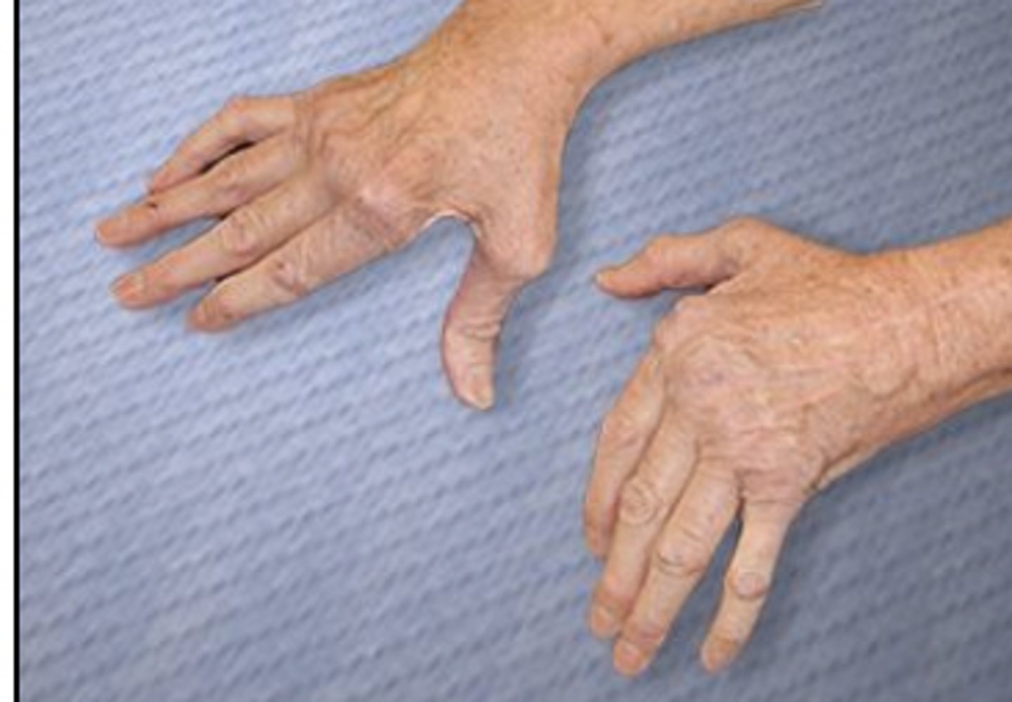
A patient with pain on the right side of the face also has tenderness at the right temporomandibular joint. A physical therapist notes deviation of the mandible toward the left at the end of the available range of mouth opening. Which of the following additional findings would be MOST expected?
1. Abnormal jaw reflex
2. Indentation behind the left condyle of the mandible
3. Paresthesia in the facial nerve (CN VII) distribution on the right
4. Hypermobility of the right temporomandibular joint
Rationale
1. The jaw jerk reflex is used to test the trigeminal nerve (CN V), but the question does not indicate neurological deficits (p. 1366).
2. The left temporomandibular joint is not hypermobile, so an indentation would not be expected (p. 1361).
3. Facial nerve paresthesias are not expected with temporomandibular joint hypermobility. The temporomandibular joint is primarily supplied by three nerves that are part of the mandibular division of the trigeminal nerve (CN V). (p. 1347)
4. A deviation during opening is associated with hypomobility toward the temporomandibular joint deviation and hypermobility contralaterally (p. 1361).
*Which of the following structures provides thermoregulation for the body by means of insulation?
1. Epidermis
2. Subcutaneous tissue
3. Dermis
4. Deep fascia
Rationale
1. The epidermis is the most superficial layer of the skin and has no fat storage (p. 12). Fat is the main insulator for the body and provides significant thermoregulation (p. 13).
2. Subcutaneous tissue, or superficial fascia, contains loose connective tissue and provides for storage of most of the body's fat, which is the main insulator for the body and provides significant thermoregulation (p. 13).
3. Dermis is the layer of skin between the epidermis and subcutaneous tissue. Dermis is made of collagen and elastic fibers and does not contain fat, but it does contain sweat glands and arterioles. It contributes to thermoregulation through sweating and arteriole dilation and constriction (pp. 12-13) but does not provide for thermoregulation through insulation.
4. Deep fascia underlies the superficial fascia and is not technically a part of the skin. It does not contain fat, which is the main insulator for the body. (p. 16)
A physical therapist is instructing a patient's care provider on how to perform a bed-to-wheelchair transfer. Which of the following teaching strategies would be MOST appropriate?
1. Have the care provider practice transferring the therapist.
2. Have the care provider observe the therapist perform the transfer with the patient.
3. Explain the transfer while demonstrating and have the care provider practice transferring the therapist.
4. Explain the transfer while demonstrating and have the care provider practice transferring the patient.
Rationale
1. Having the care provider perform without an explanation of the transfer would be trial-and-error and not the best way to learn (p. 36).
2. Demonstrating a transfer without an explanation would skip the cognitive stage of learning a psychomotor skill (p. 29).
3. The closer the learning experience is to the actual transfer, the more likely that the skill will be learned. Given that transfer skills are very specific to the individual being transferred, it is important that practice with the patient take place. (pp. 36)
4. Motor learning occurs first in the cognitive domain. Explaining the task allows for processing of the skill on a cognitive level. In order to progress to the associative phase of learning, where the goal is to fine-tune the skill, one must perform and practice that skill. Given that transfer skills are very specific to the individual being transferred, it is important that practice with the patient take place. (pp. 29, 36)
What is the MAXIMUM current density that should be used to initiate iontophoresis when the current amplitude is 10 milliamperes and the conductive surface area is 20 cm2?
1. 0.2 milliamperes/cm2
2. 0.5 milliamperes/cm2
3. 5 milliamperes/cm2
4. 20 milliamperes/cm2
Rationale
To calculate current density, the current amplitude is divided by conductive surface area; therefore, 10/20 = 0.5.
An 80-year-old patient who has left hemiparesis relies heavily on the right extremities for support. The patient has shoulder pain when the left upper extremity is elevated above 60°. Which of the following positions is BEST to facilitate simultaneous upper and lower extremity weight-bearing for the patient?
1. Modified plantigrade
2. Standing with both hands on a wall
3. Bridging
4. Quadruped
Rationale
1. Modified plantigrade is an ideal early standing posture in which to develop upper extremity and lower extremity control. Affected extremities are weight-bearing out of synergy patterns. This position is easily tolerated by elderly patients. (pic shown)
2. The patient will get limited weight-bearing through the upper extremity and is likely to have pain in the left shoulder in this position.
3. The bridging position may be difficult for an older patient to tolerate and does not involve weight-bearing through the upper extremities.
4. The quadruped position may be difficult for an elderly patient to tolerate and does not involve weight-bearing through the entire lower extremity.

Which of the following exercise programs is MOST appropriate for a patient who is at Stage IV on the Hoehn and Yahr Classification of Disability scale?
1. Treadmill
2. Elliptical machine
3. Stationary bicycle
4. Stair-climbing machine
Rationale
1. The Hoehn and Yahr Classification of Disability Stage IV describes a patient who has Parkinson disease with disability in the moderate to severe range (pp. 815-817). Postural instability and an increased risk of falling rule out a treadmill as a safe means of exercise for a patient who has moderate Parkinson disease (p. 842).
2. Postural instability and an increased risk of falling rules out an elliptical machine as a safe means of exercise for a patient who has moderate Parkinson disease (p. 842).
3. Exercise on a stationary bicycle allows for cardiovascular conditioning in a safe environment, because the patient is seated on a stationary surface (p. 842).
4. Postural instability and an increased risk of falling rule out a stair-stepping machine as a safe means of exercise for a patient who has moderate Parkinson disease (p. 842).
The Hoehn and Yahr scale is used to describe the symptom progression of Parkinson disease. The scale was originally described in 1967 and included stages 1 through 5.
A patient has significant swelling around the lateral ankle. Radiographs of the ankle reveal no evidence of bone injury. The examination shown in the photograph has a positive result. Which of the following structures is MOST likely injured?
1. Achilles tendon
2. Fibularis (peroneus) brevis tendon
3. Calcaneofibular ligament
4. Anterior talofibular ligament
Rationale
1. The photograph shows the anterior drawer test of the ankle (Magee, pp. 932-933). The Thompson (Simmonds) test is used to assess for tears of the Achilles tendon (Magee, p. 940).
2. Palpation of the proximal (base) fifth metatarsal is used to rule out avulsion fracture from a fibularis (peroneus) brevis tendon pull (Giangarra, p. 257).
3. The talar tilt test is used to determine whether the calcaneofibular ligament is torn (Magee, pp. 936-937).
4. The photograph shows the anterior drawer test of the ankle (Magee, pp. 932-933). A positive result may be obtained on the anterior drawer test only if the anterior talofibular ligament is torn; however, anterior translation is greater if both the anterior talofibular ligament and calcaneofibular ligaments are torn (Magee, pp. 932-933).
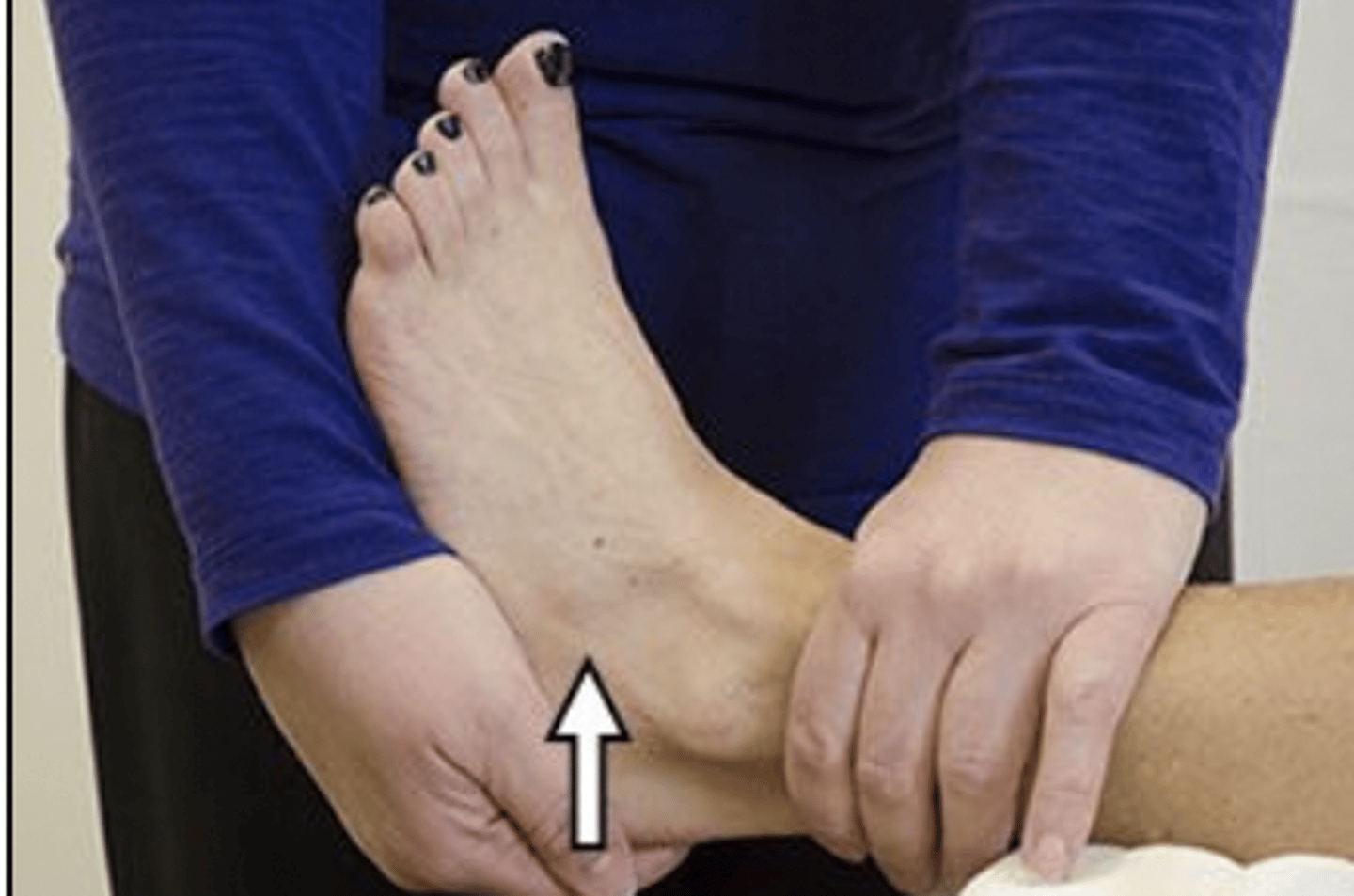
A physical therapist is developing an exercise program for a patient who has upper extremity lymphedema. Which of the following exercises should the patient perform LAST?
1. Elbow flexion
2. Cervical rotation
3. Wrist circumduction
4. Shoulder circumduction
Rationale
1. Active elbow exercise is indicated but would be performed before exercises for the wrist because the elbow is a proximal segment relative to the wrist (pp. 975-976).
2. The exercise program for lymphedema should begin with total body relaxation. Cervical rotation is an example of this initial exercise. Exercises are prescribed from proximal to distal areas. (p. 976)
3. Exercises are performed from proximal to distal areas (p. 976). Wrist circumduction is performed last because the wrist is the most distal segment (pp. 975-976).
4. Shoulder circumduction represents proximal clearance (p. 977).
A patient reports pain in the posterior lower leg during the test performed in the photograph. Which of the following conditions is MOST likely present?
1. Herniated disc in the lumbar spine
2. Idiopathic lumbar scoliosis
3. Right-sided lumbar paraspinal muscle strain
4. Osteoarthritis of the lumbar facet joints
Rationale
1. Leg pain elicited with straight leg raise may indicate lateral disc herniations.
2. Idiopathic lumbar scoliosis would not be affected by a straight leg raise test.
3. A straight leg raise test would not produce posterior lower leg pain in the presence of right-sided lumbar paraspinal muscle strain but would cause pain over the right lumbar paraspinals.
4. A straight leg raise test would not produce posterior lower leg pain in the presence of osteoarthritis of the lumbar facet joints. Flexion of the hip would cause lumbar flexion, which decreases pressure on the facet joints and would decrease pain in the lumbar spine.
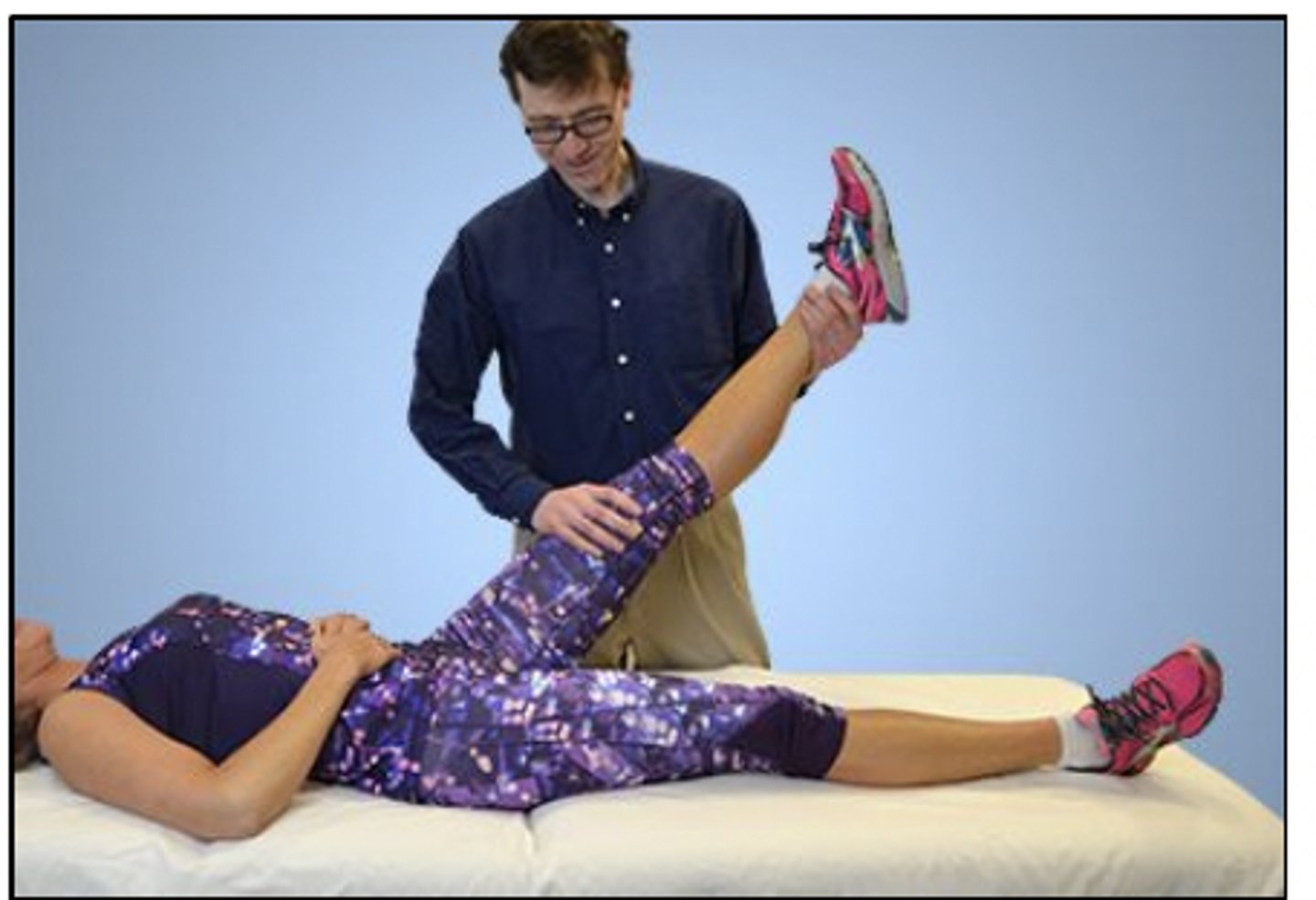
*A patient has an irregular heartbeat of greater than 100 bpm. Which of the following methods is MOST accurate for measuring the patient's heart rate?
1. Auscultate the apical heart rate for at least 60 seconds.
2. Take the radial pulse for 15 seconds and multiply by 4.
3. Measure the carotid pulse for 30 seconds and multiply by 2.
4. Take the radial pulse for 30 seconds and multiply by 2.
Rationale
With an irregular heart rate greater than 100 bpm, auscultation is the most accurate method of measuring the heart rate. Taking pulses for less than a minute would not be as accurate since irregular heartbeats could be missed, or could be present within only that 15 or 30 seconds of measurement. Rates faster than 100 bpm or slower than 60 bpm should be measured the full minute.
A physical therapist is using lumbar mechanical traction with the patient in supine position for a patient who weighs 180 lb (81.6 kg). The traction weight is initially set to 60 lb (27.2 kg). The patient reports not being able to feel any pull. Which of the following actions is MOST appropriate for the therapist to take?
1. Increase the pull to approximately 50% of the patient's body weight.
2. Switch to manual traction.
3. Substitute 40 lb (18.1 kg) of pull in prone position.
4. Discontinue the treatment.
Rationale
1. A traction force of 30% to 50% of the patient's weight is necessary to cause effective segmental vertebral separation. This patient could have a force up to 90 lb (40.8 kg).
2. Manual traction would be less likely to produce the needed force.
3. More weight, not less weight, is needed. Prone position could be considered, depending on the examination findings.
4. Increasing the traction force should be considered prior to discontinuing the treatment.
*Which of the following findings is MOST consistent with the presence of rebound tenderness noted during palpation of the abdomen of a patient who has low back pain?
1. Muscle guarding
2. Superficial reflex
3. Muscle soreness
4. Peritoneal irritation
Rationale
1. Rebound tenderness at the abdomen is not consistent with muscle guarding (pp. 340, 342).
2. Rebound tenderness is not consistent with a superficial reflex of the umbilicus when assessing for a neurologic impairment (pp. 201-203).
3. Rebound tenderness is not consistent with muscle soreness but rather is a classic sign of peritonitis (p. 341).
4. Pain on release of pressure confirms rebound tenderness, a reliable sign of peritoneal inflammation (p. 342).
A 24-month-old child who has a genetic disorder and developmental delay is able to sit independently but can easily be displaced and demonstrates emerging protective reactions and slow trunk-righting reactions in sitting position. Which of the following functional activities is BEST for short-term physical therapy for the child?
1. Pulling up to standing position
2. Obtaining a toy when placed out of reach
3. Holding a toy at midline with both hands
4. Transitioning from sitting to quadruped position
Rationale
1. Pulling to stand is too difficult for a short-term physical therapy goal for this child, who is functioning at a 5-month to 6-month level. This activity would require intact protective extension and trunk righting. (p. 61)
2. This child is at the second stage of sitting (around 5 months) and would benefit from activities that challenge trunk righting, sitting balance, and protective reactions. These are within the child's ability to achieve, because they are items in the third stage of sitting, which usually occurs around age 6-7 months. (pp. 56-57)
3. No position is noted. This task could be achieved in a supine position. Holding a toy within the center of mass in sitting position is also a skill that is likely already achieved in the second stage of sitting, at age 5-6 months. (p. 57)
4. Transitioning from sitting to quadruped position is a task that is generally achieved in the third quarter of the first year, so it is not an appropriate goal of short-term physical therapy for a child who is in the 5-month to 6-month range of development, which is consistent with second stage of sitting. (p. 61)
A 4-year-old child who has a plantar flexion contracture is referred for serial casting. The child is tearful and combative, and the parents are frustrated. Which of the following actions is MOST appropriate for the physical
1. Ask the physician to sedate the child prior to the physical therapy session.
2. Allow the child to cast a doll's leg while the therapist applies a cast to the child.
3. Ask an aide to hold the child down so the therapist can apply the cast.
4. Ask the parents to leave the room while the therapist applies the cast.
Rationale
1. Preventative interventions are directed toward minimizing potential problems that would limit a patient's participation in therapy. Therapists should choose interventions most likely to achieve successful outcomes (O'Sullivan, p. 12). Since the child will be coming for multiple appointments, and the child's independent participation in therapy is desired, this option is not the most appropriate choice.
2. Appropriate play can be important in easing tension related to the health care setting. Giving the child power in a role can be helpful. (Purtilo, p. 284)
3. Restraining the child would not demonstrate respect for the child and would not foster the child's independent participation in therapy (Purtilo, p. 284).
4. The physical therapist should not exclude the family from the health care interaction and should foster a supportive family context for the child (Purtilo, p. 288).
*A patient who has pleural effusion is performing segmental breathing exercises. Where should manual counterpressure be applied to encourage expansion of the posterior basal segments of the patient's lower lobes?
1. Lower lateral costal area
2. Posterior lower ribs
3. Anterior midchest
4. Anterior lower ribs
Rationale
1. Segmental breathing combines breathing control with manual cues to specific areas of the chest wall. The lateral costal area would be a position to treat the lateral basal segments of the lower lobes, not the posterior basal segments.
2. Proper hand placement to encourage posterior basal expansion is over the posterior aspect of the lower ribs.
3. Midchest would be used to treat the superior segments of the lower lobe, not the posterior basal segments.
4. The anterior lower ribs would be a position to treat the anterior basal segments of the lower lobes, not the posterior basal segments.
Segmental breathing AKA localized breathing or thoracic expansion exercise; intended to improve regional ventilation and prevent/treat pulmonary complications after surgery; based on the presumption that asymmetrical chest wall motion may coincide with underlying pathology (pneumonia, pleuritic chest wall pain, retained secretions, etc) and that inspired air can be directed to a particular area by facilitation or inhibition of chest wall movt through proper hand placements, verbal cues, or coordination of breathing à augments localized lung expansion for chest hypomobility (dec lung compliance, dec intrathoracic lung volume)
During gait evaluation, a physical therapist notes that a patient demonstrates a shorter step length with the right lower extremity. Which of the following problems is MOST likely the cause of the gait dysfunction?
1. Right iliopsoas contracture
2. Painful left knee
3. Decreased ankle pronation on the right
4. Left gluteus medius weakness
1. Right iliopsoas contracture may cause a shorter step length with the left lower extremity but not with the right lower extremity (Mansfield, p. 356).
2. Left knee pain will cause the patient to spend less time in left-sided stance, because the patient will try to minimize the time spent in stance (weight-bearing on the knee) to minimize the pain. Therefore, the patient will take a shorter step with the right lower extremity. (Magee, p. 1007).
3. Decreased ankle pronation would not have an effect on right-sided step length (Magee, p. 1005).
4. Gluteus medius weakness would be seen as an increase in lateral pelvic tilt, not step length (Mansfield, p. 357).
A physical therapist can BEST assess for dysdiadochokinesia by asking the patient to:
1. identify a spoon, a toothbrush, and a comb by touch.
2. bring a toothbrush or a spoon to the mouth repeatedly, using rapid brisk motions.
3. demonstrate how to use a toothbrush, a spoon, or a comb.
4. name familiar objects, such as a toothbrush, a spoon, and a comb.
Rationale
1. Inability to identify objects by touch is astereognosis (p. 1256).
2. Dysdiadochokinesia is the impaired ability to perform rapid alternating movements, which could be demonstrated by the repeated elbow flexion and extension in repetitively bringing a utensil to the mouth (p. 211).
3. Apraxia is characterized by the inability to perform purposeful movements, such as using some type of utensil (p. 1257).
4. Aphasia (expressive) is a communication disorder characterized by impaired language formation and use (pp. 652, 1450), which would impair the ability to name objects.
A patient who has a C5 spinal cord injury (ASIA Impairment Scale A) suddenly reports light-headedness and ringing in the ears while sitting upright in a wheelchair. Which of the following conditions is MOST likely present?
1. Angina pectoris
2. Deep vein thrombosis
3. Orthostatic hypotension
4. Autonomic dysreflexia
Rationale
1. Angina is cardiac-related chest pain due to ischemia; it is unrelated to ringing in the ears (p. 534).
2. The hallmark sign of deep vein thrombosis is rapid onset of unilateral leg swelling, erythema, and heat (p. 901).
3. Orthostatic hypotension is a common complication in patients who have an acute cervical injury. It manifests with dizziness or light-headedness and ringing in the ears when in a vertical position, such as sitting or standing. (pp. 72, 896)
4. Autonomic dysreflexia is common in patients who have a spinal cord injury at T6 or above and results in significantly increased blood pressure and a pounding headache (p. 895).