Microbiology 2060
1/66
Earn XP
Description and Tags
Note in here: SI units
Name | Mastery | Learn | Test | Matching | Spaced |
|---|
No study sessions yet.
67 Terms
Main Principle of Modern Science
all natural phenomena have natural causes
Jansen
late 1500s
developed first compound microscope but it had poor magnification
Hooke
improved upon Jansen’s design using better lenses, he used reflective light
Van Leeuwenhoek
introduced transmitted and focused light
crafted lenses that could magnify up to 270x
believed to be the first human to observe individual bacteria
called them animalcules
Van Leeuwenhoek’s Observation
“animalcules” from biological scrappings
“animalcules’” division
bacterial reproduction
Abiogenesis
aka Spontaneous Generation
the ability of “lesser organisms” to spontaneously form out of nonliving material
relies on the belief of “Vital Force” of abiotic structures that allow production of organisms
Not supported by modern science
Vitalism
the belief that there are vital energy forces that govern our health and well-being
Disproving Abiogenesis: Redi’s Jar
Francesco Redi Experiment:
jar containing raw meat and no lid
“produce” flies
jar containing raw meat and lid
does not “produce” flies
jar containing raw meat with a porous cloth covering
flies appear on cloth outside the jar but not on meat
was not enough to sway public opinion
Infusoria Community
the collections of microbes produced by soaking HAY in water
microorganisms from the hay are flushed into the water
a highly diverse microbial community
Used to disprove/study VITALISM
Infusion
the liquid produced by soaking plant material in water
ex. tea
John Needham Infusoria Experiment
poured infusion into a flask and boiled it (killing microbes) and then sealed it
Result: RETURN of Infusoria
Larazzo Spallanzani Infusoria Experiment
poured infusion into flask, sealed it and then boiled it (killing microbes)
Result: continued ABSENCE of infusoria
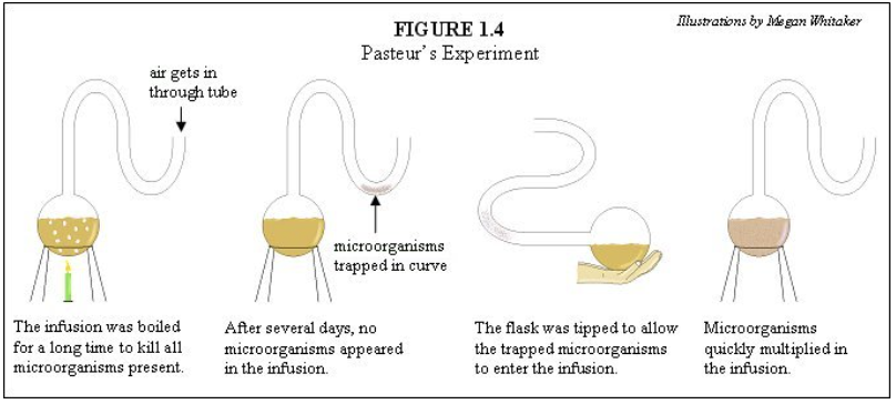
Louis Pasteur Infusoria Experiment
poured beef broth into long necked flask
heated the neck and bent it into and S-shape
boiled the bottom
Result: no microorganisms in cooled solution
finally and fully disproved abiogenesis and vitalism
S-Flask Experiment Details
the curve of the neck prevents reinfection of the broth by blocking dust from easily transporting microbes to the broth
Germ Theory
“that microorganisms are the causative agent of infectious disease”
disproves belief that disease is caused by karma, possession or the Wrath of God
first suggested by VARRO REATINUS around 80 BCE Rome
couldn’t be proven/demonstrated
Agostino Bassi
first formed the connection between microbes and the health of organisms via SILKWORMS
demonstrated mold contributed to silkworm health
Ignaz Semmelweis
made the connection between microbes and humans (indirectly)
“the elimination of microbes eliminated disease”
directed his nurse WASH their HANDS prior to attending labouring patients
reduced childbirth fever deaths drastically
Joseph Lister
extended Semmelweis’ idea by CLEANING SURGICAL INSTRUMENTS in a phenol solution prior to operations
Direct Connection between Microbes and Humans
LOUIS PASTEUR + ROBERT KOCH
developed Koch’s Postulates
concluded that a specific disease is caused by a specific microbe
etiology is the first step to treatment and prevention
Koch’s Postulates
A series of steps, as follows:
isolate microorganisms from a suspended diseased or dead animal
grow a pure culture of said microorganism
identify microorganism
inject microorganism into a healthy laboratory animal
the disease will reproduce in lab animal
repeat steps 1 and 2 using sample from laboratory animal
microorganism of concern is identified
are used to establish proof of direct connection between microbes and health
Edward Jenner
developed the first live “vaccine”
observed those infected by cowpox are resistant to smallpox
began infecting people with cowpox to prevent severe infection of smallpox
this worked due to similar enough structure for compatible antibodies
Louis Pasteur
developed the first “intention” vaccine
observed that old microbes would lose their ability to cause disease while retaining ability to induce resistance to disease they cause
his beta test Rabies vaccine on a child
Paul Ehrlich
1910
developed a synthetic compound called SALAVARSAN to treat syphillis and sleeping sickness
Alexander Flemming
discovered that the Penicillium fungus secreted an antibiotic compound (penicillin)
Antimicrobial Resistance
overuse and improper uses of antibiotics has led to many pathogenic microbes being resistant to treatment
bacteria can achieve resistance via a variety of ways
Molecular Biology in Microbiology
genetic profiles are now key in testing for the presence of certain microbes
also used for identification
microbes can be easily genetically modified using Recombinant DNA
Paul Berg
1960s
developed basis for modifying microbes
Herb Boyer + Stanley Cohen
1973
founded first biotech company:
Genentech
3 Factors in Microscopy Image Quality
Magnification
Contrast
Resolution
Magnification: BENDING LIGHT
two types:
reflection
refraction
when light enters a specimen it will both reflect and refract
occurrence due to light passing from one medium to another when the mediums have different indecies
human eye perceives the refracted light differently
usually enough to create sufficient contrast to see microbes
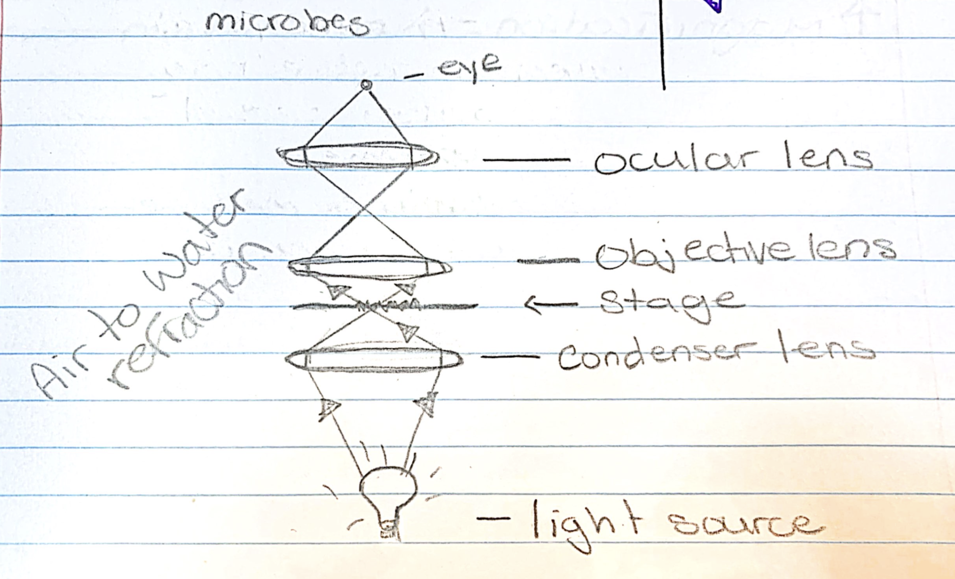
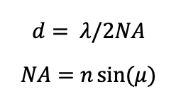
Resolution: RESOLVING POWER (d)
the ability of a lens system to allow you to see two points as being distinct
the lower the value of ‘d’ the better the resolving power
maximizing resolving power/reducing d value can be done by:
using a shorter wavelength of light (reduce wavelength)
reduces contrast
using a higher magnification
shorter focal length of lens = closer
increase angle (larger angle)
altering refractive index
make it more closely match the specimen to capture more refracted light
oil immersion
has a refractive index closer to water in specimen
Resolving Power Formulas
NA = numerical apperature
n = refractive index between specimen and lens
u = angle from specimen to outer edge of objective lens
wavelength is the wave thingy
Oil Immersion
can only be done a higher magnification (100x objective)
oil bust be held in place by surface tension (oil replace air)
lens must have short focal length
only 100x lens short enough
Focal Length
the space between the specimen and objective lens
short = close to specimen
long = far from specimen
Contrast: Staining
live cells are transparent with no significant contrast with surrounding medium
contrast can be added via:
changing illumination
adding colour (stain)
Biological stains have 2 main categories
basic
acid
Biological Stains: BASIC STAINS
the coloured portion (CHROMOPHORE) is cationic
colour binds to cells’ negative charge
simple stains use only basic stains
example → crystal violet
Biological Stain: ACIDIC STAINS
the coloured portion (CHROMOPHORE) is anionic
colour is repelled by cells’ negative charge
around the cells - stars in night sky
negative stains use acidic dyes - does not need heat fix or wash
example → nigrosin
Basic Slide Preparation
smear the sample onto a slide
needs to be a thin layer, not too thick/heavy
apply a LITTLE but of heat
this heat fixes specimen to the slide
apply appropriate staining technique
Differential/Special Stain Protocols
target specific features that differ between different bacterial types
includes use of
mordants (enhance stains)
counterstains
heat
examples:
Gram Stain
Acid-Fast Stain
Endospore Stain
Important Staining Techniques
need to know:
Gram Stain
Acid-Fast Stain
also good to know:
Endospore Stain
Flagella Stain
Gram Stain Protocol
targets an important difference in bacterial cell walls
gram+ or gram-
4 steps:
stain the fixed seam with CRYSTAL VIOLET
wash away excess
apply a MORDANT (iodine for example)
this binds to the crystal violet to form a larger complex (iodine-crystal violet complex) that will help distinguish cells
wash away excess
apply DECOLOURIZER
this removes the ICV complex from gram- cells
apply COUNTERSTAIN (safranin for example)
allows us to see gram- cells/washed cells
different colour
Iodine-Crystal Violet Complex
too big to pass through gram positive cells walls (stuck inside) but are able to pass through gram negative cell walls
Acid-Fast Stain Protocol
oriented toward bacteria with WAXY MYCOLIC ACIDS in their cell walls
often Mycobacteria
4 Steps:
stain bacteria with CARBOL FUSCHIN
if the cell wall contains mycolic acids it will bind to the Carbol Fuschin tightly
heat the slide
allows enhanced penetration of dye in cell wall
apply an ACID-ALCOHOL. RINSE
cells with mycolic acid will remain dyed and remove dye from non-mycolic acid cells
apply COUNTERSTAIN
allows you to see the unstained cells
Endospore Stain Protocol
looks/targets the formation of endospores
uses MALACHTE GREEN
4 Steps:
apply MALACHTE GREEN to the heat fixed slide
add additional heat to allow dye to penetrate endospore wall
rinse with water to remove excess dye in non-endospore cells
apply COUNTERSTAIN
so you can see vegetative cells
Endospores
are immobile form of cells that are created in response to environmental pressure
only two Genera do this
Bacillus
Clostridium
Steps to Endospore Formation
newly replicated DNA along with a small amount of cytoplasm are isolated
membrane closes around DNA package
spore septum surrounds package forming FORESPORE
peptidoglycan and spore coat form
endospore is freed from the cell
Flagella Stain Protocol
goal is to make flagella thicker so they can be seen
to do this we use a MORDANT
careful not to add too much, could cause breakage
once thickened you carefully stain the flagella
Types of Microscopy
Bright Field
Dark Field
Fluorescence
Confocal
Electron
Scanning
Transmission
Brightfield Microscopy
light from the source is focused on the specimen
image is produced by refracted light
often lacks significant contrast between refracted and background (unrefracted) light
Darkfield Microscopy
an opaque disk is placed in the path of the sources light cause the light to form a ring shape
causes the light that does not refract or reflect to miss the objective lenses
creates and image only using the light reflected/refracted via the specimen
no background light
usually poor resolution
Fluorescence Microscopy
often used to identify specific types of microorganisms in a mixed background
does not use visible light
instead uses Ultra Violet to detect antibodies
antibodies are combined with fluorochrome then introduced and stick to the specific surface proteins
Confocal Microscopy
the manipulation of specific wavelengths of light that are focused on a specimen at different depths of the specimen
specimen is treated so different parts are stained differently
essentially creates slices you can reassemble digitally
produces a 3D image of specimen
Electron Microscopy
uses an electron beam instead of light
beam has VERY short wavelength (0.0005nm)
which has a very high frequency giving it amazing resolving power and magnification (100x better)
two types
Scanning Electron Microscopy (SEM)
surface of specimen is coated in very thin layer of “electron dense medium” (usually gold)
electron beam knocks electrons off the surface of the specimen, they are collected by detectors to create an image
lots of surface detail - electron beam cannot penetrate surface
Transmission Electron Microscopy (TEM)
the specimen is fixed in resin and cut VERY thin
a time consuming and finicky process that can produce artifacts
special stains can be used to increase contrast
detailed image of internal structures
Prokaryotic Plasma Membrane Functions
permeability and transport
respiratory reactions
synthesis of the cell wall
photosynthetic reactions
Peptidoglycan
the material used to create the bacterial cell wall
2 primary structural components
long polysaccharide stands
NAG and NAM amino sugars
short peptides
join the NAG/NAM strands
composed of side chains and cross-bridges
N - acetylglucosamine (NAG)
amino sugar
modified amino group on #2 carbon
joined by glycosidic bond
N - Acetylmuramic Acid (NAM)
amino sugar
modified amino group on #2 carbon
joined by glycosidic bond
Gram Positive Bacteria
THICK layer of peptidoglycan
contiain TEICHOIC ACID in its membrane (substance regulates cell wall growth)
Gram Negative Bacteria
THIN layer of peptidoglycan
have a second membrane (outer membrane)
contains:
Lipopolysaccharides
Pourin Proteins
Lipopolysaccharides (LPs)
a large complex molecule that contains lipids and carbohydrates
3 major components:
Lipid A
O-polysaccharide
Core Polysaccharide
Lipid A
the lipid portion of LPs embedded in the top layer of the outer membrane
O-Polysaccharide
extends outward from the core polysaccharide in LPs
functions as an antigen (sugar based)
Core Polysaccharide
attach to Lipid A and contains unusual sugars
provides stability and structure to LPs
Antibiotics and How They Work
5 main types:
CELL WALL INHIBITOR
prevents the synthesis of the cell wall/peptidoglycan subunit
PROTEIN SYNTHESIS INHIBITOR
occurs inside the cell typically during mRNA translation
NUCLEIC ACID INHIBITOR
prevents proper nucleic acid (DNA/RNA) replication and transcription
PLASMA MEMBRANE ATTACKER
damage the plasma membrane to make in dysfunctional (starves the cell out by preventing photosynthesis)
METABOLIC INHIBITOR
act as a competitive or non-competitive inhibitor to vital enzymes in important metabolic pathways