anaatomy final (complete) nmu
1/211
There's no tags or description
Looks like no tags are added yet.
Name | Mastery | Learn | Test | Matching | Spaced | Call with Kai |
|---|
No analytics yet
Send a link to your students to track their progress
212 Terms
What are bones?
Calcified Connective tissues
bones function
1) Support the human body.
2) Protection of soft vital structures e.g. the skull
protects the brain.
3) Motion through joints and muscles attached to it.
4) Formation of blood cells in red bone marrow.
5) Stores and releases calcium & phosphorus.
bones are classified according to 3 ?
A. Site (Position).
B. Structure.
C. Shape
bones are classified according to position into 2 types and what are their content ?
1) Axial skeleton: includes the skull, vertebral column and thoracic cage.
2) Appendicular skeleton: includes bones of the upper and lower limbs.
the is skull ? consists of ? protects ?
The skull is the skeleton of the head region.
Consists of cranium and mandible.
Protects the brain, eyes and ears.
a) vertebral column is made of ?
b) its parts are ? 5
a) The vertebral column is
made up of vertebrae.
b) The vertebral column has 32-
33 vertebrae, divided into:
7 cervical vertebrae.
12 thoracic vertebrae.
5 lumbar vertebrae.
5 fused sacral vertebrae (Sacrum)
3-4 fused coccygeal vertebrae (Coccyx)
the thoracic cage protects ? consists of ?3
It protects the heart and lungs.
Consists of:
1. The sternum: anteriorly.
2. The ribs: 12 pairs laterally.
3. Thoracic vertebrae: 12 vertebrae posteriorly.
upper limb (هقول المكان وانت تقول اسم العضم اللي فيه)
girdle ?
proximal segment ?
middle segment ?
distal segment ?
clavicle & scapula
humerus
laterally radius proximally ulna
carpals (8)
metacarpals (5)
phalanges 3 for each finger & 2 for the thumb
lower limb (هقول المكان وانت تقول اسم العضم اللي فيه)
girdle ?
proximal segment ?
middle segment ?
distal segment ?
hip bone
femur
laterally fibula
medially tipia
tarsals (7)
metatarsals (5)
phalanges 3 for each toe and 2 for the big toe
bones are classified acc to shape 6
1- long bones
2- short bones
3- flat bones
4- irregular bones
5- pneumatic bones
6- sesamoid bones
long bones found in ? مكانهم فين يعني المنتشر عشان الامثله كتير
found in proximal and intermediate segments of limps
short bones examples ?
carpals and tarsals
flat bones examples ?
scapula rips and skull cap
irregular bones examples ?
as the vertebrae and hip bone
pneumatic bones shape and examples ?
contain air-filled spaces as bones of the face
sesamoid bones shape and examples ?
small bones, they are developed inside tendons as patella (largest sesamoid)
بعيد عن شكل السوال بس عشان تبقى رابط المعلومه
bone classification according to structure 2 ? and describe each
1) Compact bone:
Dense & ivory-like.
Example: cortex of a long bone.
2) Cancellous (spongy) bone:
Network of bone trabeculae separated by spaces
containing bone marrow
Example the epiphyses of long bones
parts of the long bones are 5 ?
1-epiphysis
2-epiphyseal cartilage (plate)
3-metaphysis
4-diaphysis
5-periosteum
كدا مترتبين اسهل في الحفظ
epiphysis is ?
the end of long bones
diaphysis is ?
The shaft formed of compact bone enclosing a medullary cavity filled with bone marrow.
epiphyseal cartilage is ? and its properties (3 points total)
1-plate of hyaline cartilage separate the end from the shaft
2-site of growth of bone in length
3-it ossifies at a certain age
periosteum is ?
a fibrous membrane covers the shaft
metaphysis is ?
region of the shaft close to epiphyseal cartilage
blood supply of bones ? 4
1-epiphyseal arteries
2-metaphyseala arteries
3-nutrient artery
4-periosteal artery
joints are ?
they are classified according to the tissue between the bones into ? 3
articulation between two or more bones
and they are classified into
1-fibrous
2-cartilaginous
3-synovial
fibrous joints
characters ؟(يعني العضمتين بينهم اي (اه والله))
movement ?
types 3?
the articulating bones are connected together by fibrous tissue
no movement or very limited movement
types are :
1-suture
2-gomphosis
3-syndesmosis
suture joints are ?
a type of fibrous joints between skull and it ossifies at old age
gomphosis joints are ?
fibrous joints between the roots of the teeth and their sockets
syndesmosis joints are ?
a type of fibrous joints wheree bones are connected by strong ligaments as interosseous membrane between tibia and fibula
cartilaginous joints
characters ?
movement ?
types ?
the articulating bones are connected together by cartilage
no movement or very limited movement allowed
types are :
1-primary cartilaginous joints
2-secondary cartilaginous joints
compare between 1ry and 2ry cart. joints
1ry and 2ry caart.
a- bones are connected by ?
b- joints ossify ?
c- movement ?
d- examples ?
answer respectively pleaaaaaaaaaaaase
a- 1ry:hyaline cartilage
2ry:fibrocartilage
b- 1ry:ossifies by age
2ry:doesnt ossify by age
c- 1ry:no movement
2ry:limited movement
d- 1ry: 1st sternocostal cartilage , eppiphyseal plate of long bones
2ry: intervertebral discs, synphysis pubis, manubriosternal joint
characters of synovial joints
mobility ?
articular surface covered by ?
articulating bones separated by ?
joint surrounded by ?
capsule lined by ?
secretion?
1) They are freely mobile joints (allow wide range of movement).
2) Articular surfaces of bones are covered by thin layer of hyaline cartilage.
3) The articulating bones are separated by a joint cavity.
4) The joint is surrounded by a fibrous tissue capsule.
5) The capsule is lined by a synovial membrane.
6) The synovial membrane secrets synovial fluid, which fills the joint cavity.
functions of the synovial fluid ? 2
✓ Lubrication and nutrition of articular cartilage.
✓ Allows free movement of the joint.
structures inside some synovial joints may be ? 3
1-cartilaginous structures like: discs, meniscus (as in knee), labrum : (as in the hip & shoulder) joints
2-ligaments (as cruciate ligament in knee)
3- tendon of muscle
types of synovial joints according to number of articulating bones 3 ?
1) Simple: consists of 2 articulating bones (e.g.: shoulder).
2) Compound: consists of more than 2 articulating bones (e.g.: elbow)
3) Complex: has intra-articular disc or menisci (e.g.: knee).
types of synovial joints according to the shape of articulating bones 7 ?
1-plane synovial joints
2-hinge joints
3-pivot
4-ellipsoid
5-bicondyloid (condylar)
6-saddle
7-ball & socket
plane synovial joints
✓ The articular surfaces are flat.
✓ Allow gliding movement in any direction.
✓ Examples: intercarpal & intertarsal
hinge joints
like the hinge of the door
example: elbow, ankle
pivot joints
✓ Have a central axis rotate in a ring.
✓ Examples: superior radioulnar
ellipsoid
✓ Have one convex surface fitting in an elliptical concavity.
✓ Examples: wrist
bichondy
Have two convex surface fitting in two concavities.
✓ Examples: knee
saddle joints
✓ The articulating surfaces are alternatively concavoconvex.
✓ Examples: carpometacarbal of thumb
ball and socket joints
✓ Have a rounded head fitting in a cup-shaped concavity.
✓ Examples: hip joint
CVS consists of ?2 and describe them
1) Heart: hollow muscular organ which pumps the blood.
2) Blood Vessels: include arteries, veins and capillaries.
the heart size ?
about the size of an individual's closed fist
heart shape?
the heart is conical in shape it has the apex and the base
the heart base formed of ? direction ?
its formed of both atria (right and left atriums)
directed posteriorly (بتبص ل ورا )
the heart apex is formed of ? direction ?
the heart apex is formed by the left ventricle
and its directed anteriorly
the heart position is ? enclosed by ? the heart champers ?
the heart lies obliquely in the middle part of the thoracic cavity
enclosed by a fibro-serous sac called pericardium
the heart is formed of 4 champers two atria and two ventricles
right atrium
type of blood ?
blood enter ?
blood leave ?
non oxygenated
superior and inferior venae cavae
tricuspied orifice to the right ventricle
left atrium
type of blood ?
blood enter ?
blood leave ?
oxygenated
4 pulmonary veins (2 from each lung)
mitral orifice to the left ventricle
right ventricle
type of blood ?
blood enter ?
blood leave ?
non oxygenated
tricuspied orifice
pulmonary trunk to both lungs
left ventricle
type of blood ?
blood enter ?
blood leave ?
oxygenated
mitral orifice from the left atrium
aorta to the body
compare between the 4 champers of the heart
valves of the heart 4 ?
mitral valve , tricuspid valve
aortic valve amd pulmonary valve
mitral valve site ?
between the left atrium and the left ventricle
tricuspid valve site ?
between the right atrium and the right ventricle
aortic valve site ?
between the left ventricle and aorta
pulmonary valve site ?
between the right ventricle and pulmonary trunk
the heart arterial supply ?
the heart is supplied by 2 coronary arteries (right and left) which arise from the ascending aorta
the heart venous drainage ?
the heart is drained by 3 cardiac veins (great, middle, and small)
which drain in the coronary sinus then the coronary sinus ends in the right atrium
arteries function ?
valves ?
blood type (oxygenated or non ?) exception ?
what is the name of it after branching?
transport blood away from the heart to different body tissue
they dont have valves
all arteries carry oxygenated blood except pulmonary artery
they divide into smaller branches and the small artery is called arteriole
veins function ?
valves ?
blood type (oxygenated or non ?) exception ?
what is the name of it after branching?
veins transport blood towards the heart
many of the veins have valves
all veins carry non oxygenated blood except pulmonary veins
veins have tributaries and the small veins are known as venules
types of arteries 3
1-anastamotic arteries
2-end arteries
3-wavy toortuous arteries
anastamotic arteries definition and sites ?
it is the communication between arteries
mainly around joints of the limbs and in the hand and foot
end arteries definition? and examples ?5
arteries which do not anastomose with adjacent arteries
examples : renal, retinal, splenic,
coronary and cerebral arteries.
wavy (tortuos) arteries
definition? and examples ? 4
arteries with wavy course supplying expansible or moving organs
examples: facial, lingual, uterine, and splenic arteries
capillaries are ?
capillaries are minute vessels in the form of a network connecting the arterioles with venules
and their walls are very thin to allow gas and fluid exchange between the blood and the tissue
the respiratory system is divided into ? 2 and mention that each of them consists of
▪ Upper respiratory tract:
Consists of nose, pharynx & larynx.
▪ Lower respiratory tract:
Consists of trachea, bronchial tree & lungs.
the nasal cavity into ? by ?
right and left parts by nasal septum
the nasal cavity opens anteriorly and posteriorly by ?
the anterior and posterior nasal openings
اه والله زيي زيك
the posterior nasal opening connects what with what ?
connects the nasal cavity with the pharynx
the nose has 4 functions ?
warming , moistening , and filteration of the inspired air and also smell ofc (wow)
paranasal sinuses are ?
their types ? 4
their functions ? 2
❖ Definition: Extension from nasal cavity into the bones of skull.
❖ Types:
1- Frontal air sinus
2- Maxillary air sinus
3- Sphenoidal air sinus
4- Ethmoidal air sinus
❖ Function:
• Resonance of voice.
• Lightening of the scull bone.
pharynx is ?
its parts 3 ?
function ?
❖ Definition: It is a muscular tube extending from the base of skull to the level of the 6th cervical vertebra.
❖ Parts:
• It is subdivided into 3 parts:
✓ Nasopharynx: behind the nose (contains adenoid).
✓ Oropharynx: behind the mouth (contains tonsils).
✓ Laryngopharynx: behind the larynx.
❖ Function:
• Common pathway for food & air.
larynx is ?
extension (begins and ends at ?)
continues as ?
structure ?
functions ?
❖ Definition: Fibro – cartilaginous organ connects the pharynx to the trachea.
❖ Extension:
• Begins at the level of C3
• Ends: opposite the C6 and
continues as trachea.
❖ Structure:
• It consists of cartilages
connected together by
muscles & ligaments.
❖ Functions:
• Air passage.
• Voice production
trachea
length ?
diameter ?
site ?
begins and ends at ?
its structure
❖ Length: 10 cm (4 inches)
❖ Diameter: 1 inch
❖ Site: its upper half lies in the neck while, its lower
half lies in the thorax
❖ Begins: At the level of C6 cervical vertebra as a
continuation of the larynx.
❖ Ends: at the lower border of 4th thoracic vertebra
by dividing into right and left principle bronchi.
❖ Structure: Composed of 15-20 C-shaped rings of
hyaline cartilage.
the trachea is composed of how many rings and what is it made of ?
composed of 15-20 C-shaped rings of hyaline cartilage
the difference between the right and left bronchi
inhaled foreign bodies destiny (اي شغل الدراما دا)
the right bronchi is shorter, wider and more vertical (in line with the trachea)
so inhaled foreign bodies usually pass to the right lung
compare between the right and left bronchus
jjbhbhbh
lungs shape ? apex and base direction ?
• Cone-shaped, has:
✓ Apex: directed upwards, towards the
neck.
✓ Base: directed downwards, towards
the diaphragm.
the lungs has how many surfaces ? describe each please
❖ The lung has two surfaces:
1- Medial (Mediastinal) surface:
✓ related to the heart and great
vessels.
2- Lateral (Sterno-costal) surface:
✓ related to the sternum and ribs.
compare between right and left lungs
pewpewpepwpp
pleura definition ? and layers ?
❖ Definition:
•Pleura is a closed serous sac which encloses the lung.
❖ Layers:
•It has 2 layers and pleural cavity in between:
✓ Visceral pleura: is the inner layer.
✓ Parietal pleura: is the outer layer.
pleural cavitiy is ?
•The potential space that lies between the 2 layers & contains a thin film of serous fluid
to prevent friction between the 2 layers
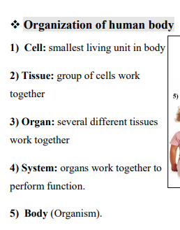
organization of human body
اول سلايد غالبا ملهاش لازمه اهي بردو
describe the anatomical position
its made of 5 points
1.The person stands erect.
2.The feet are together.
3.Arms are straight by the sides of the body.
4. Palms are facing forward.
5.The face is directed forward
لازم عالاقل اضعف الايمان تكون متخيله
midsaggital plane other name ? describe it ..
median plane
.
vertical divides the body into right and left equallllllllll halves
Parasagittal planes: other name ?
describe bro
(paramedian) plane
Vertical & parallel to midsagittal plane.
Divides body to right & left unequal parts
coronal plane
Vertical & perpendicular to midsagittal plane.
Divides body to anterior & posterior parts
transverse plane other name ?
describe it bro
horizontal plane
.
Perpendicular to midsagittal & coronal planes.
Divide the body into upper & lower parts.
. Superior / inferior:
a. Superior (above): is toward the head.
b. Inferior (below): is toward the feet.
Anterior / posterior:
a. Anterior (ventral): is toward the front of the body.
b. Posterior (dorsal): is toward the back of the body.
Median / Medial / lateral:
a. Median: At the middle line of the body.
b. Medial: Toward the midline of the body.
c. Lateral: Toward the side of the body.
Proximal / distal
a. Proximal: Toward the root of the limb
b. Distal: Away from the root of limb
Superficial / Deep
a. Superficial: External near to the surface
b. Deep = Internal away from the surface
Ipsilateral / Contralateral
a. Ipsilateral: At the same side
b. Contralateral: Opposite side
Flexion / extension:
Flexion: decreasing the angle between two bones (bending or approximation).
Extension: increasing the angle between two bones (straightening).
Abduction / adduction:
Abduction: movement away from the median plane.
Adduction: movement towards the median plane.
rotation
Medial rotation: movement of anterior surface toward the midline.
Lateral rotation: movement of anterior surface away from the midline.