Cariology Lecture 1
1/35
There's no tags or description
Looks like no tags are added yet.
Name | Mastery | Learn | Test | Matching | Spaced |
|---|
No study sessions yet.
36 Terms
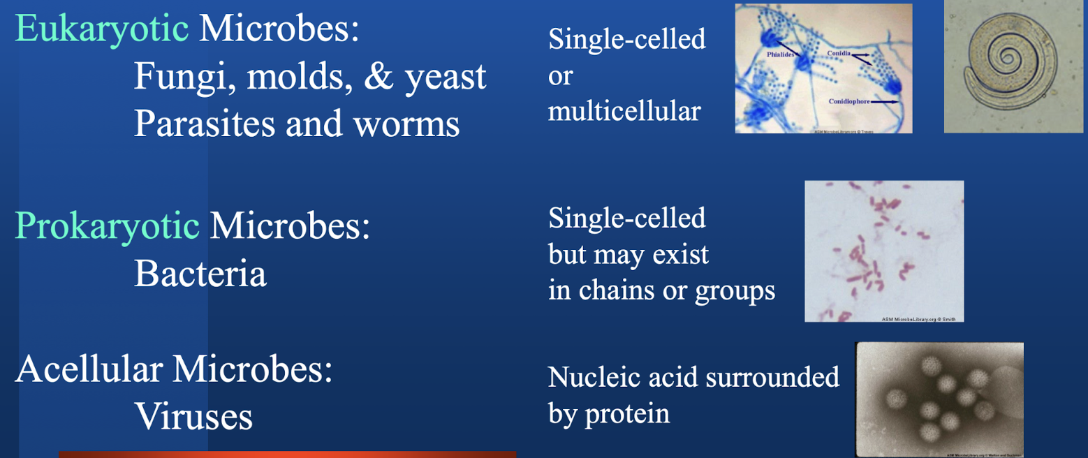
What are examples of eukaryotes, prokaryotes, and acellular microbes? Which are single-celled, multicellular and not a cell?
What is the typical eyepiece of a brightfield microscope and its objectives?

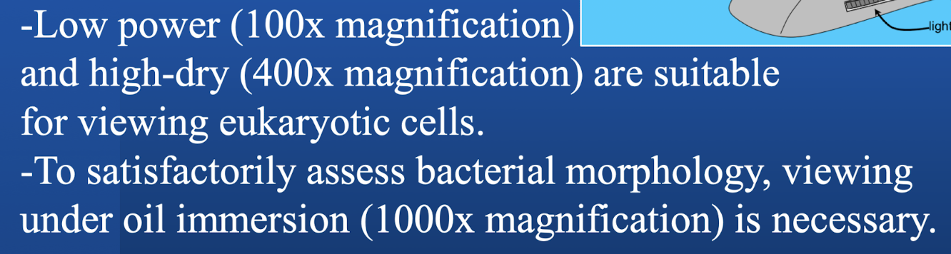
Which magnifications do you need for viewing eukaryotic cells? Prokaryotic cells? What are these techniques called?
What is the resolving power range for electron microscopy? What magnification do you need for virus viewing?

What microflora are in the oral cavity? Which are the primary inhavitants of dental plaque and causative agents of dental caries?
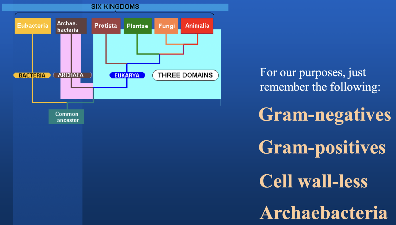
What are the four classifications of bacertia that we are worried about?
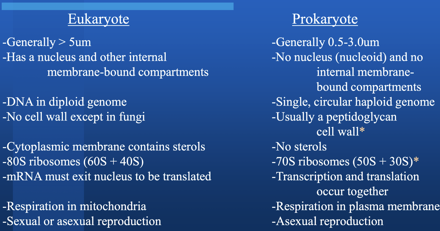
Highlight some major differences between eukaryotic and prokaryotic in terms of size, organelles/nucleus, DNA structure, walls, sterols, ribosomes, transcription/translation, respiration, and reproduction.
What are the major targets for antimicrobials?
The peptidoglycan cell wall and 70S (30S + 50S) ribosomes.
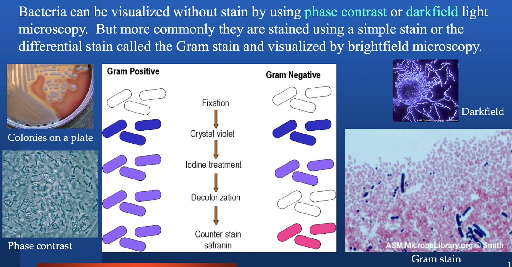
How are bacteria visualized without stain (two methods)? What is the most common method and visualization?
What are the three factors of caries?
Tooth surface
Fermentable carbohydrates (how enamel is eroded)
Oral bacteria
Why does understanding the size of the microbes matter?
Sterilization (different membranes are selective for different microorganisms)
Why is oil immersion necessary when viewing bacteria?
Oil immersion results in less scattering of light rays for greater resolution.
Bacteria in the mouth are mostly which kind? Are archaebacteria found in high levels?
Mostly gram positive, but not exclusively. Archaebacteria are not found in high levels in the mouth but are in the environment, so we can obtain those from what we eat.
What is the main benefit of unstained viewing of microbes?
Unstained, we can view viable organisms. With staining, the bacteria must be killed and fixed.
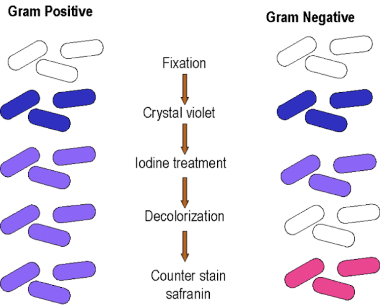
Describe the process of gram staining.
Both start colorless and are fixed. Stained with crystal violet and both will be purple. They are treated with iodine to complex with crystal violet and help keep crystal violet there for gram positive. Iodine is not sufficient to keep crystal violet in the gram negative bacteria. Decolorization occurs (rinsing with 95% ethanol or acetone sometimes) to wash out the crystal violet iodine from gram negative organisms that will stay in gram positive, resulting in purple gram positive and colorless gram negative organisms. Counter stain safranin makes gram negatives red/pink and does not affect gram positive.
If one were to forget iodine, what would the gram negative and positive bacteria appear as?
The gram positive would likely appear the same as the gram negative because the crystal violet will be washed out. Both the gram positive and negative bacteria would be decolorized. The counter stain will result in all of them appearing red/pink.

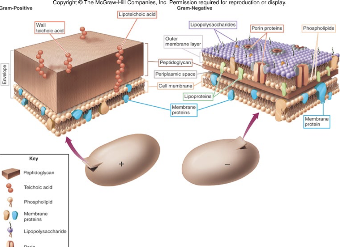
Describe the cell wall anatomy of gram positive and negative bacteria.
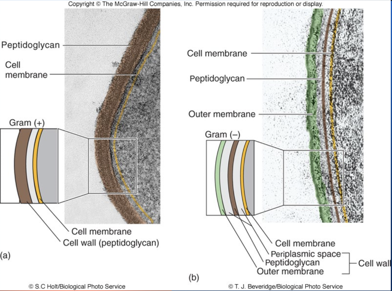
Gram positive bacteria have what kind of acids? Gram negative bacteria have what kind of protein on the outer membrane surface?
Teichoic/lipoteichoci acids; LPS
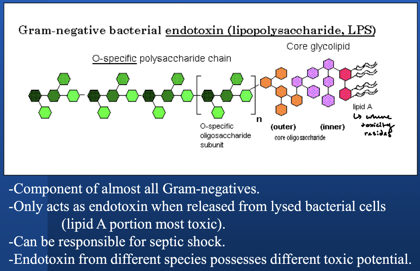
What is the endotoxin in gram negative bacteria? When is this endotoxin working as a toxin?
LPS. In tact microorganisms do not have toxic LPS. The cells have to be dead or released somehow to act as an endotoxin.
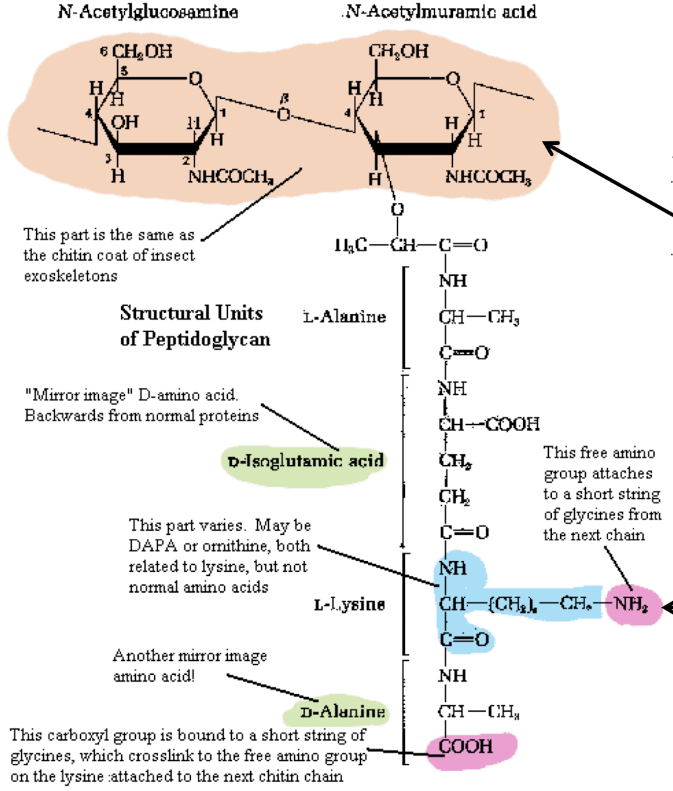
Describe the structure of petidoglycan? Where is the crosslink always attached to?
Repeating disaccharid (NAM and NAG). The crosslink is always attached to the N-acetylmuramic acid moiety (NAM).
What is cross-linking? Which (+ or -) has more extensive cross linking? Which is direct? Which one has an intermediate and what is it?
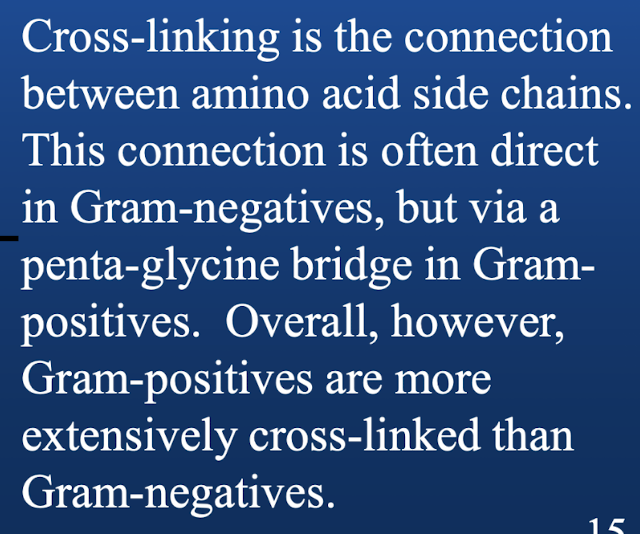
Which (gram positive or negative) is much easier to lyse/break and why?
Gram negatives are easier to lyse and break than gram positive because their layers in the peptidoglycan layer are not as extensively cross linked as are gram positives.
Which bacteria would allow the most direct access to an extracellular substance that attacks the peptidoglycan?
Gram positive because of the lack of an outer membrane.
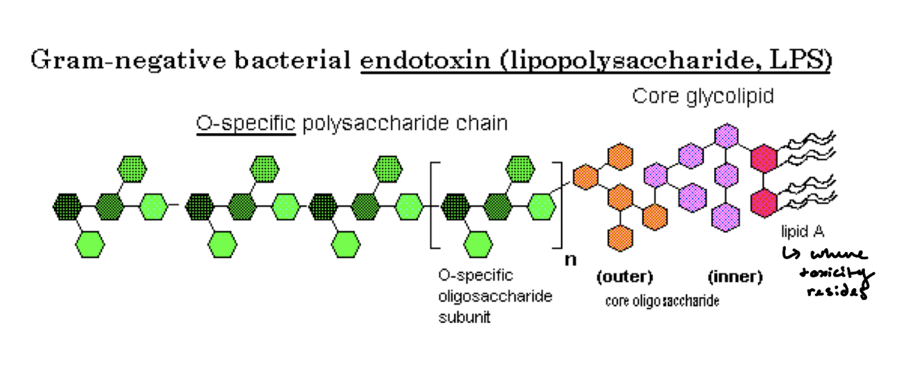
What are the regions of LPS? Which area is most toxic?
Why do we have a normal microflora? What is the association between us and our microflora in most instances? What are the three benefits to having a normal flora?
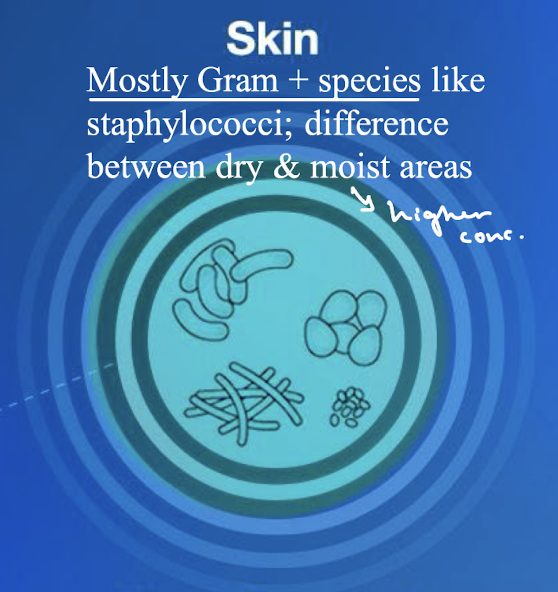
What microbes typically live in the skin?
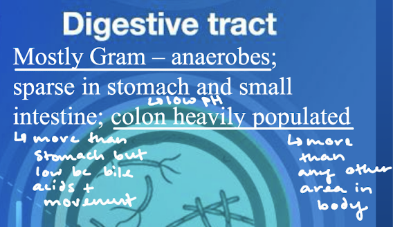
What microbes typically live in the digestive tract? Which areas of the digestive tract are more heavily populated?
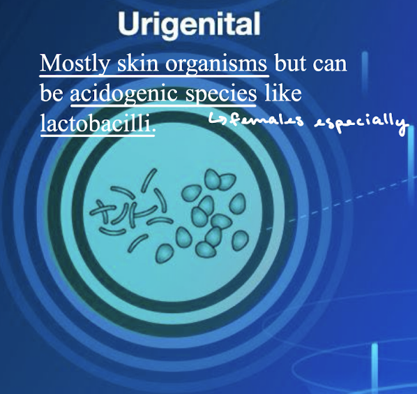
What microbes typically exist in the urigenital regions?
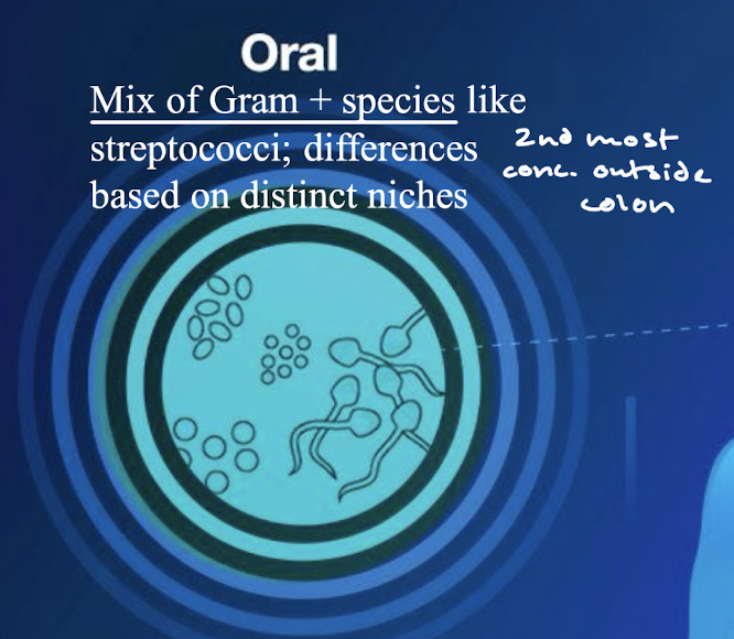
What microbes typically live in the oral cavity?
What are the five bacterial properties designed to promote colonization and propagation and what is the corresponding host response to avoid being overtaken by the bacteria?
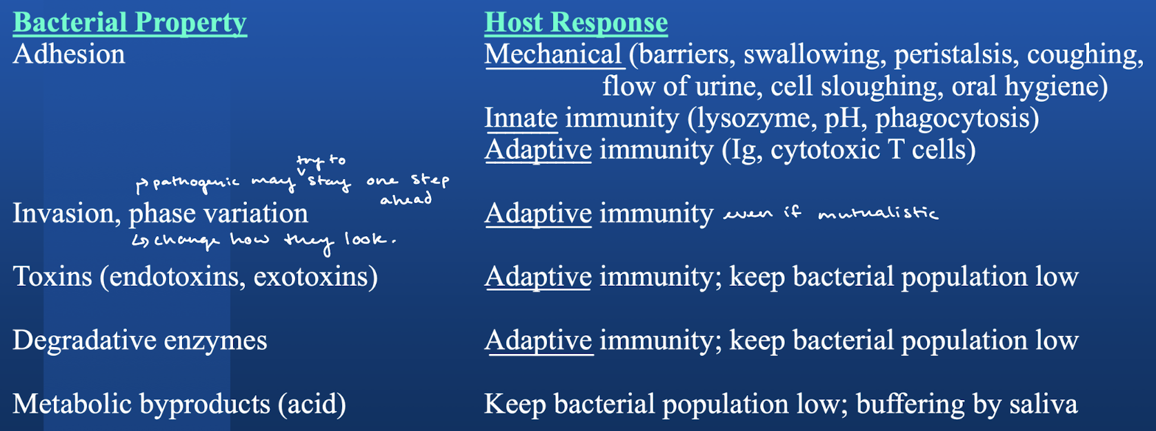
What is phase variation?
Changing the way the microbes are perceived by the immune system to try to stay one step ahead of the host response.
Bacteria that can cause disease are called ______. How do we most often think of disease coming about?
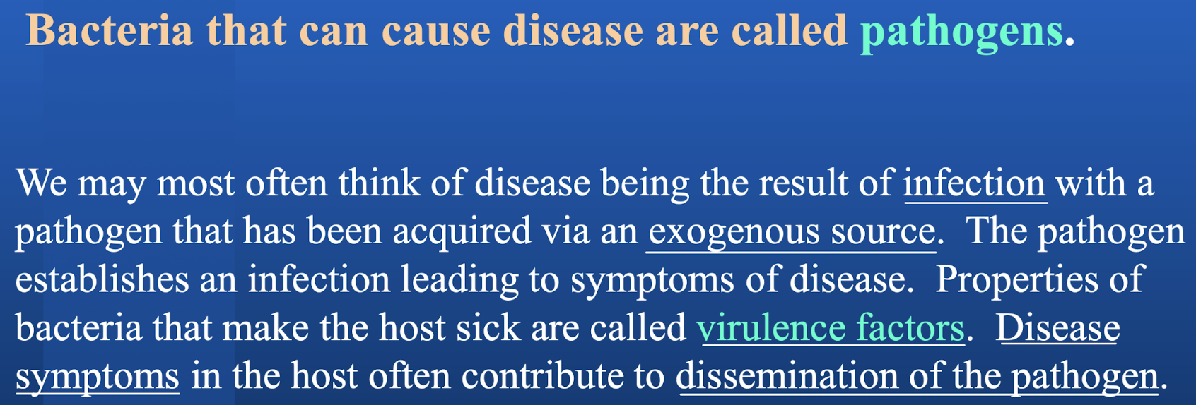
What are virulence factors? Disease symptoms in the host often contribute to what?
What is an opportunistic infection?
Overgrowth of normal flora or introduction of normal flora to sterile sites, possibly due to weakening of the host defenses.
Dental caries and periodontal disease are generally considered what kind of infections?

The altered composition of the microbiome is said to be in _______. What is an example of this?