Deck 1: Gross Anatomy + Orbits
1/97
There's no tags or description
Looks like no tags are added yet.
Name | Mastery | Learn | Test | Matching | Spaced | Call with Kai |
|---|
No study sessions yet.
98 Terms
Describe the characteristics of the eyelid.
thinnest layer of skin
has no subcutaneous fat
eyelids will never get a fatty layer, no matter how fat you get
List the functions of the eyelid.
Physical protection of the eye (dust, light)
Lubrication
What are the 3 actions of eyelids?
Reflexive — lid closes fast if an object approaches/touches the eye or lashes
Voluntary — squeezing the eyes tightly shut or winking
Involuntary — routine blink
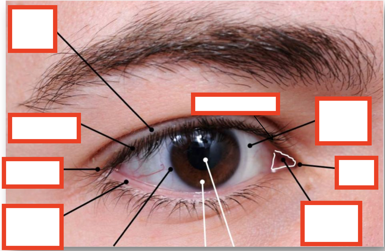
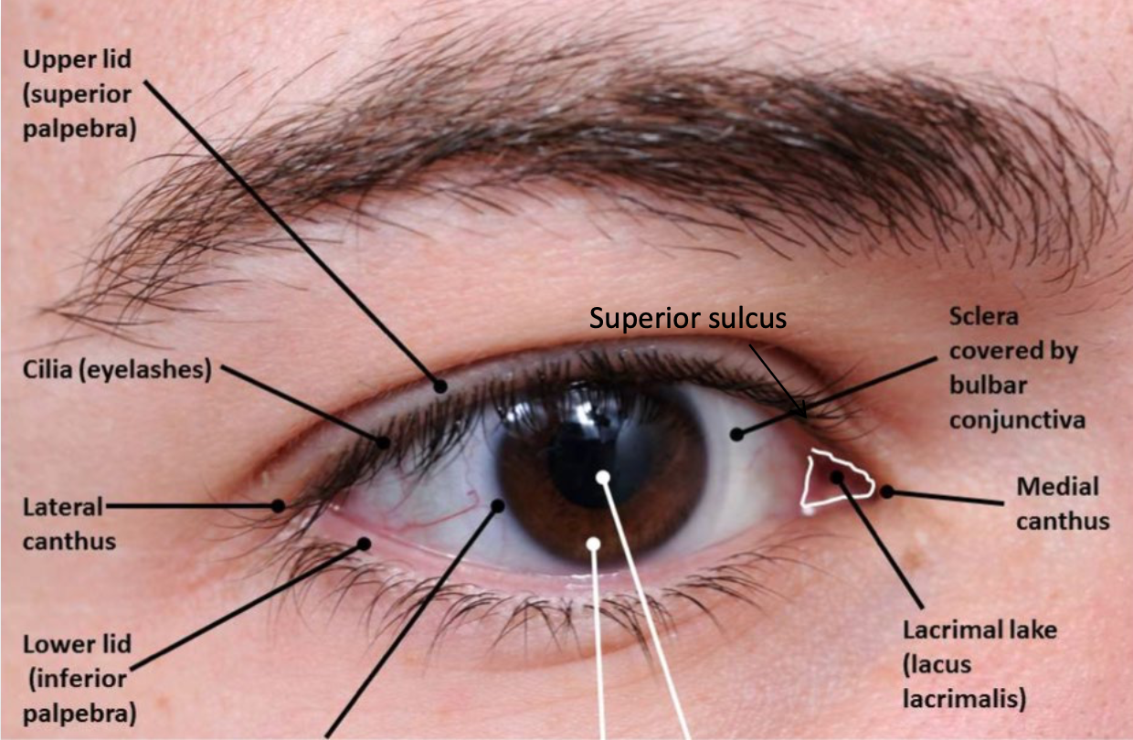
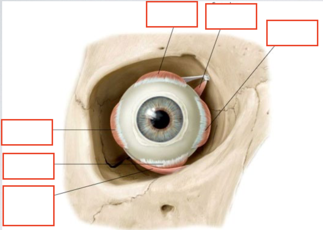
Label the Landmarks of the Eye In-situ.
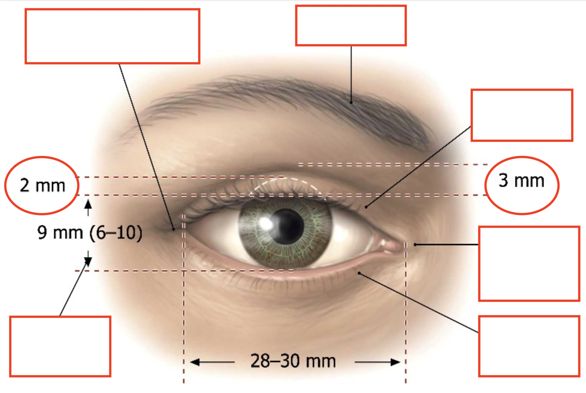
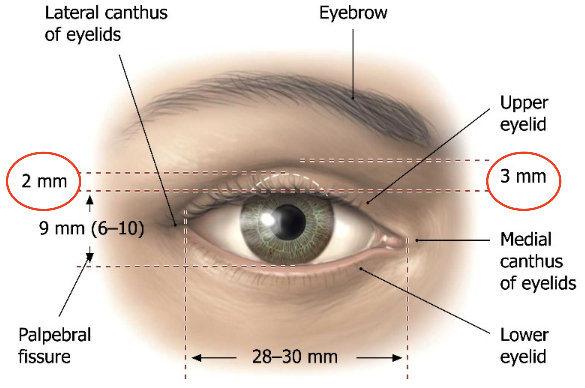
List the key features of the eyebrow.
→ Hair-bearing skin that runs parallel to the superior orbital rim (the bony edge around eye socket)
Projects from the forehead due to a fat pad
esp near the middle, pushing the hairs slightly forward over the eye
Underlying muscles move the eyebrow during facial expressions
What is the role of the stiff hairs that make up our eyebrow?
protect the eye from dust, sweat, and bright light
What is the role of the supraorbital notch?
passageway for the supraorbital nerve and vessels which supply sensation to upper eyelid, forehead & scalp
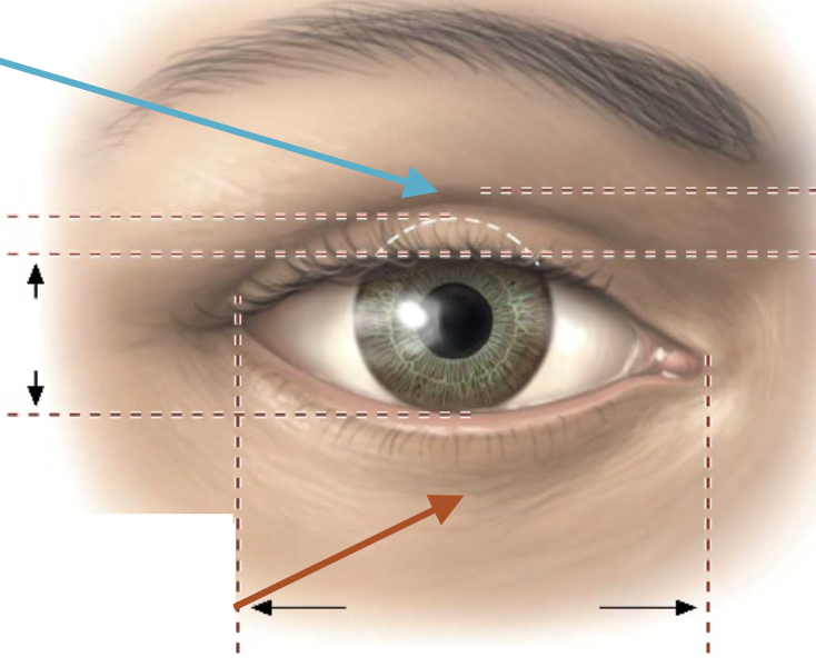
What is the Palpebral Fissure?
horizontal opening b/w the upper and lower eyelid margins
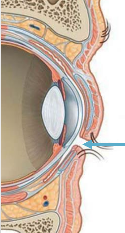
How do the eyelids move when open and closed?
Open and looking straight ahead:
Upper eyelid → covers the top 2–3 mm of the cornea
Lower eyelid → rests just below the corneoscleral junction (limbus)
When closed:
Upper eyelid → moves down to cover the entire cornea
Lower eyelid → moves very little b/c it has less muscle movement
Label.
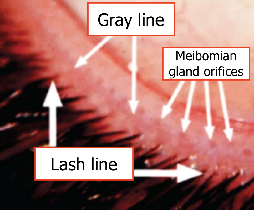
Label the components of the Palpebral Margins
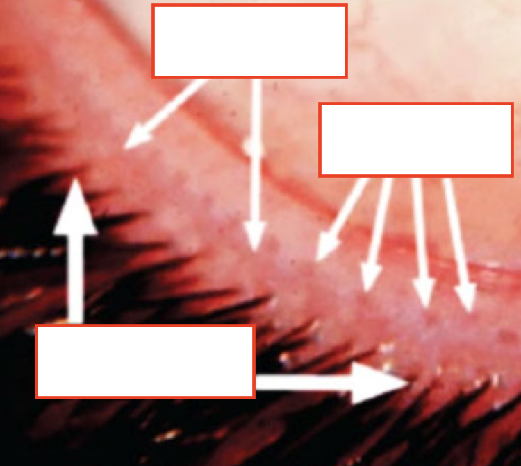
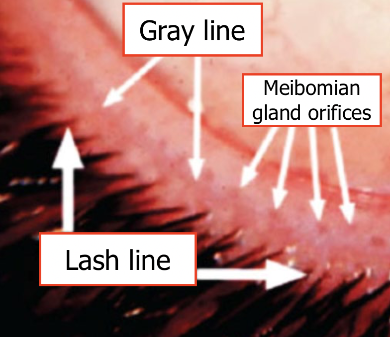
List the key features of the Palpebral Margins.
Lash line
found in superior and inferior margins
Gray line
Meibomian gland orifices
Mucocutaneous junction (i.e., Marx’s line)
line where the skin of the eyelid ends & conjunctiva begins
What are the key features of the lash line and the eyelashes on each eyelid?
→ 2 rows of cilia ‘eyelashes’ on each lid
Superior - longer & curl upward
Inferior - shorter & curl downward
-Lash Life cycle = 5 to 11 months
Which nerves are responsible for the sensory innervation that triggers reflex eyelid closure?
Reflex = sensory
Upper eyelid = ophthalmic division of the trigeminal nerve (CN V1)
Lower eyelid = maxillary division of the trigeminal nerve (CN V3)
What are the palpebral sulci? How do the superior and inferior palpebral sulci differ?
→ are furrows or creases that appear in the eyelids when the eyes are open
Superior - forms a distinct upper eyelid crease with a fold
Inferior - forms a less prominent crease with no fold
What are the palpebral commissures? How do the medial and lateral palpebral commissures differ?
→ junctions where the upper and lower eyelids meet
Medial - at the inner corner of the eye
Lateral - at the outer corner of the eye

What do both medial and lateral palpebral commissures define?
Canthi — aka the medial and lateral angles of the eye
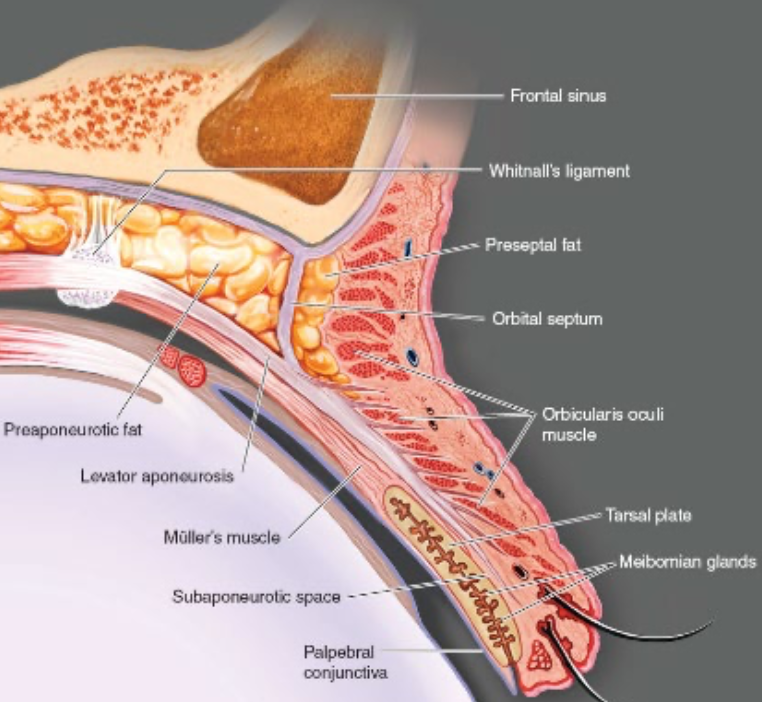
What are the major structures of the upper eyelid from anterior to posterior in cross section?
Skin
Orbicularis oculi muscle
Orbital septum
Levator Aponeurosis
Tarsal plate + sebaceous glands (of Zeis/Meibomian)
Müller’s muscle (superior tarsal muscle)
Palpebral conjunctiva + its accessory lacrimal glands of Krause and Wolfring
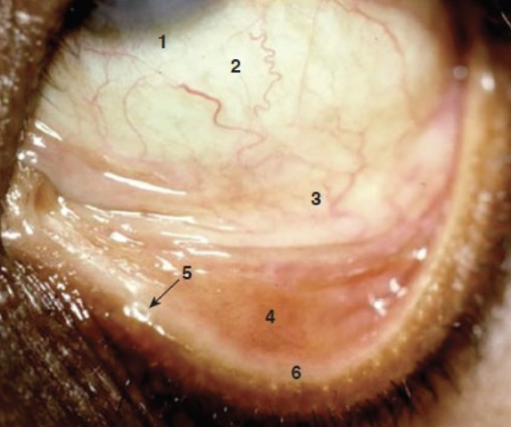
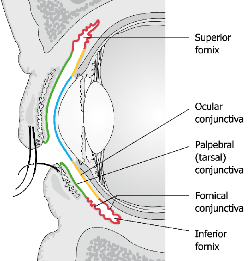
What is the conjunctiva? What is it made up of?
→ entire mucous membrane that lines the inside of the eyelids and covers part of the eyeball
Bulbar conjunctiva: begins at the limbus (1) and loosely covers the anterior 1/3 of the sclera (visible)
Note: remaining 2/3 of the sclera is under the eyelids and is still the bulbar conjunctiva
Palpebral conjunctiva: pinkish tissue (4) tightly lining the inner surface of the lower eyelid, continuous with eyelids
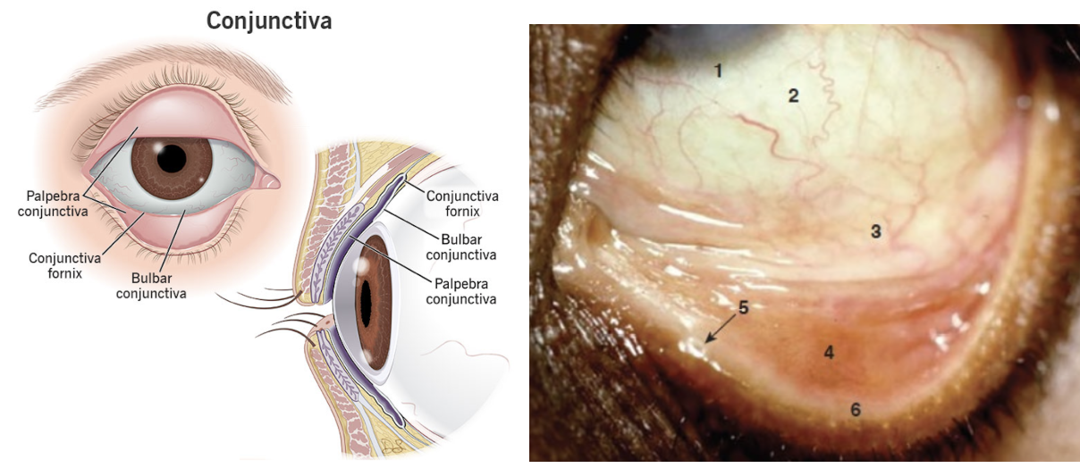
What happens to the bulbar conjunctiva during massive allergic reactions?
it can billow out beyond the margins of the eyelids
Recall: it’s loosely attached to the sclera
What is the Conjunctival fornix? What’s another name for it?
→ fold or “blind pocket” created where the bulbar conjunctiva reflects onto the palpebral conjunctiva (3)
aka “cul-de-sac”
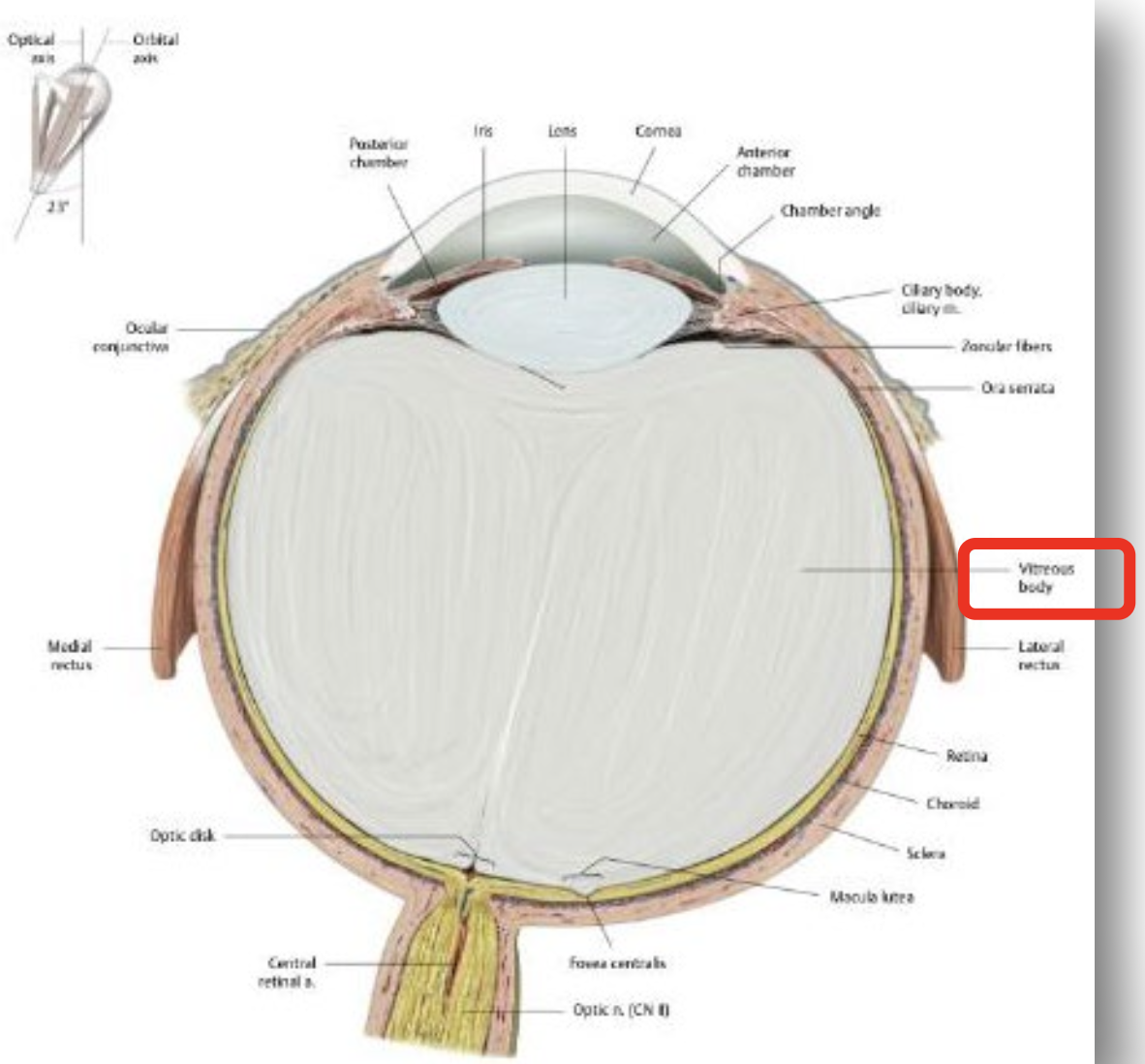
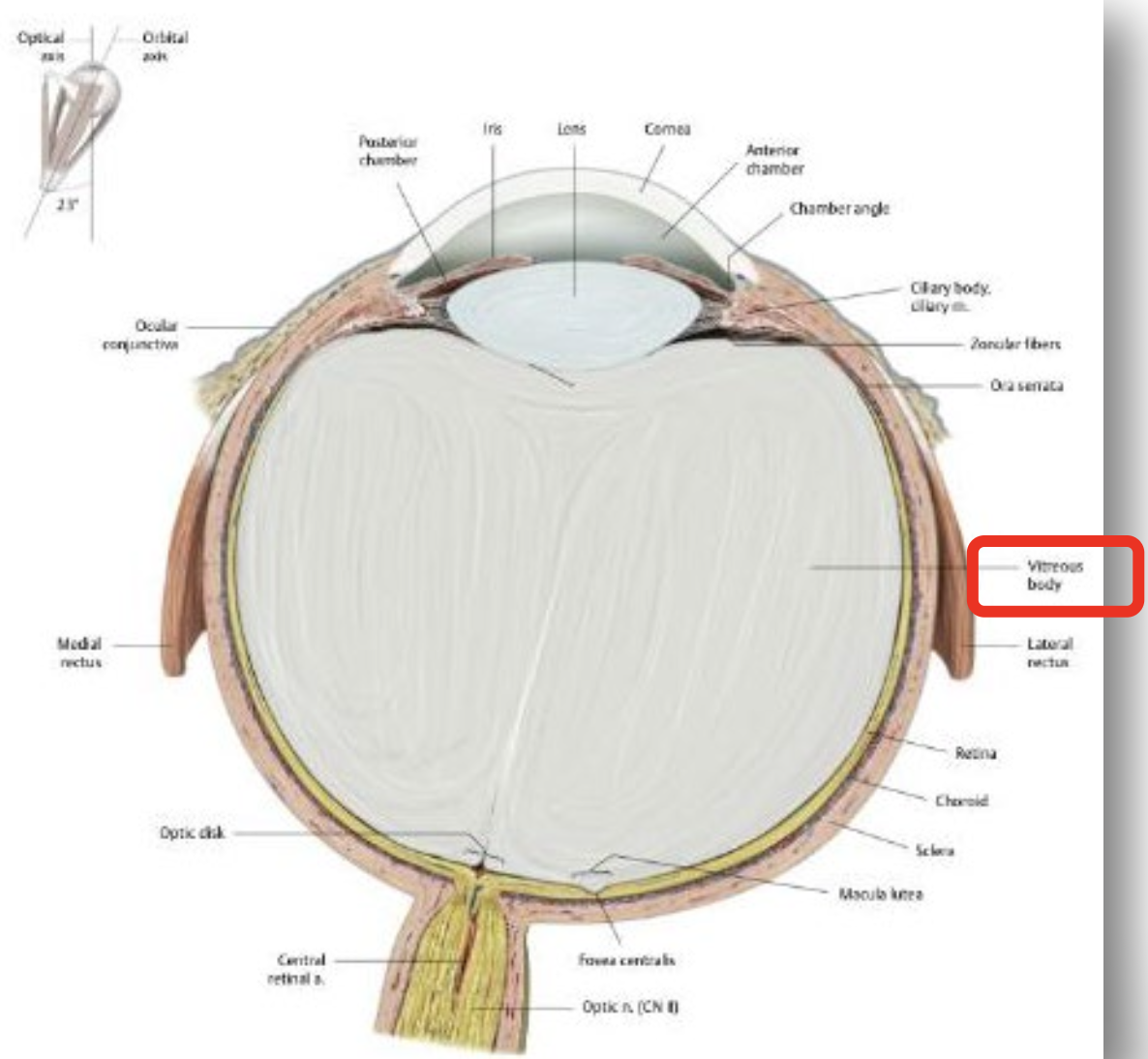
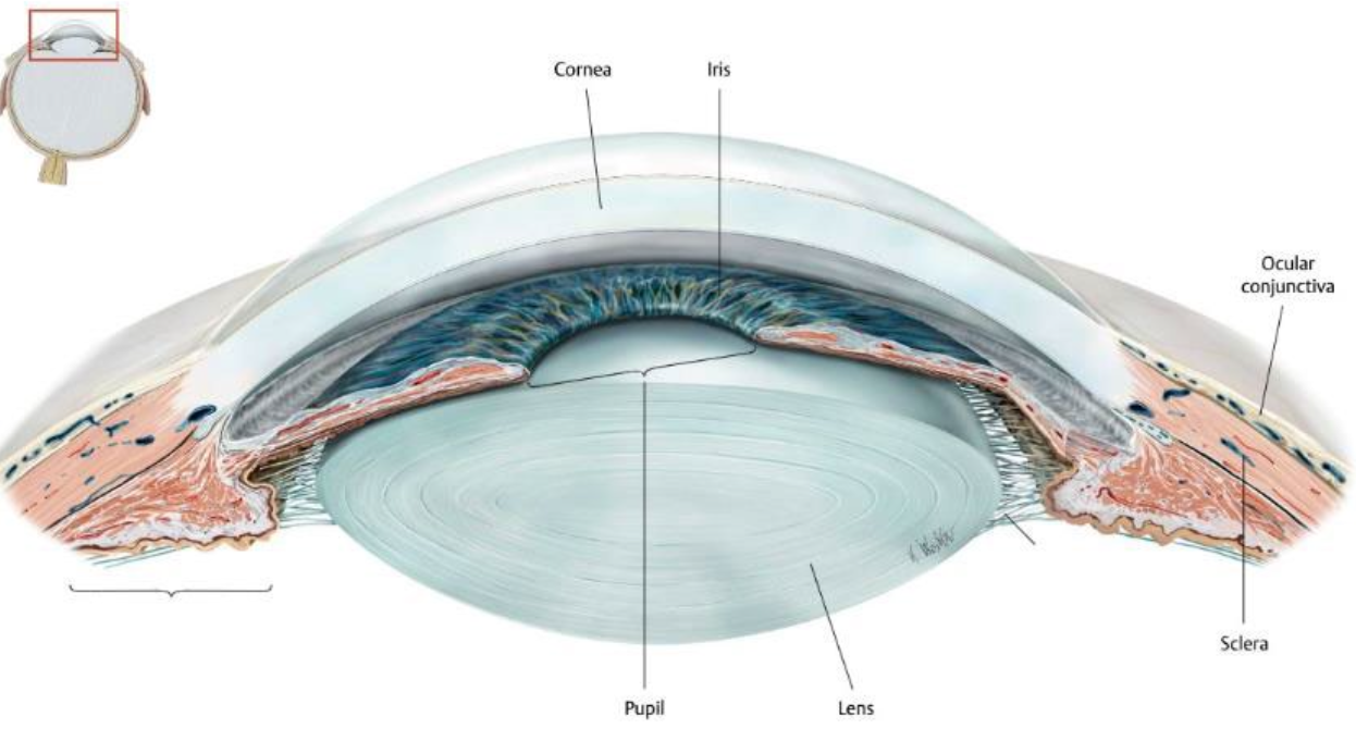
Label the sagittal view of the eyeball.
What are the key features and functions of the eyeball?
Role: focuses light onto photosensitive cells that convert photons → neural signals for visual processing
has the optical apparatus of the visual system
occupies most of the anterior portion of the orbit
25 mm in diameter
all anatomical structures within the eyeball have a circular or spherical arrangement
suspended by 6 EOMS
Describe the anterior and posterior poles of the eye.
Anterior pole = apex of cornea
Posterior pole = the point on the posterior sclera transected by a line normal to the corneal apex at the horizontal equator of the eye
Note: doesn’t correspond to the position of the optic nerve
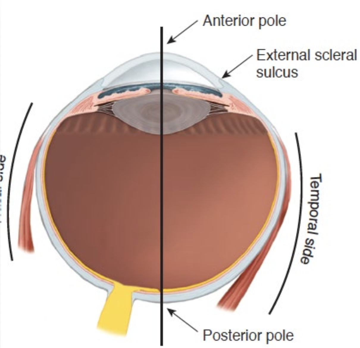
Why is the temporal side of the eye is longer than the nasal side?
b/c the optic nerve doesn’t exit exactly at the back center of the eyeball
exits slightly toward the nasal side
What are the average dimensions of the human eyeball and its clinical range?
Vertical diameter (height): 23.2 mm (average)
Horizontal diameter: 24.0 mm (average)
Antero-posterior (axial) length: 24.3 mm avg
Range: 22 mm (very hyperopic) – 27 mm (very myopic)
Note: Can be measured clinically
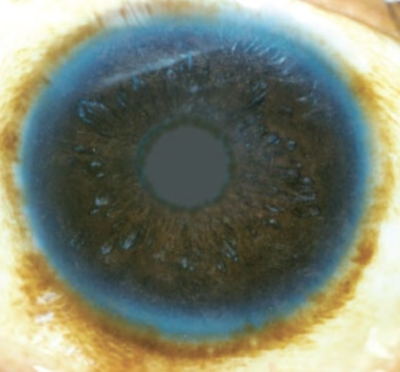
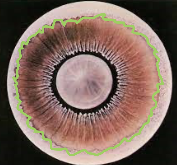
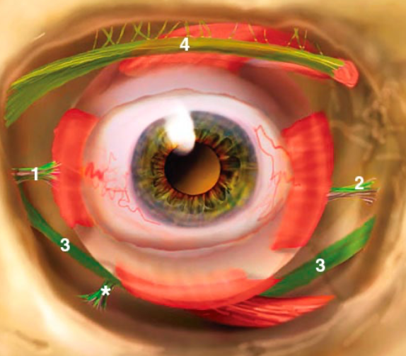
What are the key anterior external landmarks of the eye?
Limbus
Iris
Pupil
When eye is removed: EOM attachments are visible
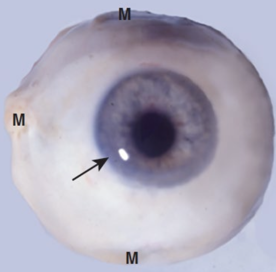
What is the limbus? What it’s significance in the eye?
→ junction b/w clear cornea & white sclera
has corneal stem cells
Limbal pigment (melanocytes) protects these continuously dividing cells
Genetic/ancestral adaptation — People from high-sun areas often have a pigmented ring at the limbus
due to increased melanocyte production in response to high UV
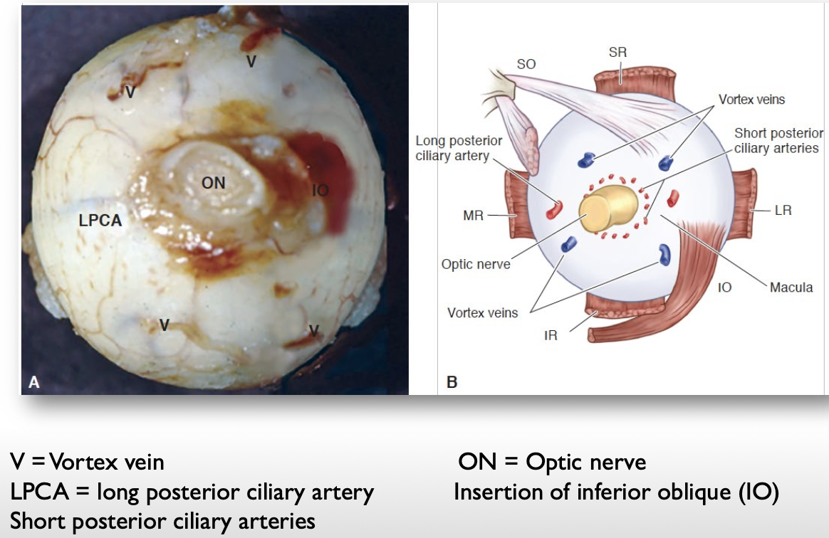
List the key posterior landmarks of the eye.
**Not visible in-situ
Optic nerve
Dura mater blends with sclera
4 vortex veins (1 vein/quadrant)
IO muscle attachment site
LPCA (medial to optic nerve)
SPCA
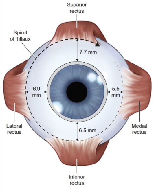
What is the order and distance of rectus muscle insertions from the limbus farthest to closest? What’s this anatomical arrangement known as?
Superior = 7.7mm
Lateral = 6.9mm
Inferior = 6.5mm
Medial = 5.5mm
→ This anatomical arrangement = Spiral of Tillaux
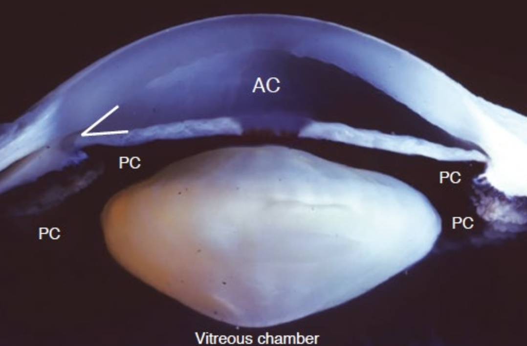
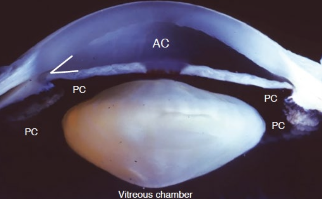
List the 3 compartments the eye in made up of.
What liquid is found in each compartment?
Anterior chamber
Posterior chamber
Vitreous chamber
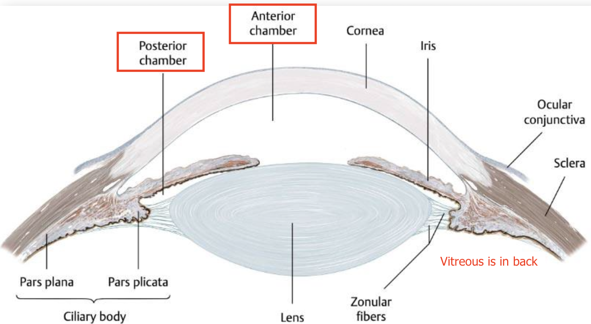
Aq humour in anterior & posterior
Vitreous humour in vitreous chamber
Where is the Anterior chamber located?
→ found b/w the cornea & iris and pupil/lens
contains the anterior chamber angle at its lateral edges
What is the anterior chamber angle?
→ allows for aq humour drainage
contains TM, Schlemm canal
What is the aqueous humour?
→ clear, nutritive fluid
Function: Meets the metabolic needs of the avascular tissues bordering the anterior chamber, including:
Cornea
Lens
TM
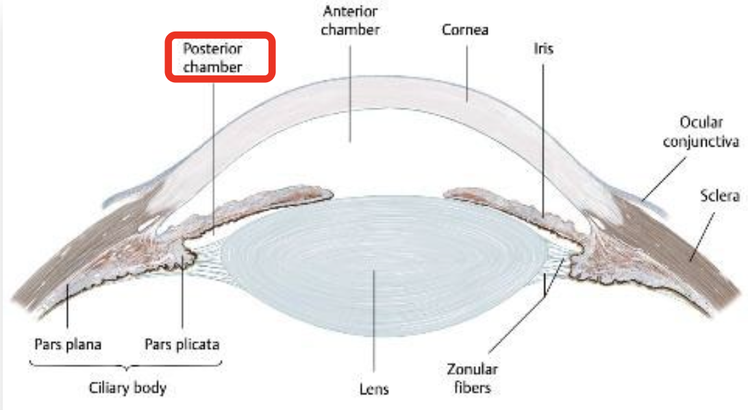
What is the Posterior chamber?
Donut-shaped space
Surrounds the crystalline lens
found b/w the posterior surface of the iris and the inner surface of the ciliary body
What is the Vitreous Chamber?
→ Largest of the spaces within the eye
Found posterior to the lens
contains the vitreous body (which is made up of vitreous humour)
What is the vitreous humour?
→ gel-like substance that:
is 99% water by weight
has a sparse collagenous support matrix and envelope
Functions:
Transmits light
Holds the retina in place
Supports the lens
List the 3 structural layers of the eye and what it contains. (Hint: Mention if these’s structures are found anterior or posterior)
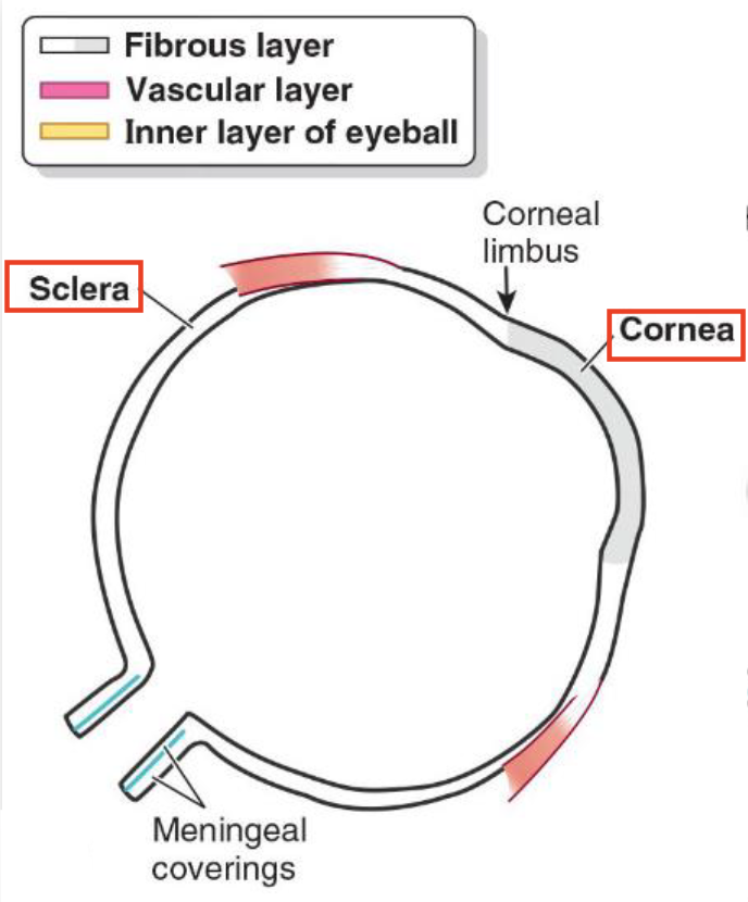
1) Fibrous layer (outer coat/tunic)
Cornea (anterior)
Sclera (posterior)
2) Vascular layer (middle coat/tunic)
Ciliary body (anterior)
Iris (anterior)
Choroid (posterior)
3) Inner layer (inner coat/tunic)
Retina - includes the optic & nonvisual part
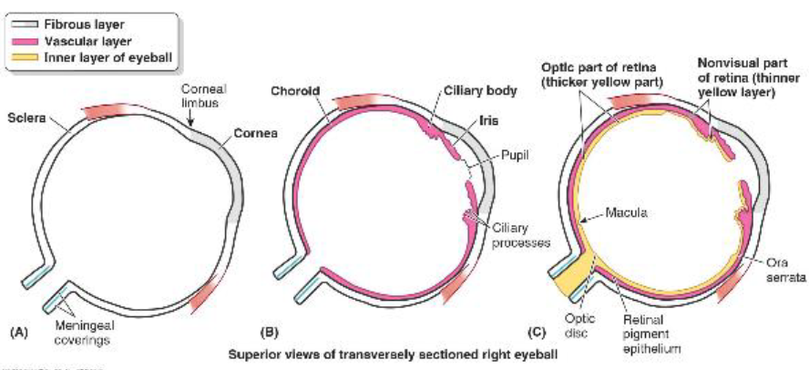
❌ What’s Not Included: CT layers that surround the eyeball (bulbar conjunctiva; tenon capsule)
Describe the Fibrous Tunic (outer eyeball layer).
→ AKA “Corneo-Scleral Coat”
“External fibrous skeleton” of the eyeball
provides shape and resistance
provides attachment site for EOMs & intrinsic muscles
Fibrous Tunic
What is the Cornea?
Transparent
avascular
covered by tear film (Thickness: ~0.5 mm)
Principal refractive surface of the eye
Fibrous Tunic
What is the Sclera?
Opaque
fibrous exoskeleton of the eye to which EOMs are attached to
Fibrous Tunic
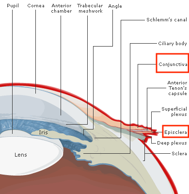
What are the structures overlying the sclera?
Episclera - thin layer of vascularized CT covers the sclera, extending from the limbus all the way to the back of the optic nerve sheath
Conjunctiva
Describe the Vascular Tunic (middle eyeball layer).
→ aka the “Uvea”
Divided into the anterior & posterior uvea
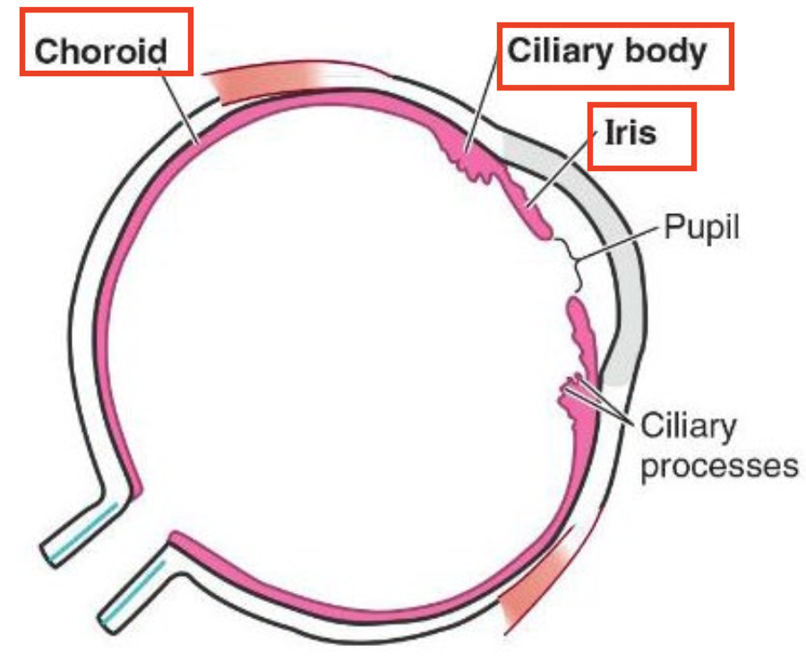
Vascular Tunic
Where do the anterior and posterior uvea meet?
Ora serrata
b/w ciliary body (end of anterior uvea) & choroid (start of posterior uvea)
Vascular Tunic
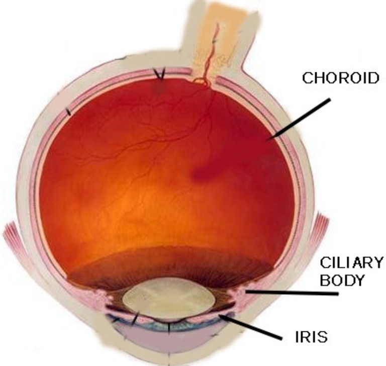
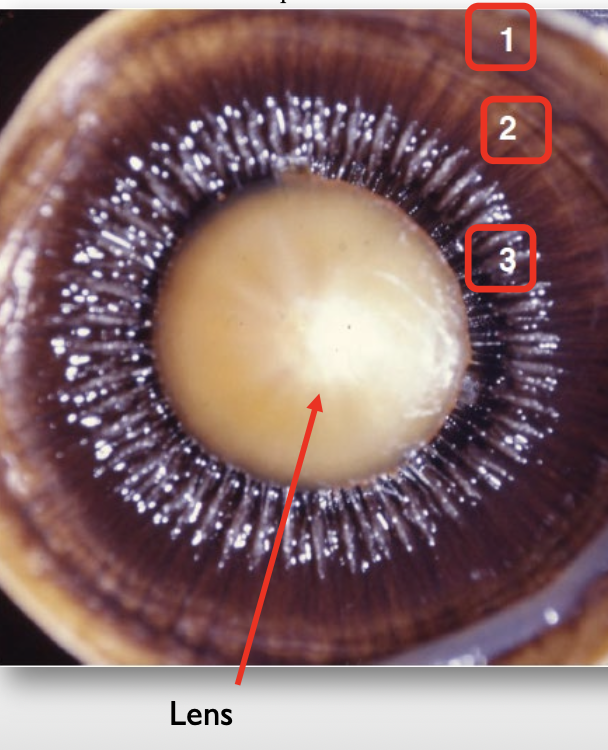
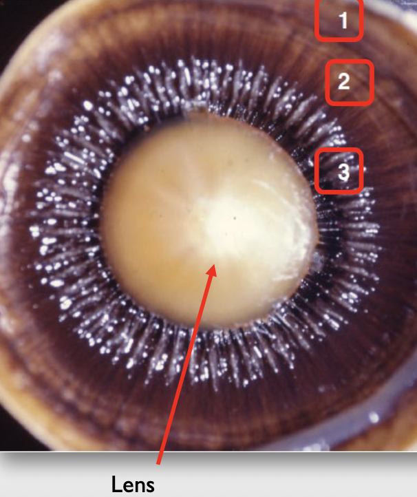
What is the Iris?
→ Thin contractile diaphragm with a central aperture- the pupil
on the anterior surface of the lens
Function: controls light entry
Vascular Tunic
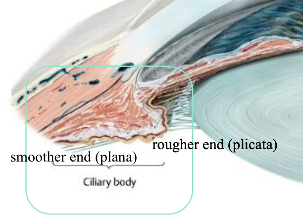
What is the ciliary body?
→ ring-like thickening of the vascular layer
found behind the corneoscleral junction
is muscular & vascular
- Divided into 2 parts:
Pars plicata (3) - rough
Pars plana (2) - smooth
Vascular Tunic
What is the junction b/w the pars plana & retina?
Ora serrata
Recall: Ciliary body is part of the uvea. Ora serrata lies at the transition from the ciliary body (end of anterior) to the choroid (start of posterior uvea)
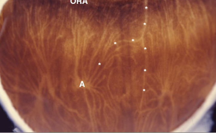
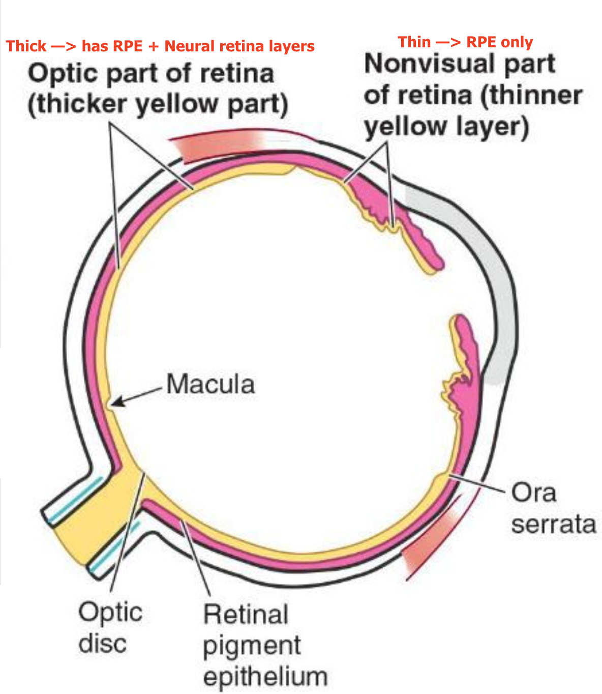
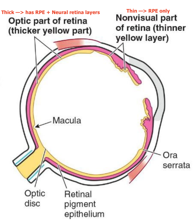
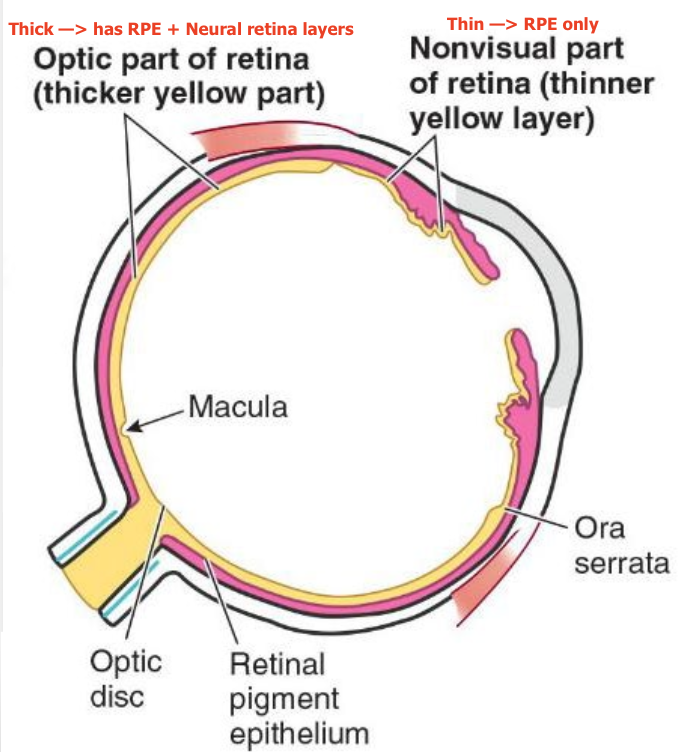
What is the Ora Serrata?
→ visible line of separation b/w the retina and the irregular posterior border of the ciliary body
marks the transition from the neural retina (visual) & RPE (non-visual)
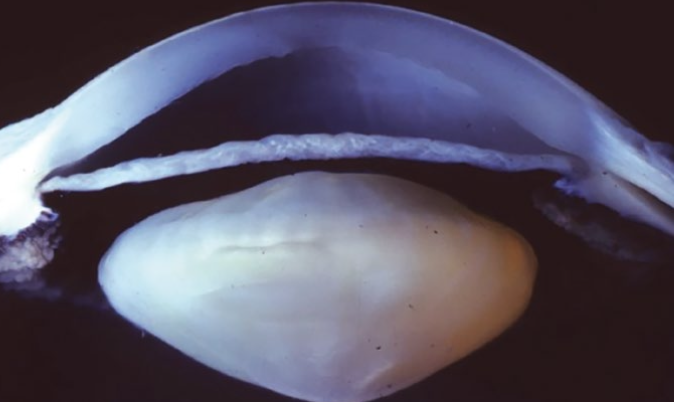
What are Crystalline Lens?
Transparent, biconvex structure enclosed in a capsule
Location: Posterior to the iris & anterior to the vitreous humor of the vitreous body
Continues growing throughout adult life → potentially impacts the iris and outflow angle for humor
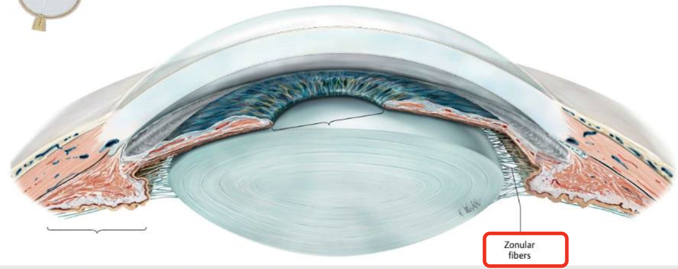
What structures suspend the crystalline lens, and where do they originate?
→ Zonules are anchoring fibrils that:
Originate from the ciliary body
Suspend the crystalline lens in place, behind the pupil
- AKA: “zonular fibers” or “suspensory ligaments”
Vascular layer
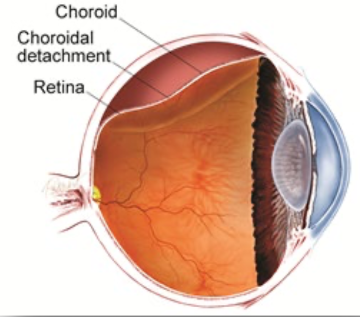
What is the Choroid?
→ densely vascularized layer that provides metabolic support for the outer half of the retina
Highly pigmented! — Dark reddish-brown layer b/w the sclera and retina
attached firmly to the pigment layer of the retina but can easily be stripped from the sclera
is continuous anteriorly with the ciliary body
Describe the Neural Tunic (retina/inner eyeball layer).
→ acts as the sensory neural layer of the eye
- Made of 2 parts:
Neural (sensory) retina - “thick visual part”
RPE (non-visual) - “thin non-visual part”
💡Think of the retina as a "flattened out nerve" — it’s an extension of the brain
Neural Tunic
What is the Neural Retina?
→ Photosensitive tissue that converts photons of light into chemical/electrical messages
originates from the optic nerve which is why it’s the “optic/visual part of retina (functional)”
Neural Tunic
What is the RPE?
Single layer of pigmented epithelial cells that nourish the outer retina
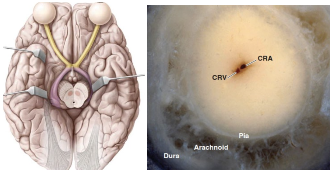
Why is the optic nerve considered a tract of the brain?
→ it’s actually flat piece of the brain (the only part of the brain that’s visible!)
Surrounded by meningeal coverings (dura, arachnoid, and pia) just like the brain
contains the CRA & CRV
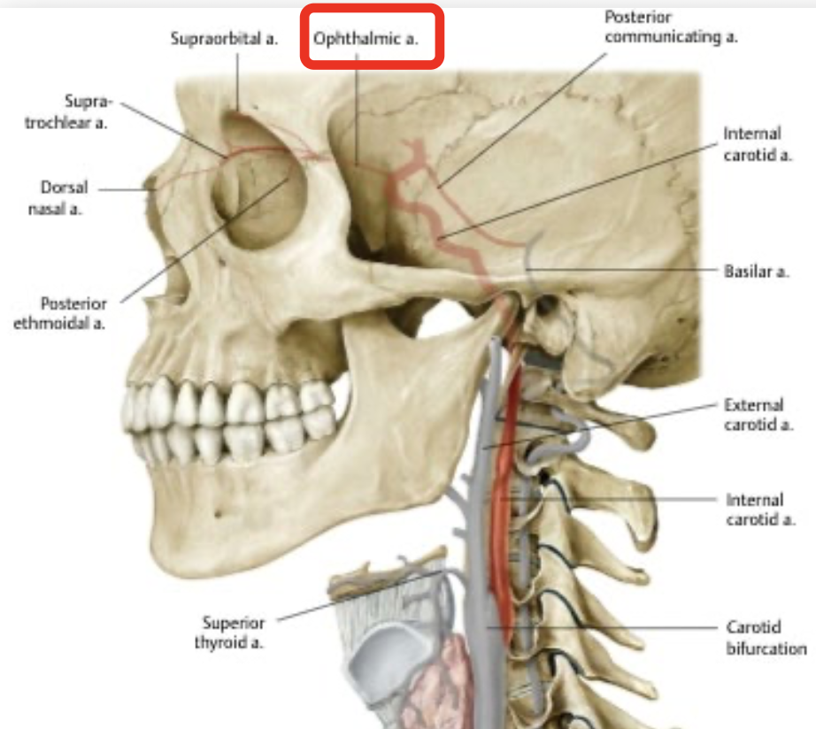
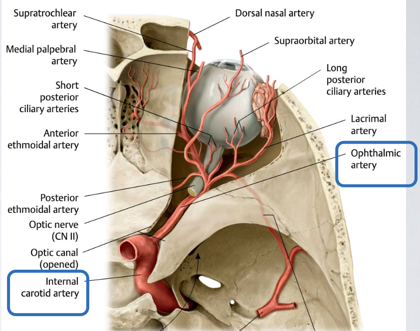
What is the main artery supplying the eye, and what are its key features?
Ophthalmic artery
Origin: First branch of the internal carotid artery
Function: Gives off many smaller branches to provide most of the eye’s blood supply
What are the smaller branches of the Ophthalmic artery group into?
Orbital group - supplies external/extracular structures
Ocular group - supplies internal ocular structures
List the key branches of the Ophthalmic Artery.
CRA: supplies the inner retina
Ciliary arteries → LPCA & SPCA
SPCA - supply the choroid & optic nerve head
LPCA - stretch anteriorly to supply the ciliary body & iris
Lacrimal artery: supplies the lacrimal gland, eyelids & conjunctiva
Muscular branches: supply the EOMs & send branches to the anterior segment of the eye (anterior ciliary arteries)
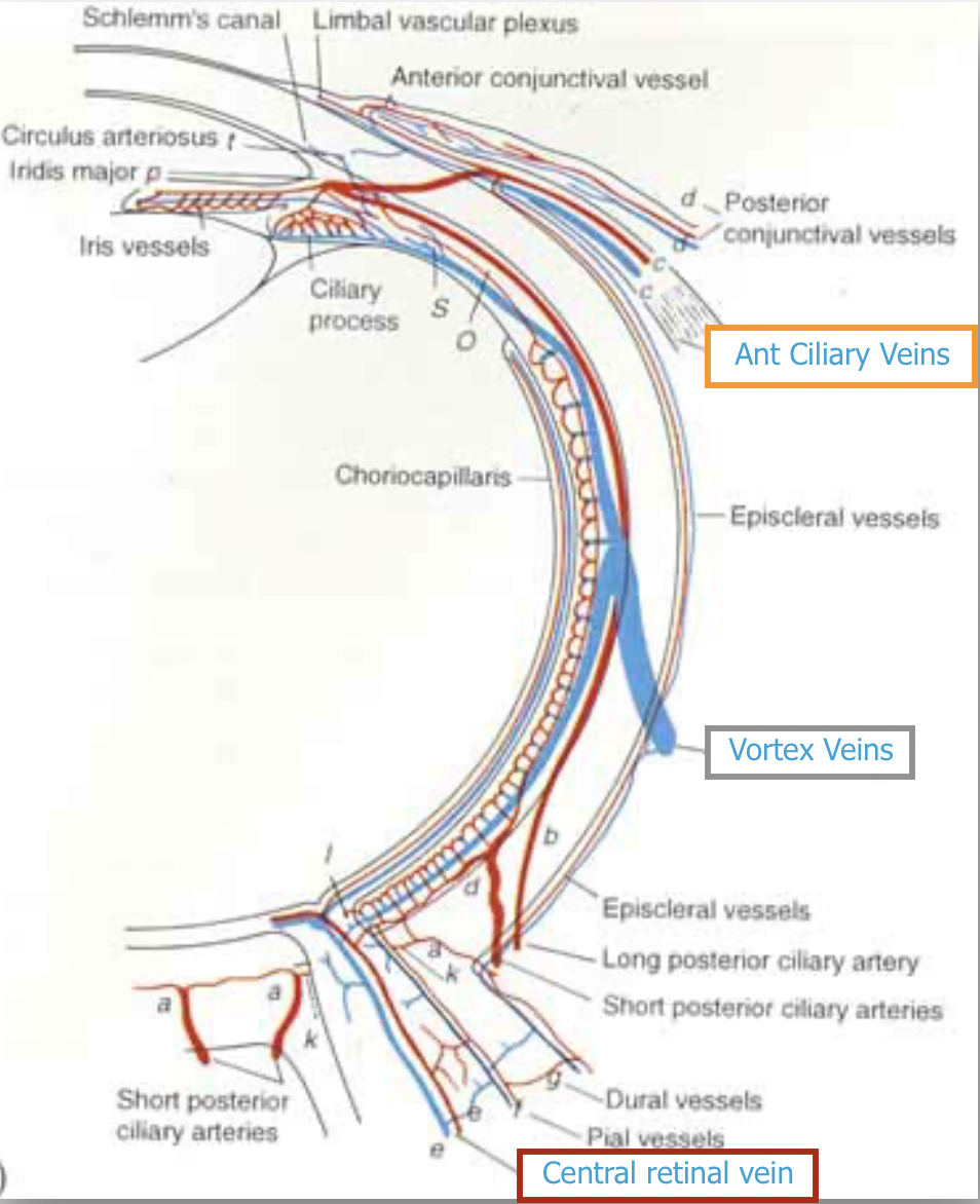
The venous blood leaves the eye via four pathways. Outline these pathways.
CRV: drains the retina, running parallel to the CRA
Vortex veins: drain the choroid; typically 4-6 veins drain into the superior & inferior ophthalmic veins
Superior & inferior ophthalmic veins: main drainage pathways of the eye into the cavernous sinus
Anterior ciliary veins: drain the anterior structures (e.g., ciliary body & iris)
What are the characteristics of the cranium/skull?
made of 22 bones
has openings and passageways like fissures, foramina, and canals which allow nerves and vessels to enter and exit the cranial vault
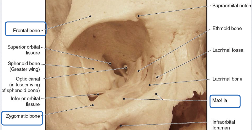
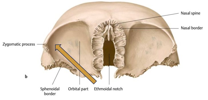
List the 7 bones of the orbit.
ELFMPZS
Ethmoid
Lacrimal
Frontal
Maxilla
Palatine
Zygomatic
Sphenoid
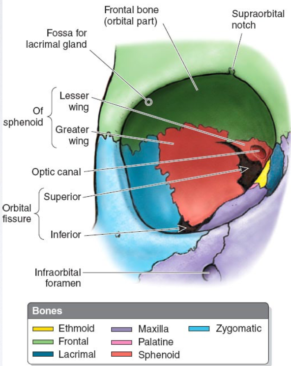
What bones form the orbital rim?
Frontal, Maxillary & Zygomatic bones
these portions are thicker than the orbital walls
BUT 40–60% of all facial fractures still involve the orbital rim
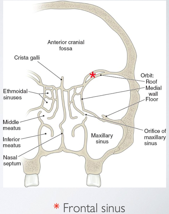
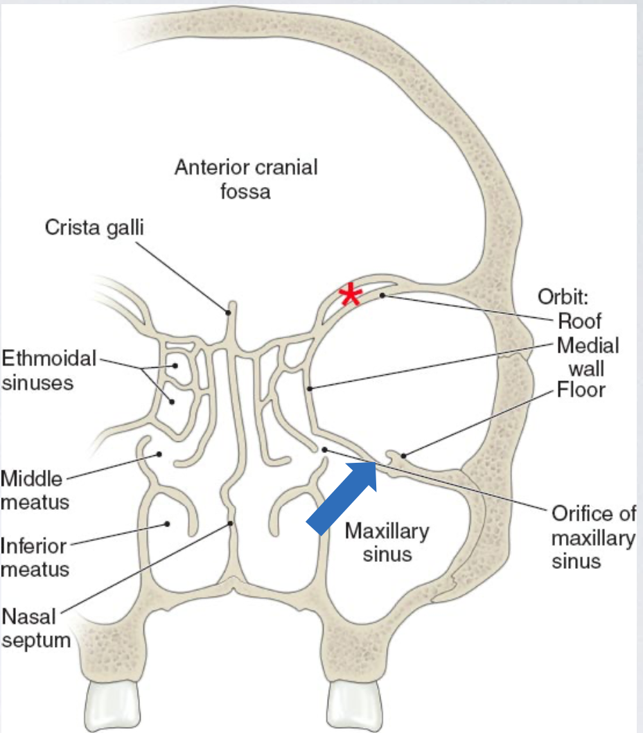
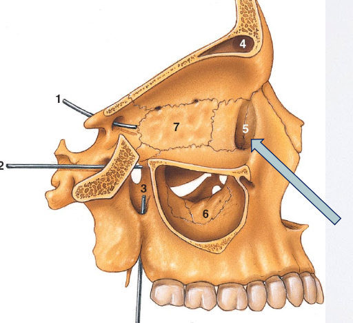
What is the Frontal Sinus?
→ a gap (air-filled cavity) within the frontal bone that makes up one of the paranasal sinuses
What are the components of the orbit?
Floor
Roof
Medial Wall
Lateral Wall
What is the orbital floor formed from?
maxilla
zygomatic
small contribution from palatine bone
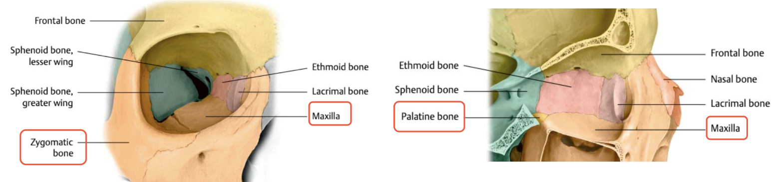
Why is the floor of the orbit commonly damaged in blunt force injuries?
Orbit floor is thicker than medial wall
BUT it’s often damaged in blunt injuries due to thinning at the infraorbital groove
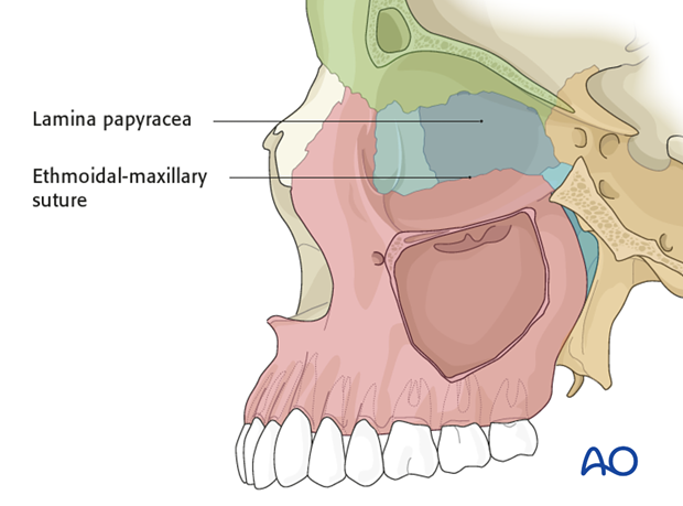
What is medial wall formed from?
lesser wing of the sphenoid bone
lamina papyracea of the ethmoid bone
lacrimal bone
maxilla
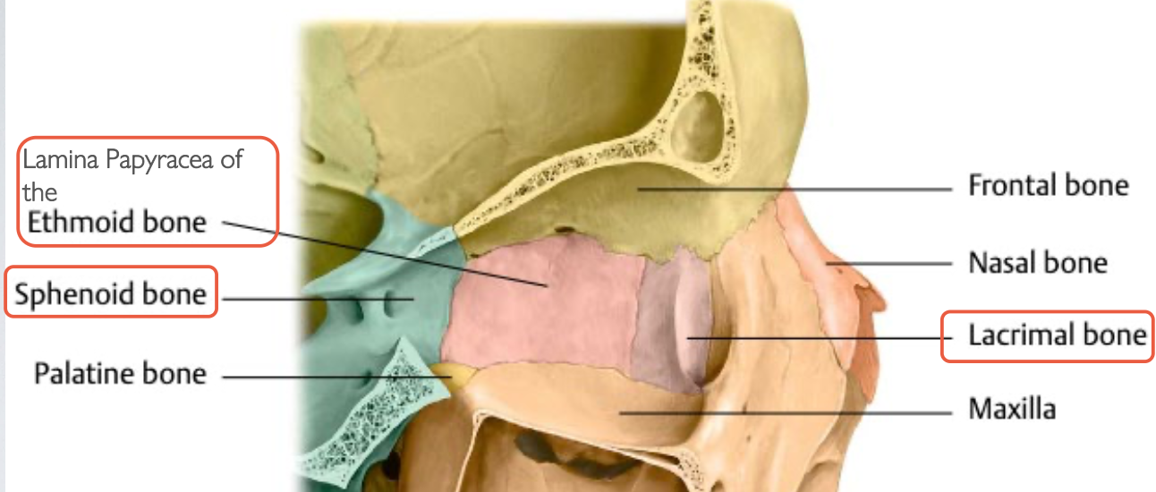
Along with the orbit floor, why is the medial wall also vulnerable to fractures?
due to the thinness of the translucent lamina papyracea
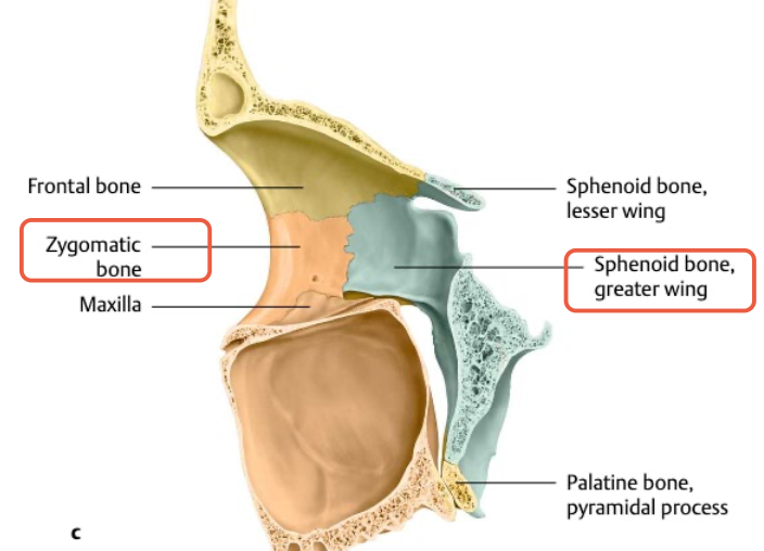
What is lateral wall formed from?
greater wing of the sphenoid bone
zygomatic bone
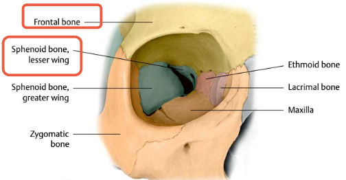
What is orbital roof formed from?
lesser wing of the sphenoid bone
frontal bone
Is the lacrimal fossa the same as the fossa for the lacrimal gland?
❌ No
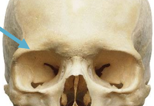
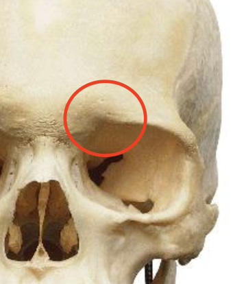
Fossa for the lacrimal gland — holds the gland (superolateral orbit)
Lacrimal fossa — holds the lacrimal sac (inferomedial orbit)
Where is the fossa for the lacrimal gland located? What’s its function?
→ posterior to the superolateral orbital rim, where the inner contour of the frontal bone rises sharply upward
accommodates/houses the lacrimal gland
Where is the lacrimal fossa located?
→ posterior to the inferomedial rim of the orbit
accommodates the lacrimal sac
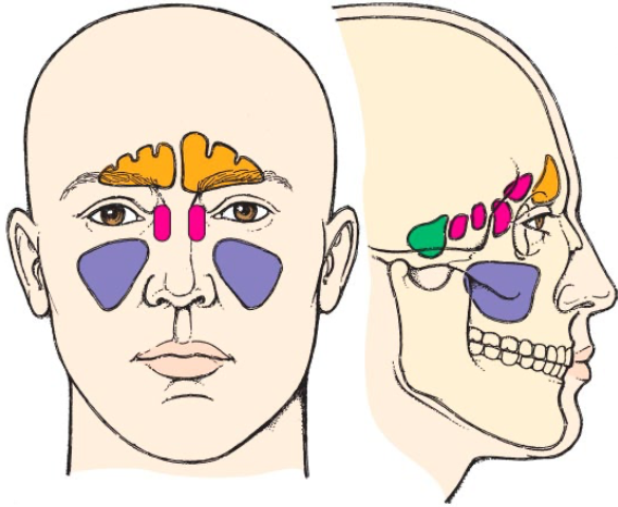
What are the paranasal sinuses and their color codes in diagrams?
Gold - Frontal sinuses
Pink - Ethmoid sinuses (air cells)
Purple - Maxillary sinuses
Green - Sphenoid sinuses
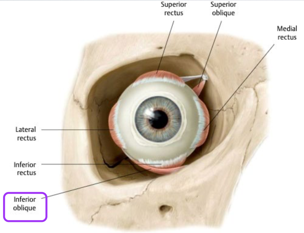
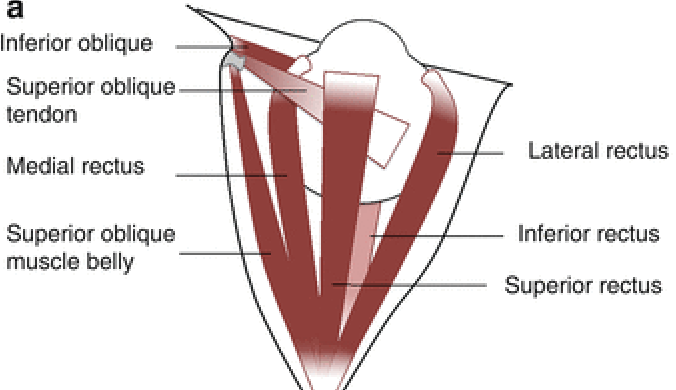
Which EOMs are strap-shaped and run a direct course from origin to insertion?
4 rectus muscles
inferior oblique
Label.
Label.
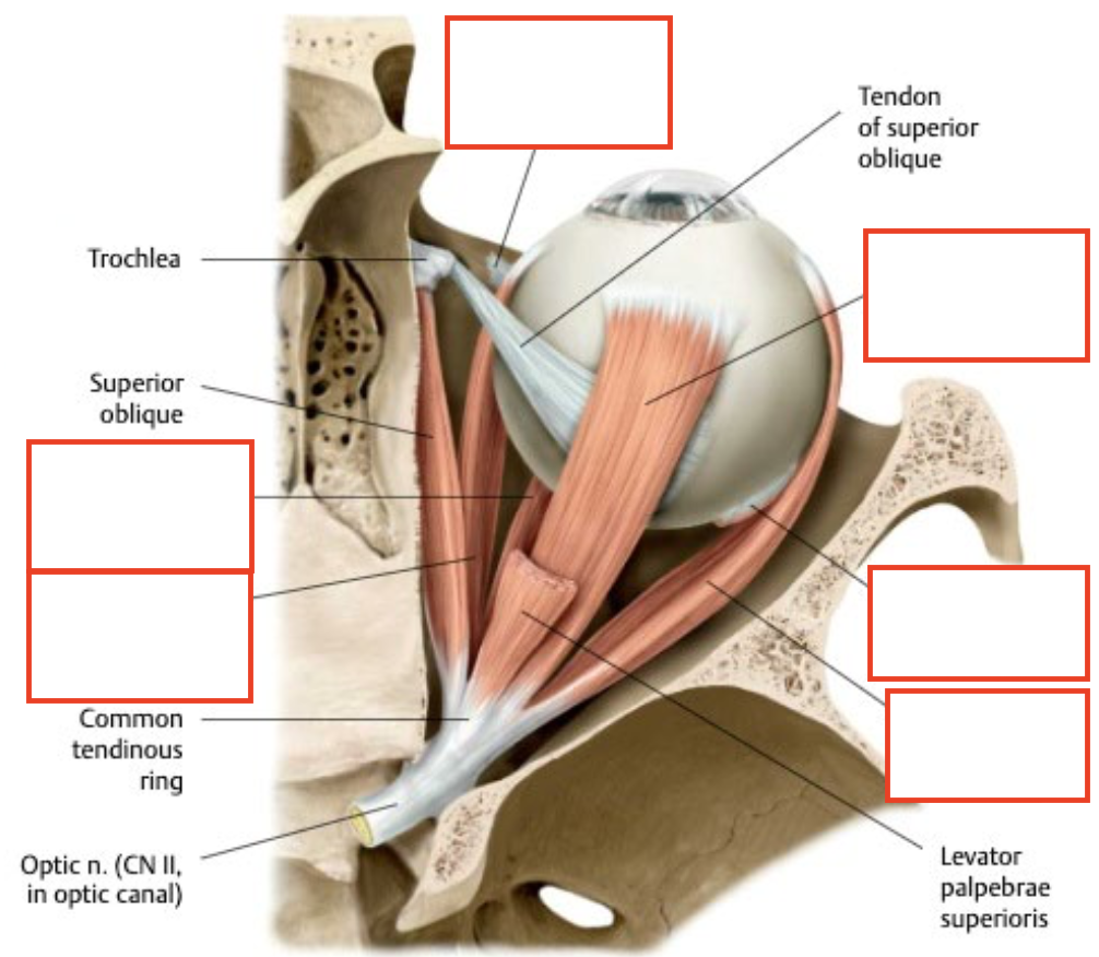
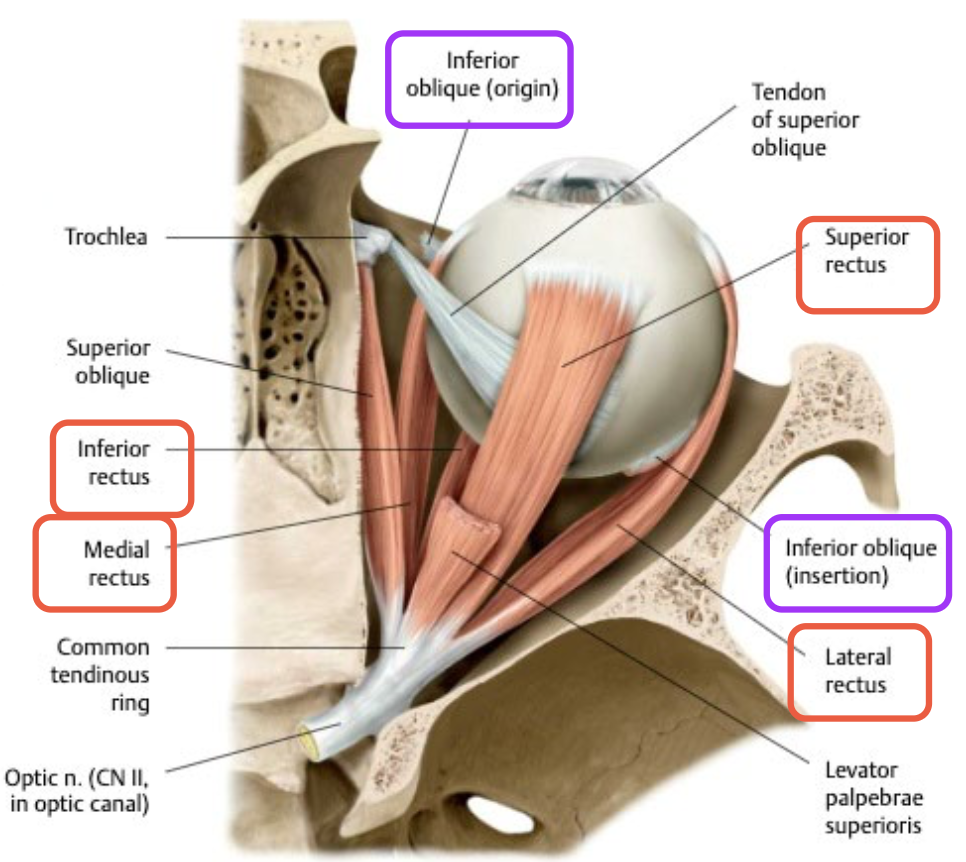
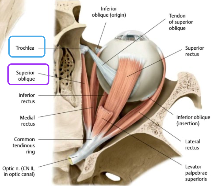
Describe the Superior Oblique.
ovoid shape
runs anteriorly, parallel to the medial orbital wall and above the medial rectus
gives rise to a long, slender tendon that runs through the pulley of the trochlea
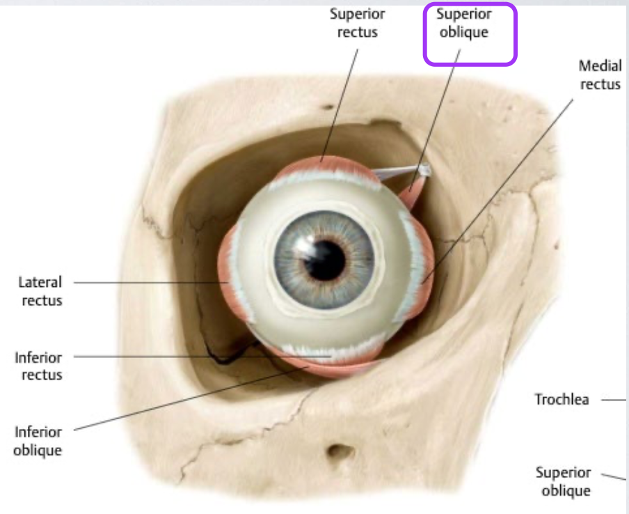
What is the trochlea?
→ cartilaginous loop that’s fixed to the superomedial orbital rim
after passing through the trochlea, the SO tendon reverses course to insert onto the superior surface of the eye, sliding beneath the SR muscle
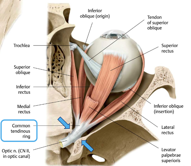
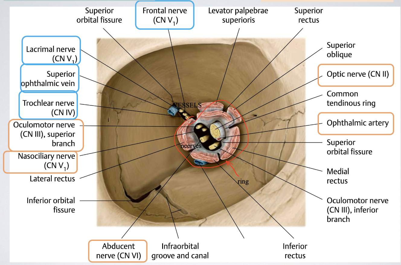
What is the annular ring?
ring that’s attached to the greater and lesser wing of the sphenoid at orbital apex
Lesser wing: small, forms roof
Greater wing: large, forms floor of middle cranial fossa
separates the SO fissure into 2 compartments (NASO & LMSFT)
4 recti + SO originate here

List the structures inside the ring.
NASO
nasociliary nerve
abducens nerve
sympathetic nerves
oculomotor nerve
optic nerve
ophthalmic artery
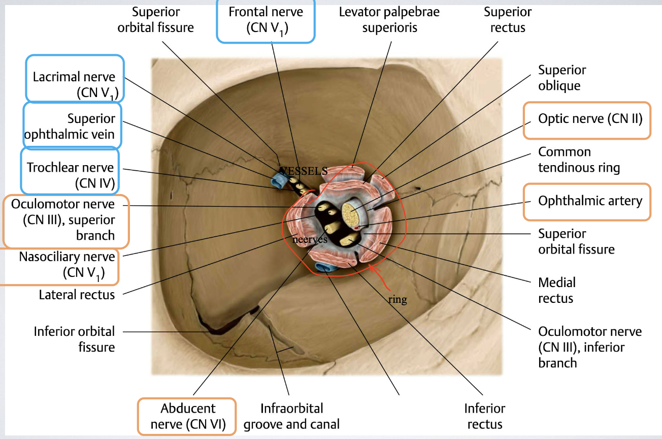
List the structures outside the ring.
LMSFT
lacrimal nerve
branch of middle meningeal artery
superior ophthalmic vein
frontal nerve
trochlear nerve
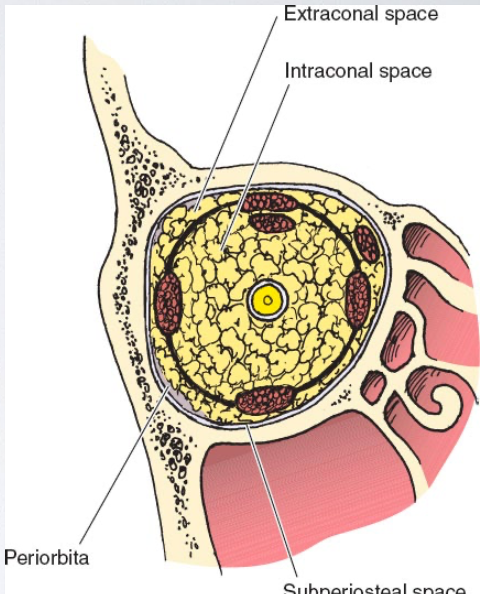
What is the Periorbita?
orbital periostium that forms the outer edge of all orbital structures and lines the inside of the orbital bones
What does the fascia surrounding the rectus muscles and levator palpebrae superioris form?
connects the edges of the rectus muscles → form the ‘muscle cone’
What are the 2 principal fascial compartments of the orbit?
Intraconal (muscle cone) space – inside the cone of rectus muscles
Extraconal space – outside the muscle cone (b/w the periorbita & cone)
What is contained in the intraconal space?
optic nerve
ophthalmic artery
ophthalmic nerves
permanent orbital fat
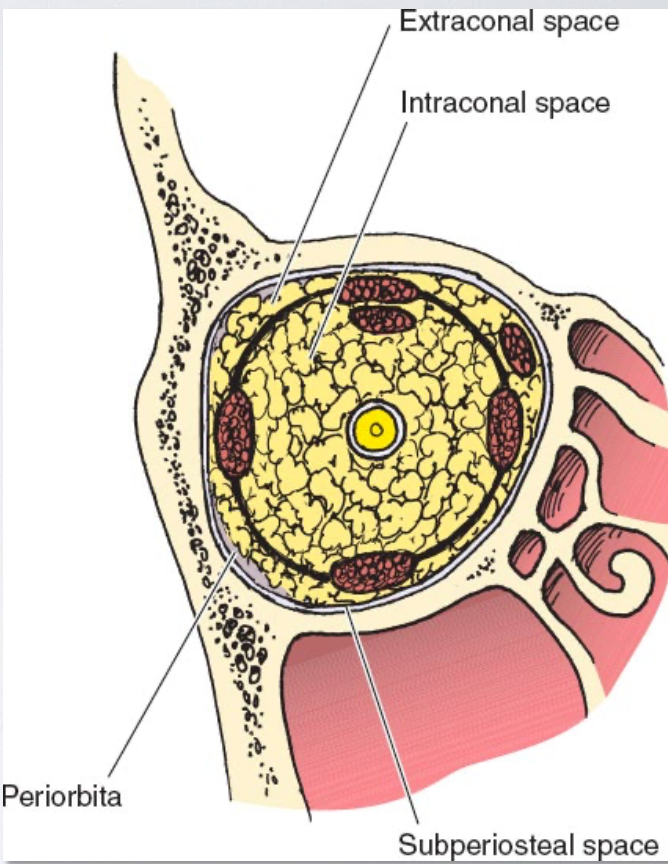
What is contained in the extraconal space?
CT
vessels
fat
What is the function of orbital fat?
→ provides cushioning and support for the eye and vasculature
helping hold the globe forward

Why is orbital fat called “permanent fat”?
→ the only fat that doesn’t get used up under normal conditions
it can decrease in severe malnutrition, causing the eye to sink back into the orbit
What do the fascial attachments in the orbit form?
→ form ligament-like structures that attach to the orbital walls (annular ring)
this helps stabilize & control eye movement
What is the function of the fascia that attach to orbital walls (e.g., orbital ligaments)?
Controls how strongly the EOMs pull on the eyeball → limits eye movement beyond the orbital aperture/eye socket
Hold the globe centered in the orbit
What are the fascial connections in the orbit called?
ligaments
List the principle ligaments.
(1) Lateral Check ligaments
(2) Medial Check ligaments
(3) Ligament of Lockwood
(4) Whitnall’s ligament
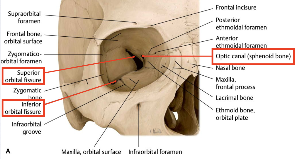
List the openings of the orbit.
Optic canal
Superior orbital fissure
Inferior orbital fissure
Note: Very little goes in through the front opening
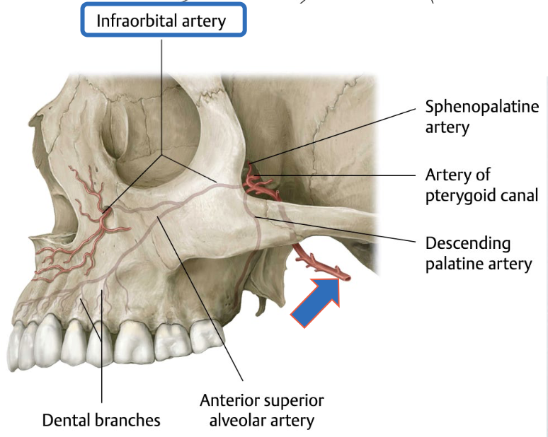
Supply to the Orbit
Which arterial systems & arteries contribute to the orbital blood supply?
Internal carotid system (via the ophthalmic artery)
External carotid system (via the infraorbital artery)
Supply to the Orbit
What is the origin of the Ophthalmic artery?
1st branch of internal carotid artery
Supply to the Orbit
What is the origin of the Infraorbital artery?
Maxillary artery - terminal branch of the external carotid artery, within the pterygopalatine fossa
Drainage of the Orbit
How does venous blood drain from the eye and surrounding structures?
→ Venous blood from the eye, orbit, eyelids, and face drains into the superior or inferior ophthalmic veins
2 veins have freely anastomosing (interconnecting) vascular fields, allowing shared drainage pathway