D652: Oral Path Unit 2 Review
1/391
There's no tags or description
Looks like no tags are added yet.
Name | Mastery | Learn | Test | Matching | Spaced | Call with Kai |
|---|
No analytics yet
Send a link to your students to track their progress
392 Terms
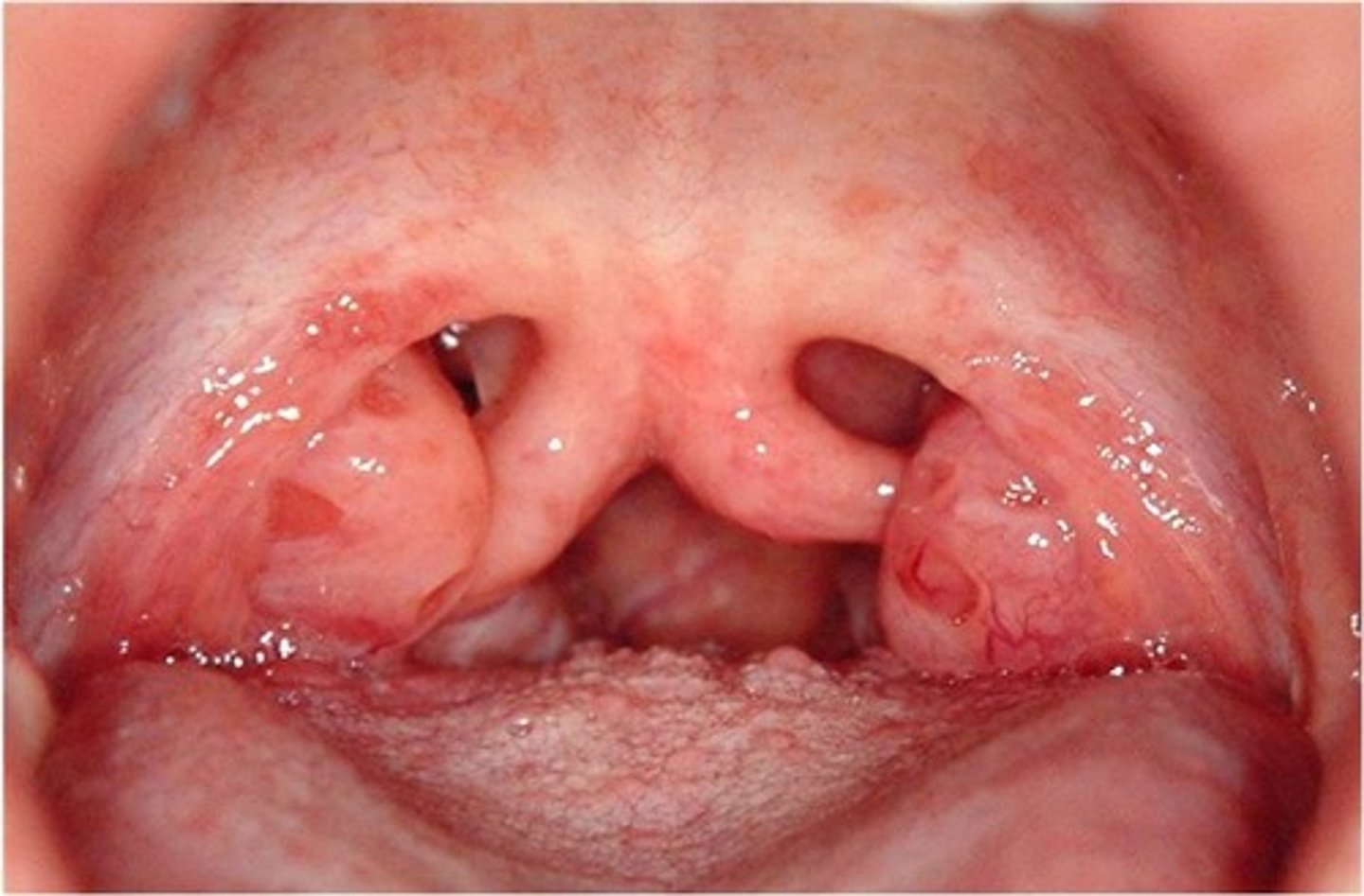
____ ___ is the most minimal manifestation of the cleft palate.
Bifid uvula

bifid uvula is the most minimal manifestation of the ___ ___.
cleft palate
Which side of the lip is MOST common for a unilateral cleft lip?
A. right
B. left
B. left (by a lot)

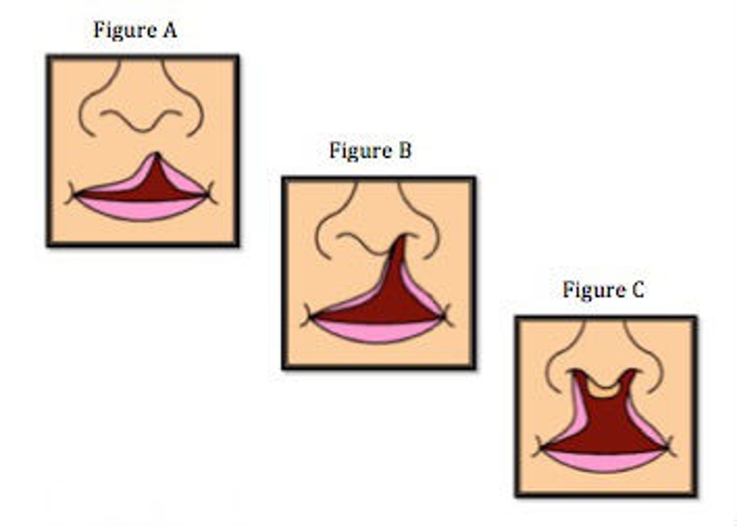
Are unilateral or bilateral cleft lips more common?
unilateral
- common congenital defect
- 80% are unilateral

T/F: incomplete cleft lips extend into the nostril
false; they do not
Which two orofacial clefts have the same etiologic process?
A. Cleft lip + cleft palate
B. Cleft palate only
C. Cleft lip only
D. A & B
E. A & C
F. B & C
E. A & C
CL + CP and CL only have the same process
T/F: patients with cleft lip or cleft palates (or both) are likely to have another syndrome
true (>400x)
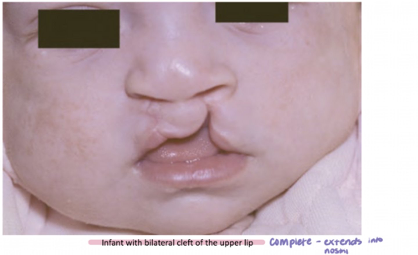
A ____ cleft lip extends up into the nostril and usually occurs between the lateral incisor and cuspid.
A. incomplete
B. complete
B. complete
A complete cleft lip extends up into the ____ and usually occurs between the _____ incisor and _____.
nostril; lateral incisor and cuspid (canine)
- also, teeth are normally missing (congenitally)
Which orofacial cleft anomaly is almost always associated with cleft palate?
A. lateral facial cleft
B. median cleft of the upper lip
C. oblique facial cleft
D. all of the above
C
Which of the following is NOT true of cleft lips?
A. they are typically unilateral
B. they are a defective fusion of the medial nasal process with the mandibular process
C. they are less common than a cleft palate alone
D. they are a common congenital defect
B.
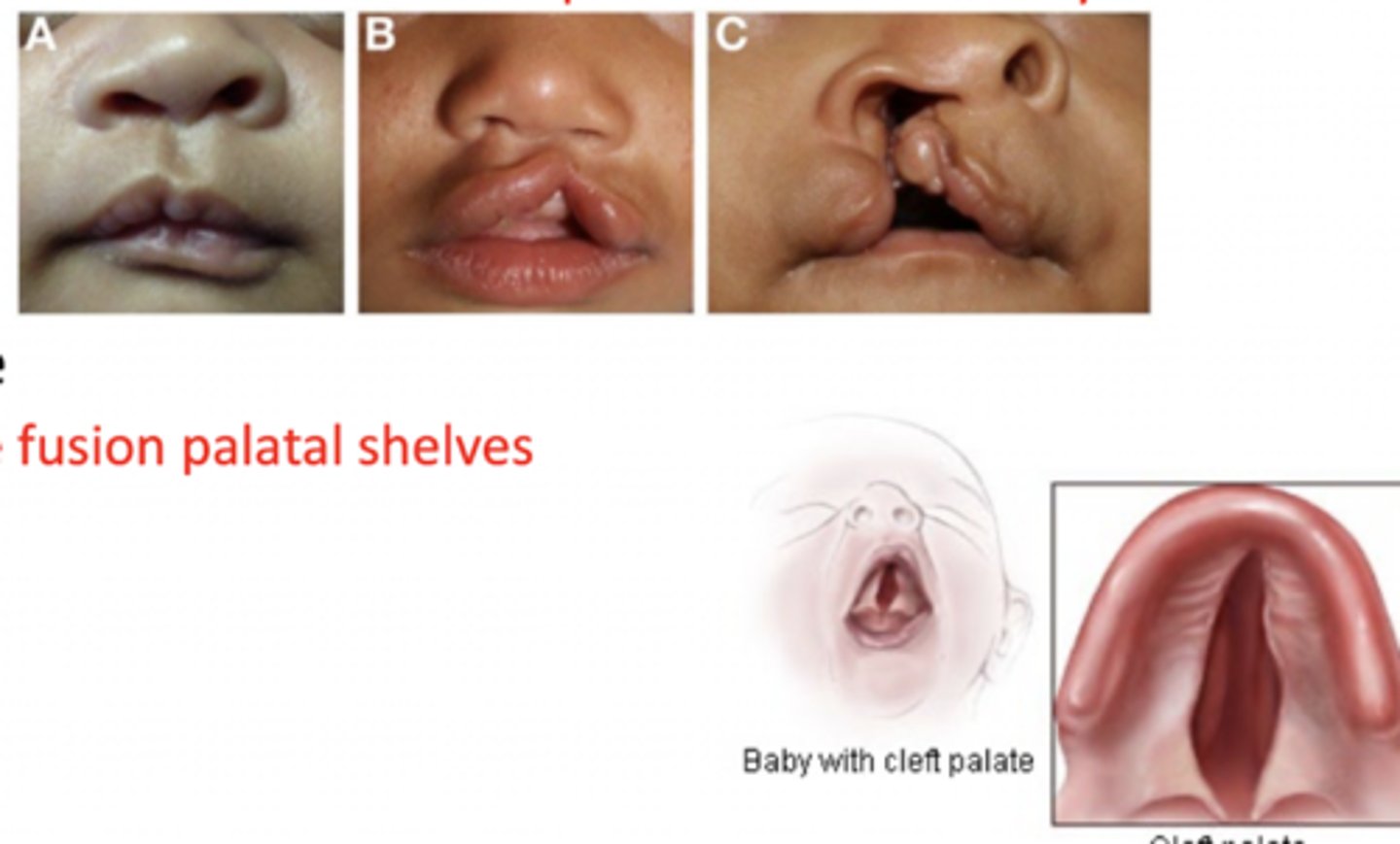
defective fusion of the medial nasal process with the maxillary process
When comparing a cleft lip to a cleft palate, which processes fail to fuse? (defective fusion locations)
Cleft palate = defective fusion of palatal shelves
Cleft lip = defective fusion of the medial nasal process with the maxillary process
Which of the following is NOT considered a feature of the Pierre Robin sequence?
A. cleft palate
B. mandibular micrognathia
C. macrodontia
D. glossoptosis
C. macrodontia
What is the triad of the Pierre Robin sequence?
cleft palate, mandibular micrognathia (small mandible), glossoptosis (posteriorly positioned tongue, feeding and swallowing issues)
T/F: commissural lip pits are associated with facial or palatal clefts
False
- manifest as blind fistulas ~1-4 mm at the vermillion border in the corners of the mouth
Which form of lip anomaly is usually inherited with cleft lip +/- cleft palate?
A. commisural lip pit
B. double lip
C. paramedian lip pit
D. none of the above
C. paramedian lip pit
_____ lip pits are the most common form of syndromic clefting and are located bilaterally at the midline of the lower lip vermillion.
paramedian
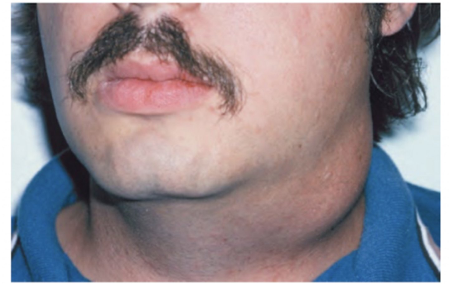
Double lip is more common in the ____ lip.
A. upper
B. lower
A. upper
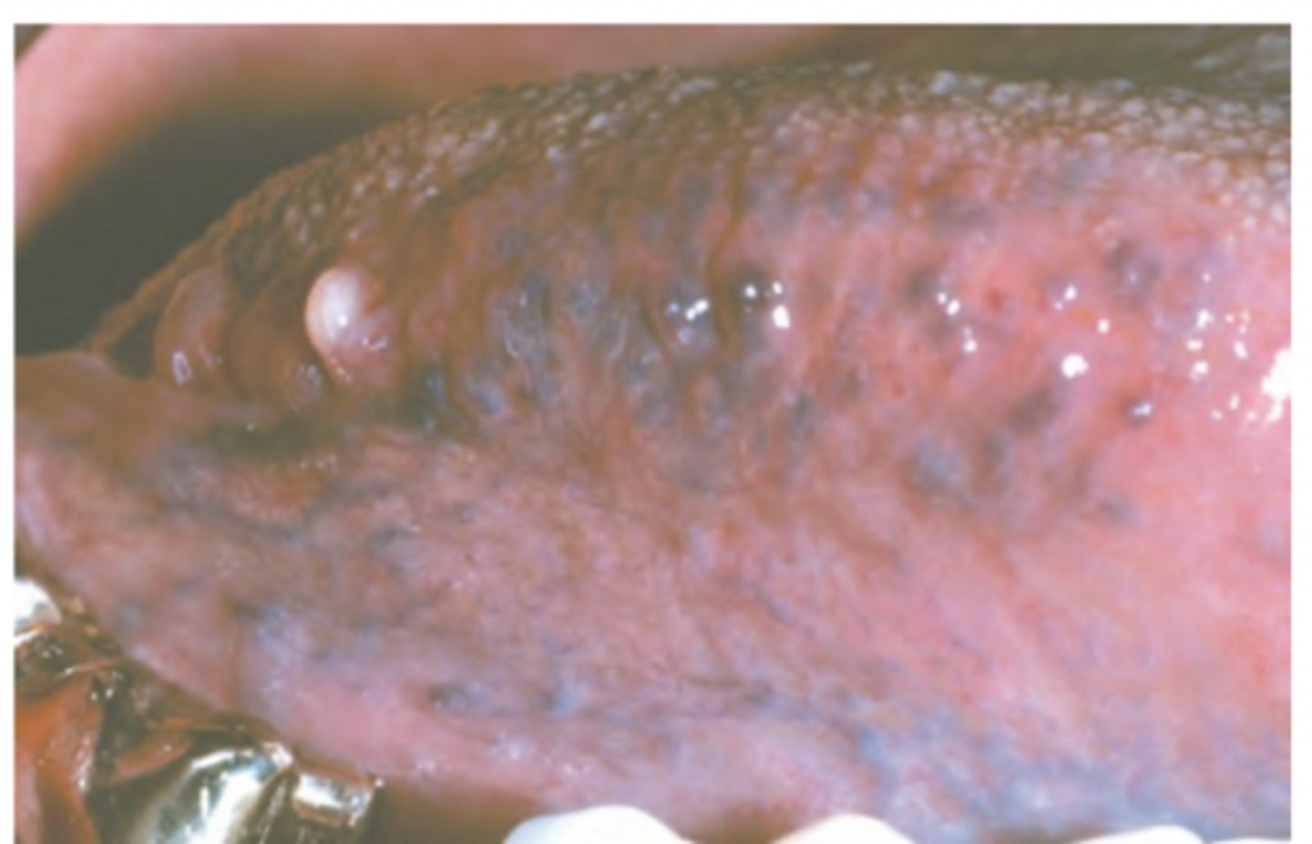
Varicosities are associated with which of the following?
A. trauma
B. chronic illness
C. advanced age
C. (loss of collagen and aging)
Where are varicosities most commonly located?
sublingually
T/F: all exostoses are the same as tori
False
An ______ is a localized bony protruberance from the cortical plate
exostoses
In general, an exostoses is defined as a localized bony protruberance from the ____ ____
cortical plate
Palatal exostoses are located only on the lingual aspect of the ____ ____.
maxillary tuberosity
- usually bilateral
- males
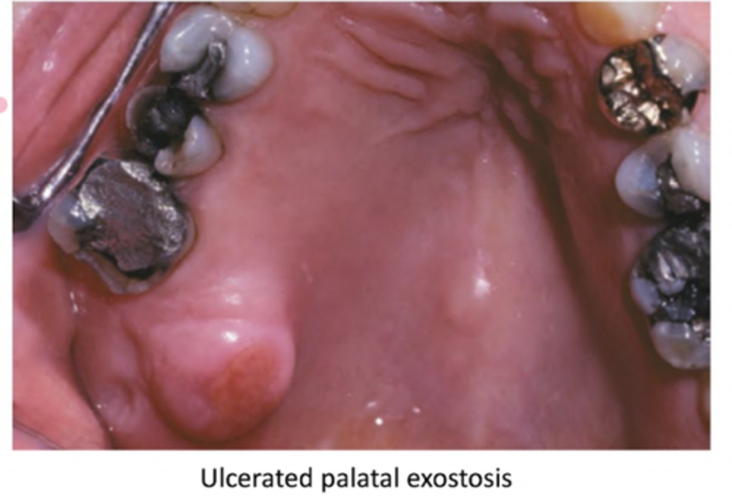
Are both the buccal and palatal exostoses usually bilateral?
yes!
Out of the two, which can be seen in a radiograph?
A. exostoses
B. palatal tori
A. exostoses (may be radiopaque)
Whihc of the following is NOT correct regarding the exostoses?
A. solitary exostoses may occur in response to local irritation
B. palatal exostoses are only located on the lingual aspect of the maxillary tuberosity
C. reactive subpontine exostosis develop beneath a pontic from the alveolar crest
D. buccal exostoses are usually symptomatic
D.
(asymptomatic)
Buccal exostoses are common in adults and are defined as a ____ row of bony nodules along the facial aspect of the maxillary and/or mandibular ____ ____.
bilateral; alveolar ridge
___ ___ exostosis develop from the alveolar crestal bone beneath the pontic.
reactive subpontine
If an exostosis is solitary, it is an indication for _____.
biopsy (I have this written in my notes)
A ____ ____ is an exostoses in the midline of the hard palate.
torus palatinus
A tous palatinus is an exostoses in the ____ of the ____ palate.
midline; hard palate
Torus palatinus can appear as
A. flat
B. spindle
C. nodular
D. lobular
E. all of the above
E
- usually less than 2 cm
- may slowly increase in size w/ age
Which of the following is prone to MRONJ?
A. torus palatinus
B. torus mandibularis
C. palatal exostoses
D. buccal exostoses
E. stafne defect
A
(only torus palatinus, not mentioned for mandibularis)
Torus mandibularis is present along the ____ aspect of the mandible.
lingual
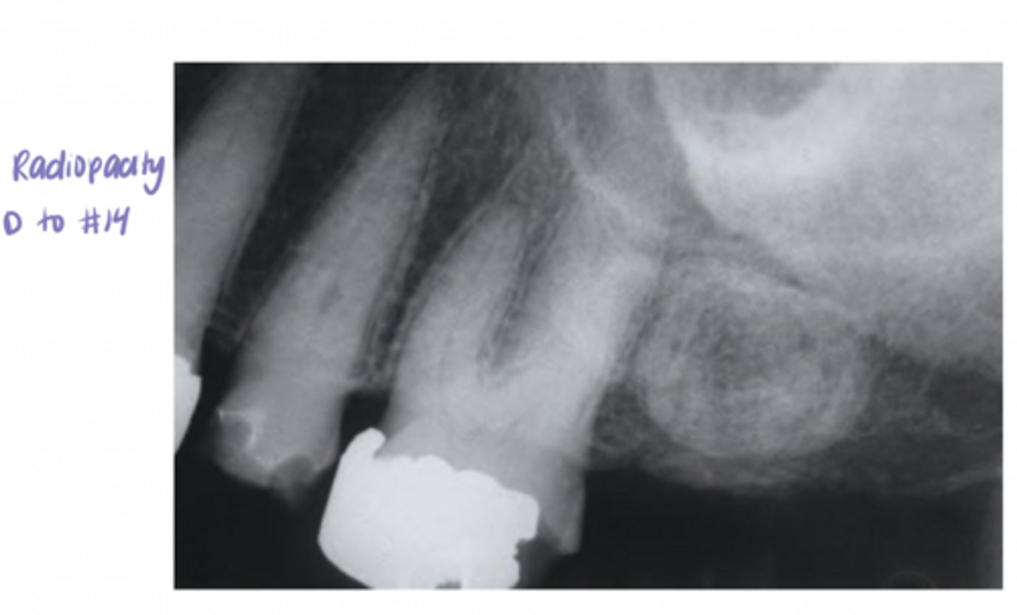
Statement 1: torus mandibularis is typically bilateral
Statement 2: torus mandibularis is most common in the premolar region
both are true
bilateral (>90%)
When torus mandibularis is evident on a radiograph, it may appear on periapical films as a radiopacity ______ on the _____.
superimposed; roots
Which is more common: torus mandibularis or torus palatinus?
palatal tori
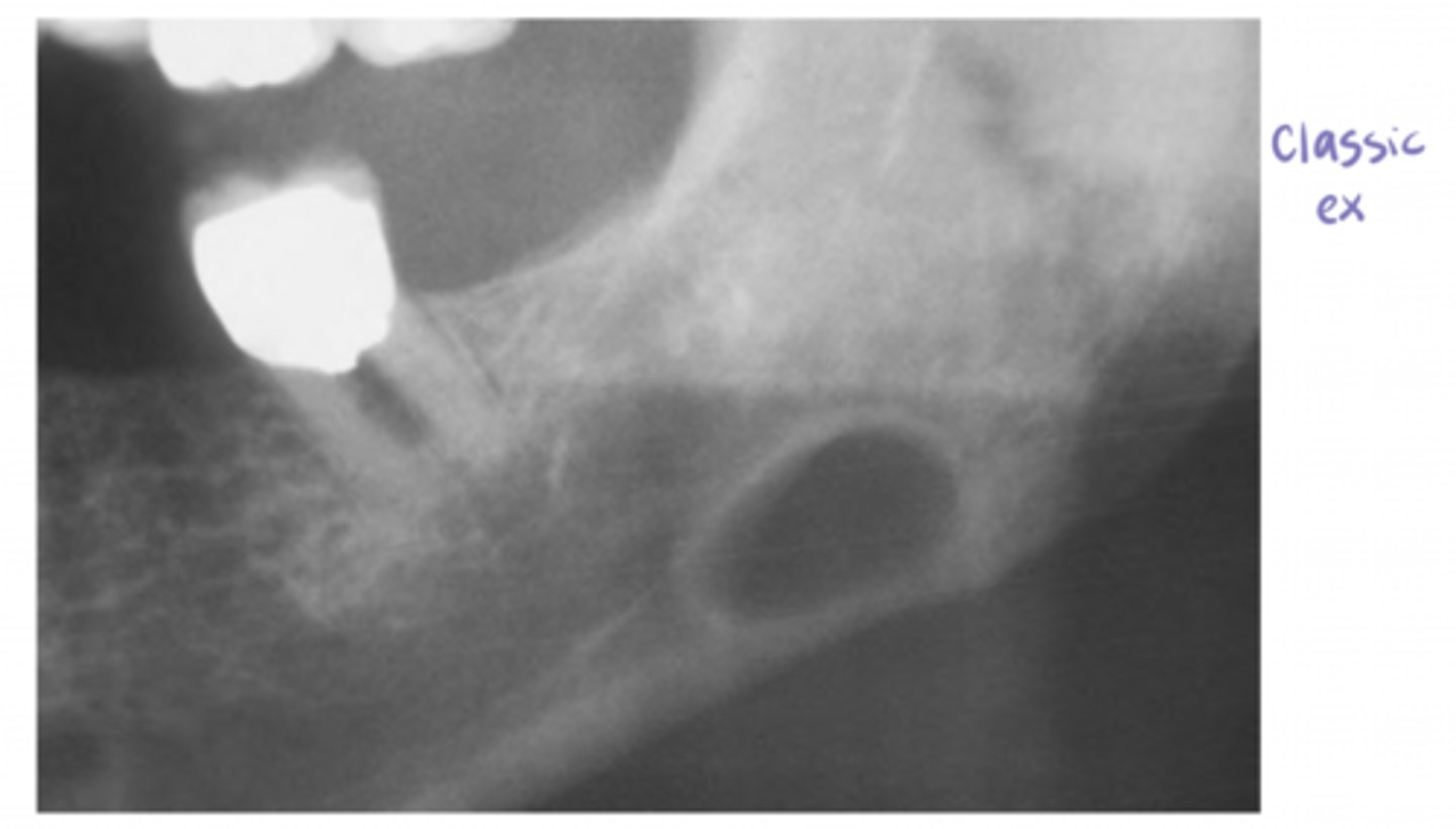
the ____ ____ represents a focal concavity of the cortical bone on the lingual surface of the mandible from the submandibular gland
stafne defect
Which gland does the stafne defect impact?
submandibular
The stafne defect will present as a radiolucency below the ____ ___ usually between the molar teeth and the angle of the mandible
mandibular canal
The stafne defect will present as a radiolucency below the mandibular canal usually between the _____ teeth and the _____ of the mandible
molar; angle
Can the stafne defect present more anterior to the molars, such as the premolar region?
Yes
T/F: the stafne defect radiolucency is always well-defined with a sclerotic border
true
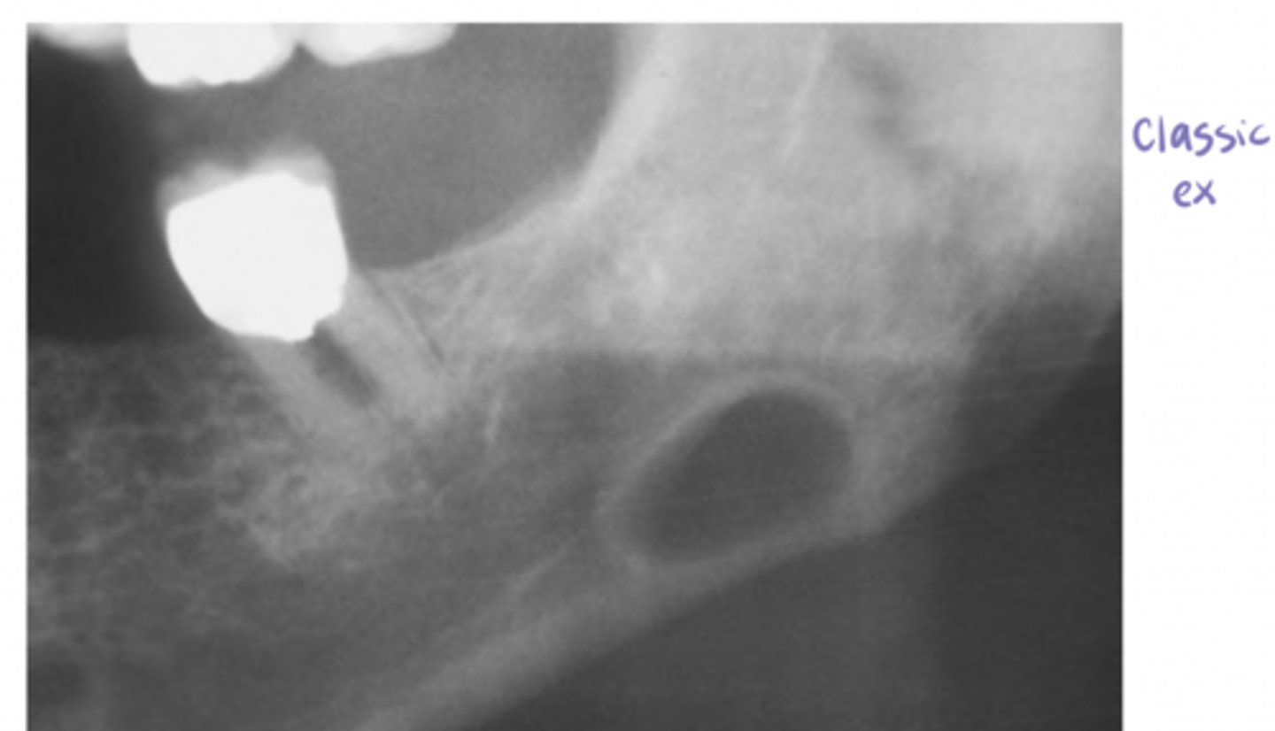
T/F: the stafne defect is pathognomonic with a radiograph
true (no biopsy needed), can confirm dx with CT or MRI
A cyst is a pathologic ____ lined by ____.
cavity; epithelium
NOT a tumor or a neoplasm
A cyst is ____ in origin (disease process)
developmental
can be non-odontogenic or odontogenic
Which cyst is self-healing and does not need treatment?
palatal cysts of the newborn
completely gone by age of 1
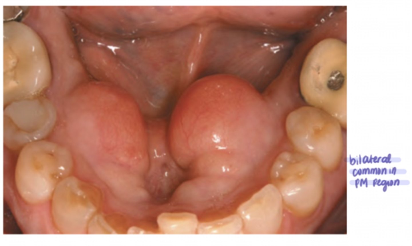
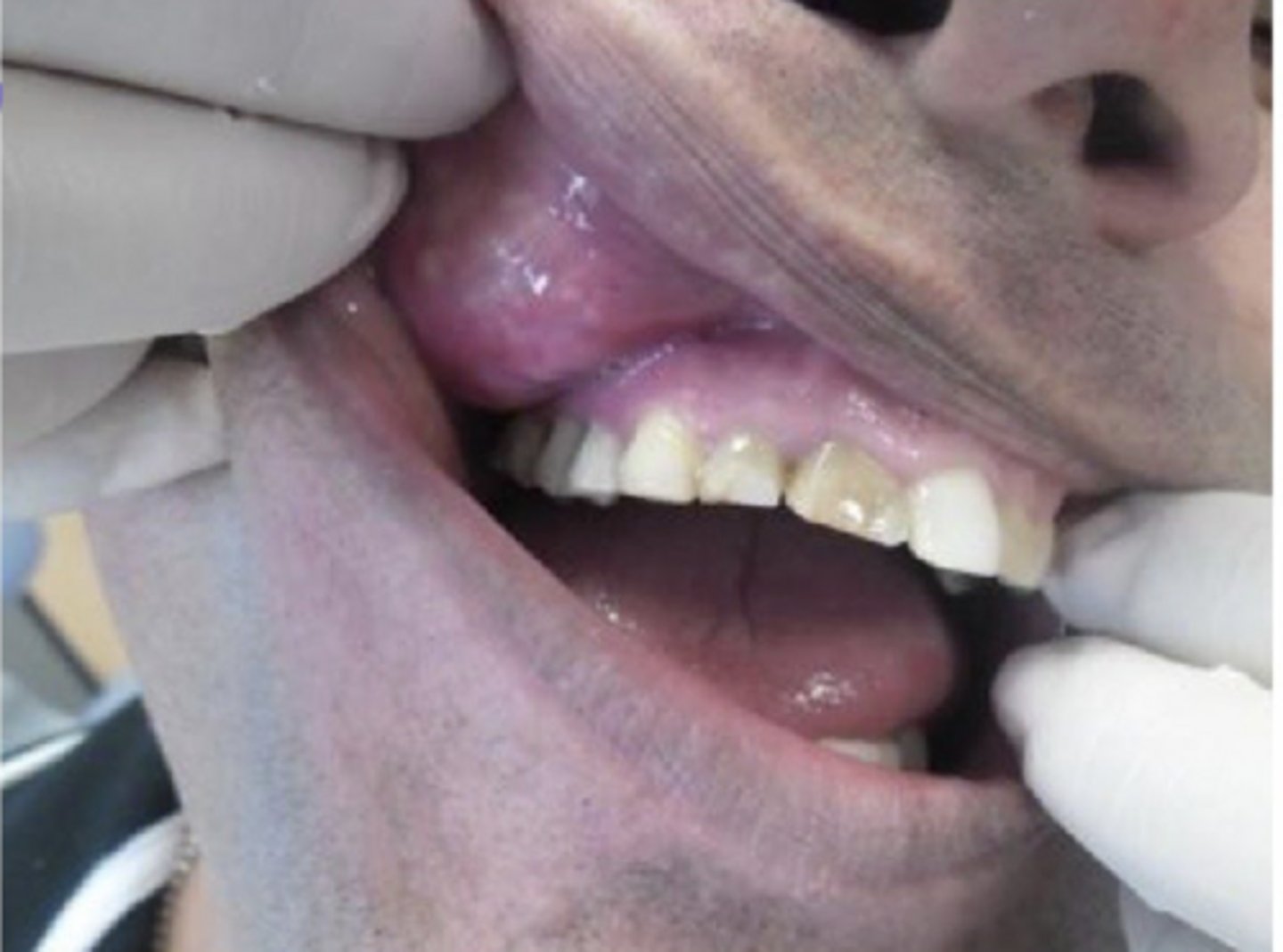
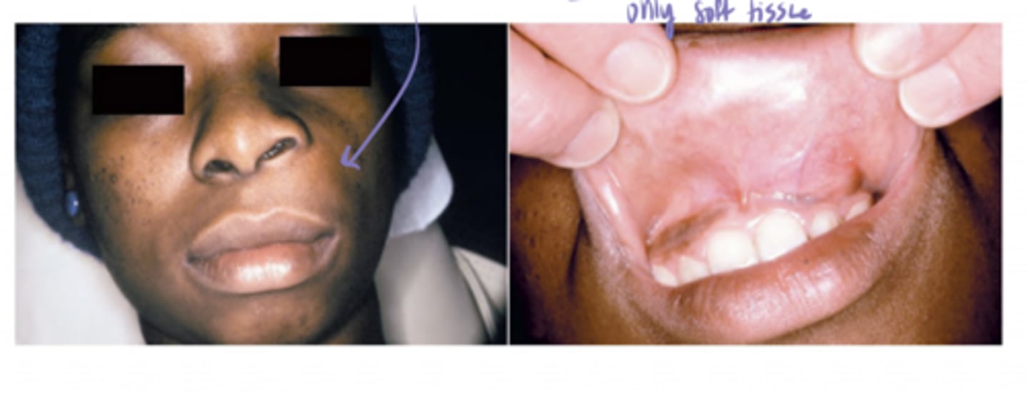
Nasolabial cysts are located in the ___ ___, ____ the midline.
A. upper lip; on
B. lower lip; on
C. upper lip; lateral to
D. lower lip; lateral to
C.
- appears as a swelling and causes elevation of the ala of the nose
Nasolabial cysts have no radiographic changes because they are formed by exclusively ____ _____.
soft tissue
Nasolabial cysts have no ____ ____ because they are formed by exclusively soft tissue.
radiographic changes
_____ cysts have no radiographic changes because they are formed by exclusively soft tissue.
nasolabial
Which cyst is the most common non-odontogenic cyst of the oral cavity?
nasopalatine duct cyst
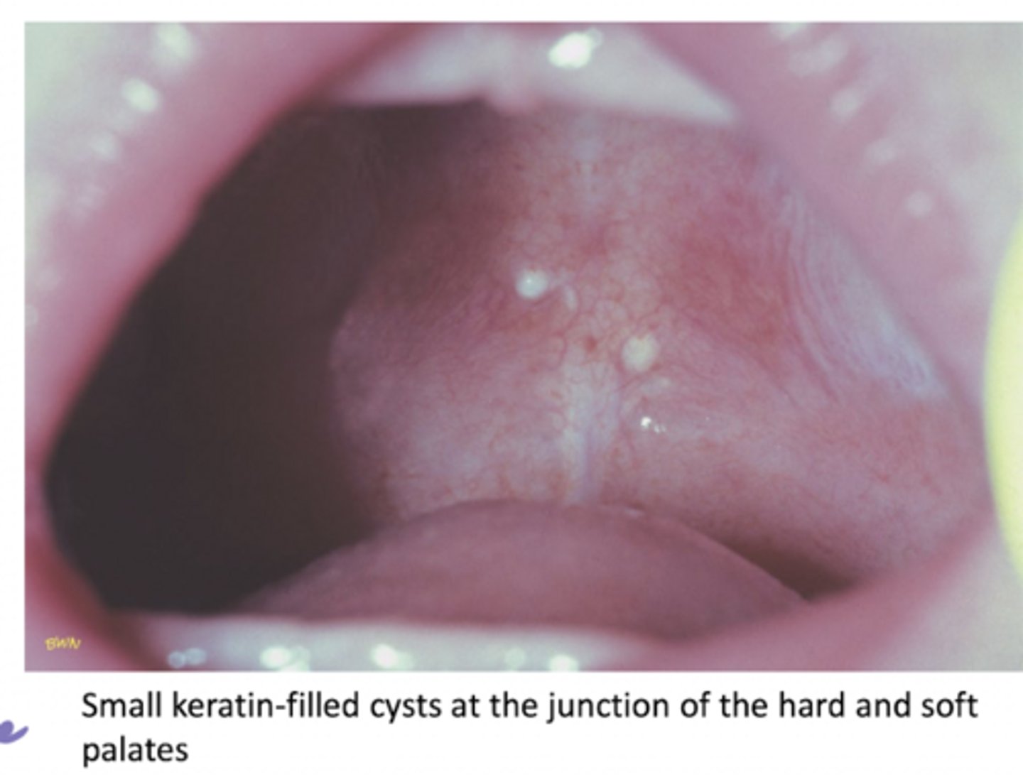
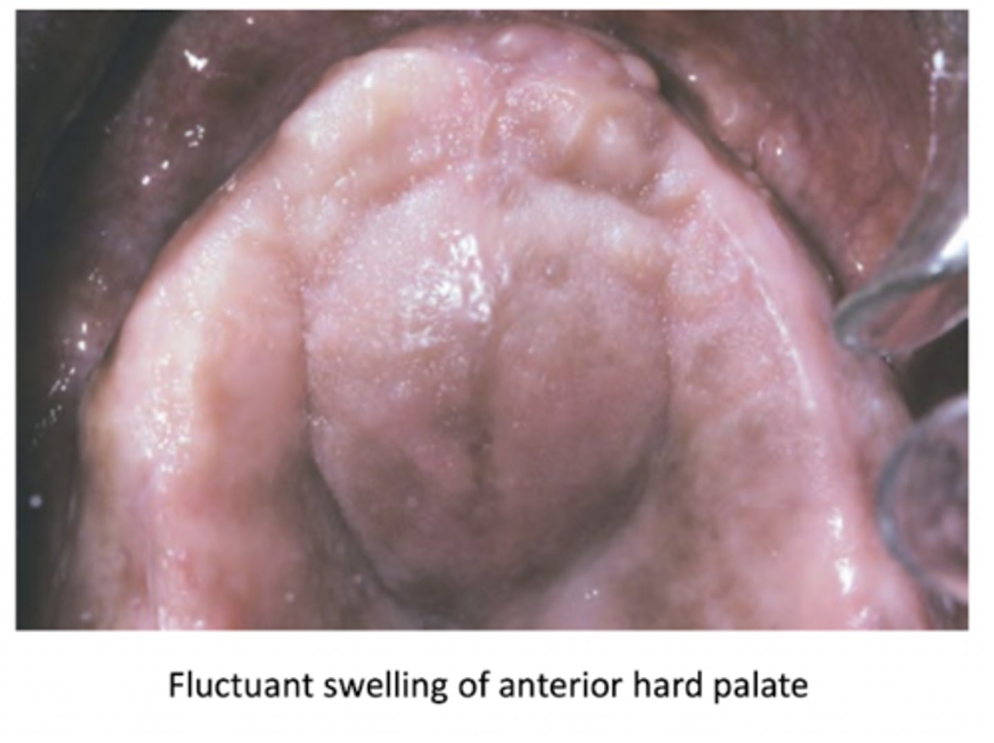
A ____ ____ ___ presents as a firm or fluctuant swelling of the midline of the hard palate posterior to the palatine papilla.
median palatal cyst
A median palatal cyst presents as a ____ or _____ ____ of the midline of the hard palate posterior to the palatine papilla.
firm or fluctuant swelling
If a nasopalatine duct cyst is ONLY composed of soft tissue, it is called a cyst of ___ ___.
incisive papilla (or nasopalatine duct cyst)
How are nasolabial cysts treated?
complete surgical excision (rare recurrence)
Nasopalatine duct cysts are normally ___ mm in size.
A. <3
B. >3
C. <6
D. >6
D.
(<6= just incisive canal)
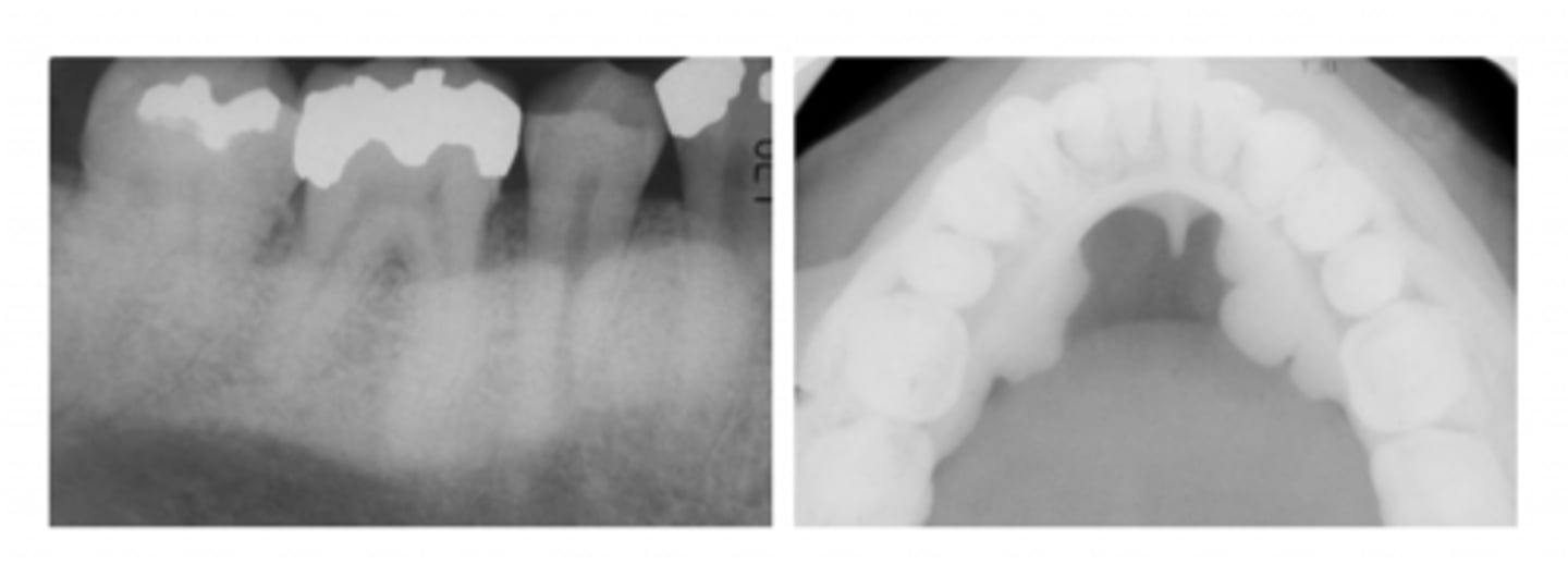
A nasopalatine duct cyst is a well-circumscribed radiolucency near the midline of the ____ ____.
anterior maxilla (ONLY)
T/F: A nasopalatine duct cyst is radiographically diagnostic
FALSE
A median palatal cyst is shown in an occlusal radiograph as which of the following?
A. ill-defined radiopacity
B. well-defined radiopacity
C. ill-defined radiolucency
D. well-defined radiolucency
D.
(nodule in the posterior palate with radiographic evidence)
What shape are median palatal cysts radiographically?
ovoid or circular
Which type of follicular cyst (on the skin) is the most common?
epidermoid
(arises after localized inflammation)
A pilar cyst is a common keratin-filled cyst located on the _____.
scalp
A ____ cyst is a common scalp cyst.
pilar
Epidermoid cysts are common in...
A. younger adults
B. males more than females
C. acne-prone individuals
D. all of the above
D.
Where are epidermoid cysts mostly located?
head, neck, and back (acne-prone)
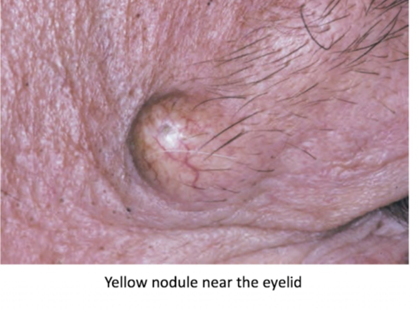
____ ____ of the skin are derived from the follicular infundibulum and arise after local inflammation.
epidermoid cysts
Epidermoid cysts are derived from the _____ _____ and arise after local inflammation.
follicular infundibulum
Epidermoid cysts are derived from the follicular infundibulum and arise after ____ ____.
local inflammation
What are follicular cysts of the skin (epidermoid, pilar) cysts filled with?
A. collagen
B. elastin
C. keratin
D. serum
C.
T/F: pilar cysts are movable
true
A ____ cyst is more common in males, while a ____ cyst is more common in females.
A. epidermoid; pilar
B. pilar; epidermoid
A
mnemonic: women have more hair -> pilar cysts are on the scalp
Cysts are cavities lined by _____ _____ _____ (tissue type)
stratified squamous epithelium
Which of the following cysts is classified as a benign cystic teratoma?
A. epidermoid cyst
B. dermoid cyst
C. pilar cyst
D. thyroglossal duct cyst
B. dermoid cyst
a dermoid cyst is considered a teratoma becuase it is composed of tissue from more than one ____ layer.
germ (endoderm, mesoderm, ectoderm)
- this would have skin appendages
Which of the following does NOT have skin appendages?
A. dermoid cyst
B. epidermoid cyst
B.
dermoid cyst = teratoma = ectoderm germ layer has skin appendages
T/F: a dermoid cyst contains one or more skin appendages in its wall (histologically)
true
A _____ ____ cyst is found anywhere along the midline from the base of the tongue to the suprasternal notch.
thyroglossal duct
A thyroglossal duct cyst is found anywhere along the ____ from the base of the tongue to the suprasternal notch.
midline
A thyroglossal duct cyst is found anywhere along the midline from the base of the _____ to the _____ _____.
base of the tongue; the supersternal notch
If a thyroglossal duct cyst is attached to the hyoid bone or the tongue, in which direction will it move during swallowing?
vertically
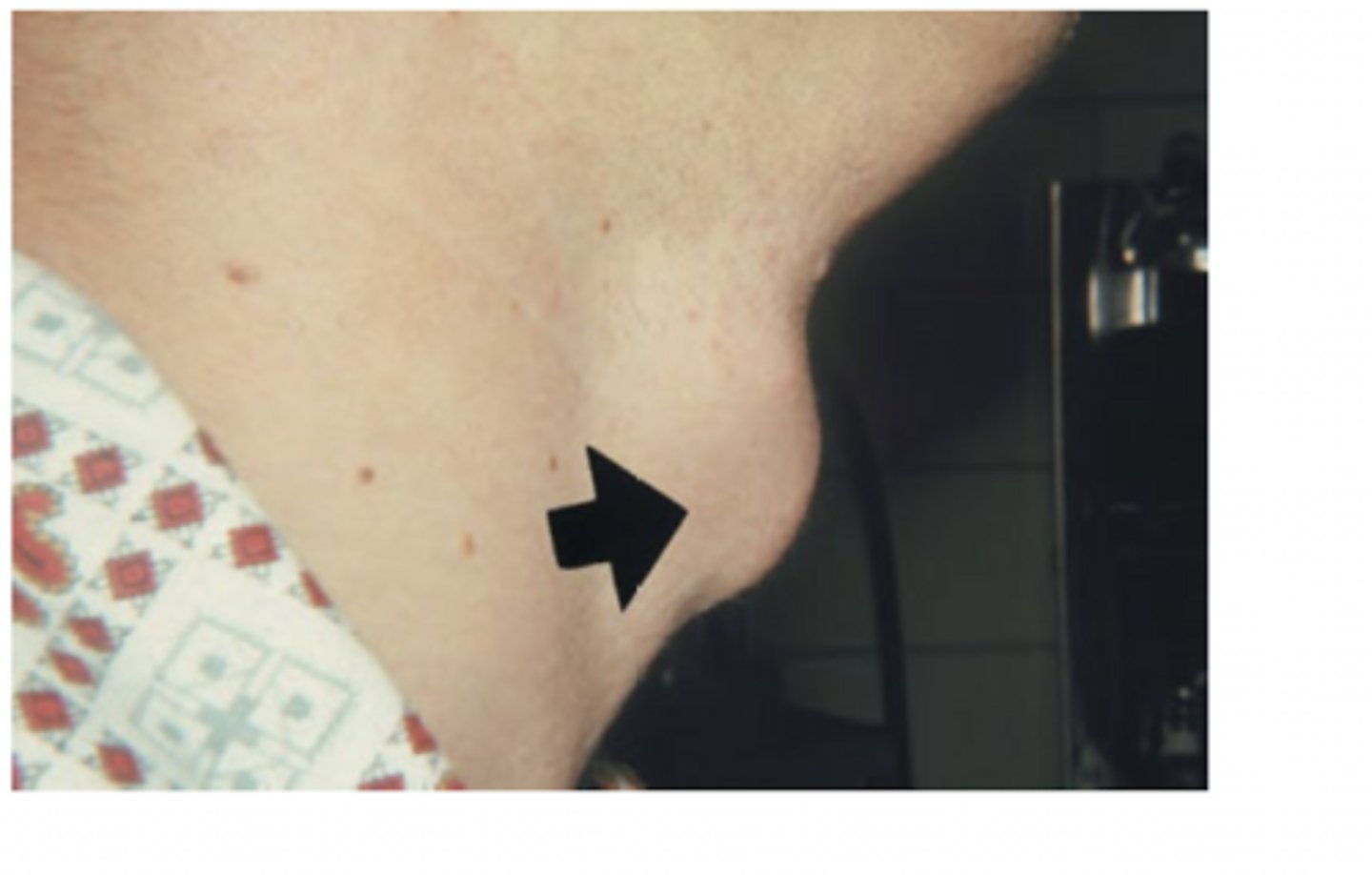
If a thyroglossal duct cyst is attached to the _____ bone or the ____, it will move vertically during swallowing.
hyoid; tongue
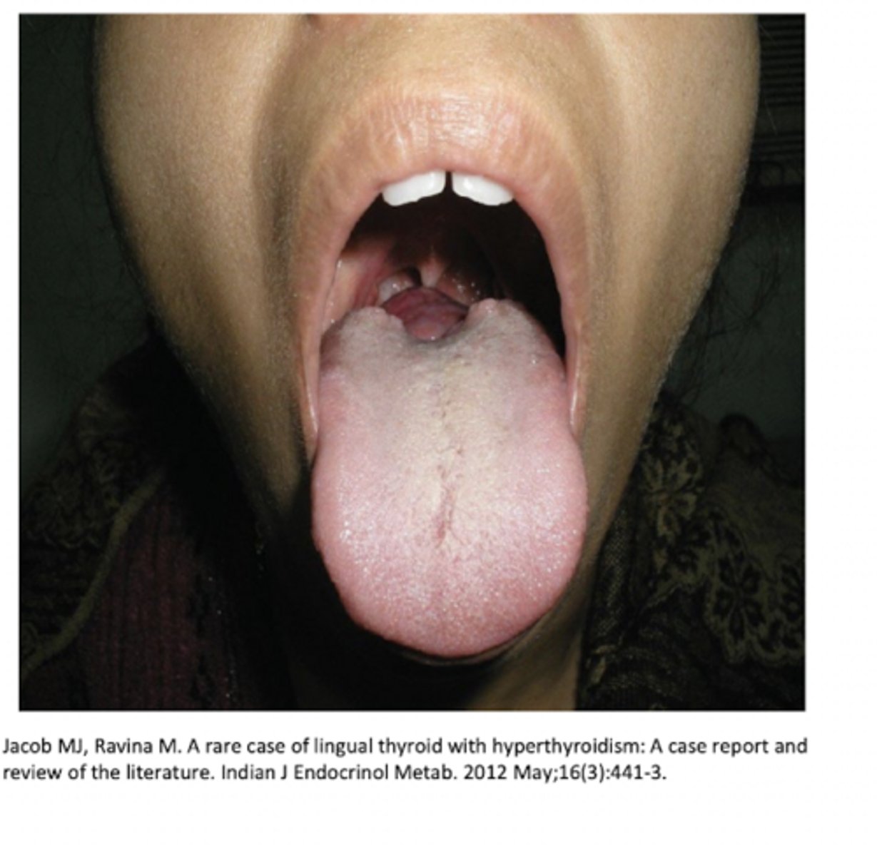
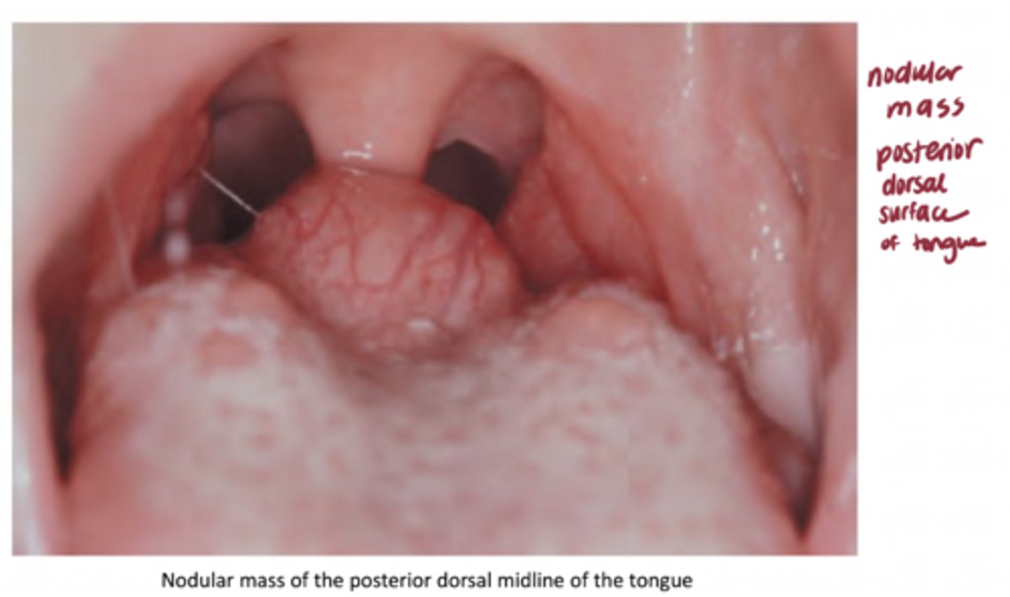
A lingual thyroid remains at the base of the tongue (only in the gland) ____% of the time.
70%
- nodular mass
- posterior dorsal surface of the tongue
What are the differential diagnoses of a lateral neck mass?
RBILM
Reactive lymphadenopathy
Branchial cleft cyst
Infectious lymphadenopathy
Lymphoma
Metastatic disease (carcinoma, melanoma)
Which of the following is NOT a differential diagnosis for a lateral neck mass?
A. metastatic disease
B. reactive lymphadenopathy
C. lipoma
D. branchial cleft cyst
C. lipoma
(lymphoma, also infectious lymphadenopathy)
A branchial cleft cyst is located in the ___ ____ neck anterior or deep to the sternocleidomastoid.
upper lateral
An oral lymphoepithelial cyst is rare, occurs in the areas of intraoral lymphoid tissue such as _____ ____ and the ____ lymphoid tissue.
Waldeyer's ring (palatine tonsils, lingual tonsils, pharyngeal adenoids) and the accessory lymphoid tissue.
What are the three main components of Waldeyer's ring?
palatine tonsils, lingual tonsils, pharyngeal adenoids
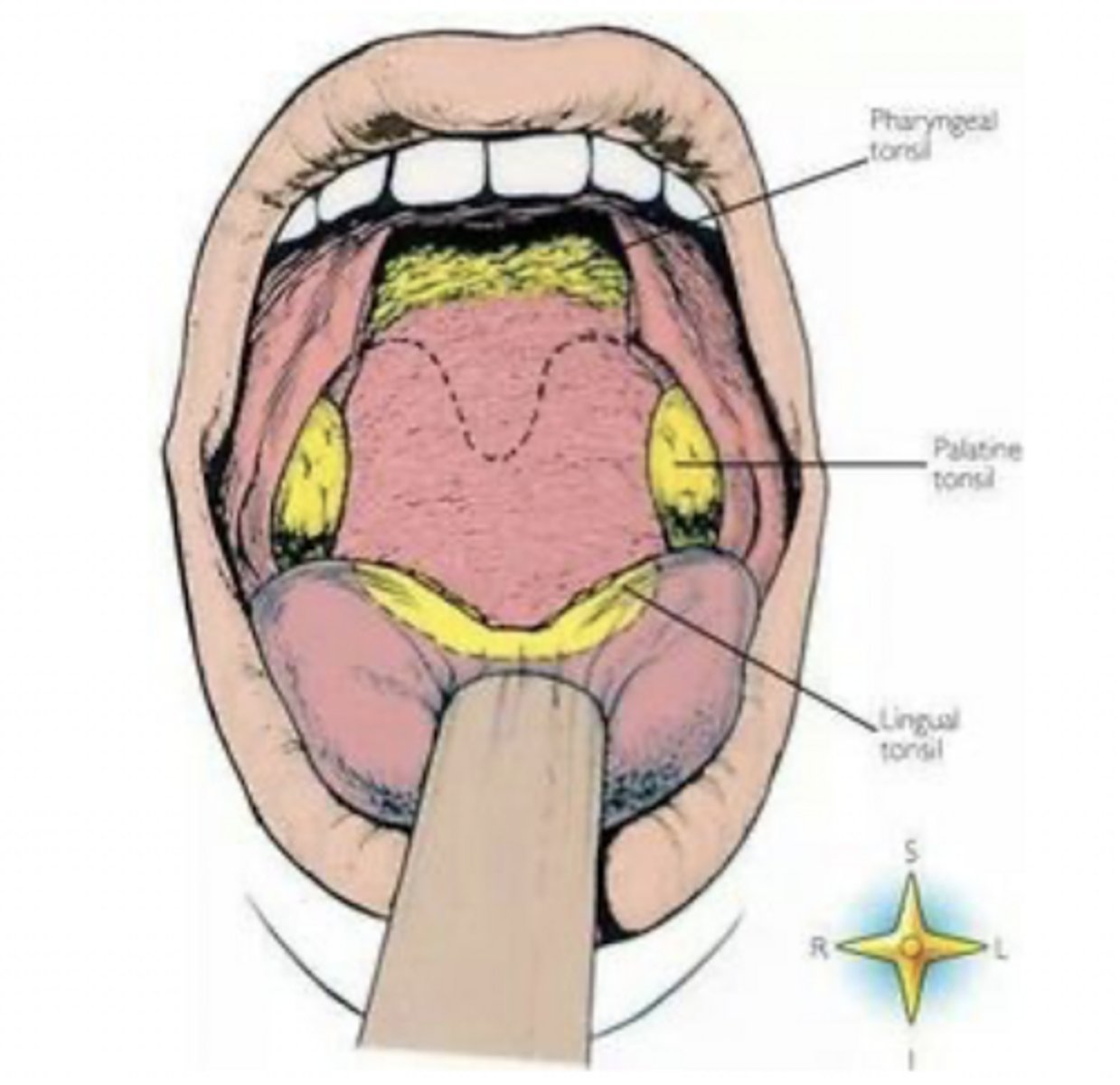
What are the accessory lymphoid tissues? (3)
floor of mouth, ventral tongue, soft palate
SVF
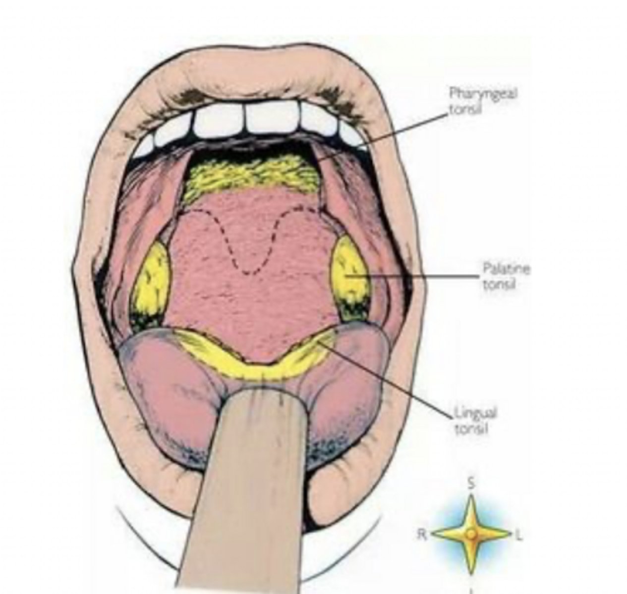
___ ____ cysts occur in areas of intraoral lymphoid tissue, such as Waldeyer's ring or accessory lymphoid tissue.
oral lymphoepithelial
Indurated means ____
hard
What are the four different terms that could describe a "rough" surface texture?
papillary, corrugated, fissured, crusted
When describing multiple lesions, the term ____ describes that they are separated from each other, while ____ means they are growing together and becoming one large lesion.
discrete; coalescing
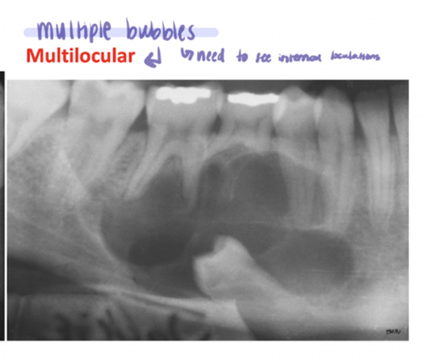
Which of the following radiographic densities can be described using unilocular or multilocular?
A. radiopaque
B. radiolucent
B. radiolucent
- NEVER used to describe radiopaque lesions
When formulating a differential diagnosis for a radiograph, you should consider the lesions effect on the ____ ___ and ____ ____.
neighboring teeth; cortical bone
a _____ radiograph contains both radiolucent & radiopaque areas
mixed
unilocular (image)
radiolucent; one bubble/cavity

multilocular (image)
radiolucent; multiple bubbles (need to see internal loculations)
If a radiographic lesion is below the IAN or non-tooth-bearing areas of the jaws, it is considered...
A. odontogenic
B. non-odontogenic
C. not related to tooth development
D. B & C
D