Vascular System & Vessels
1/42
There's no tags or description
Looks like no tags are added yet.
Name | Mastery | Learn | Test | Matching | Spaced | Call with Kai |
|---|
No analytics yet
Send a link to your students to track their progress
43 Terms
What are the 2 components of the circulatory system?
Cardiovascular system
Lymphatic system
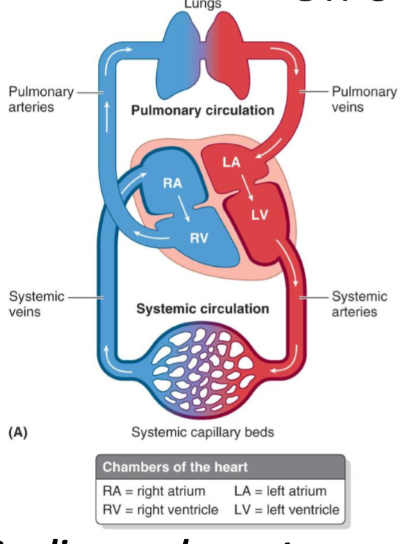
What are the 2 main events which are occurring in the cardiovascular system?
Pulmonary circulation: oxygen-poor blood travels to the lungs for oxygenation via the pulmonary arteries
Systemic circulation: oxygen-rich blood is returned to the heart via the systemic veins and is sent to the body for circulation
What are the 2 main events which are occurring in the lymphatic system?
Draining of surplus tissue fluid & plasma proteins, & removal of debris from cellular decomposition & infection
What are the 4 main constituents of the lymphatic system?
Plexuses
Vessels
Nodes
Tissues
What is the vascular system derived from & hence where is it found?
Derived from mesoderm
Only present in mesoderm-derived structures (e.g. muscles, connective tissue, & dermis)
Provide some examples of avascular & non-lymph structures
Epidermis (ectoderm)
Surface epithelium (endoderm)
Articular cartilage (exception for the mesoderm-derived cartilage)
State the 3 types of blood vessels
Arteries
Veins
Capillaries
What type of blood leaves the arteries, & where is this blood delivered to?
Blood under high pressure leaves the arteries
Oxygen-rich blood is delivered to capillaries
Except for blood leaving the right ventricle (which goes to the lungs to be oxygenated via the pulmonary arteries)
What do the pulmonary arteries carry?
De-oxygenated blood
What is lumen?
Open area that exists in the centre of the blood vessels
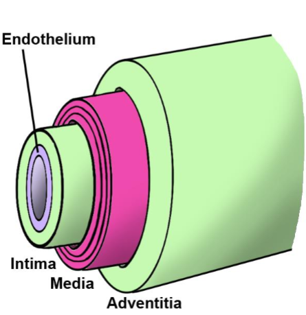
What are tunics?
The 3 layers of all blood vessels
What are the 3 tunics in blood vessels, from deep → superficial?
Tunica intima: Innermost lining, flattened epithelial cells supported by delicate connective tissue
Tunica media: the middle tunic layer which is composed of smooth muscle, and is highly variable in its thickness relative to the lumen (which determines whether it produces arteries, veins, or lymphatic ducts)
Tunica adventitia: outermost connective tissue layer
What are capillaries made of?
Tunica intima + basement membrane
Define the 3 types of arteries
Conducting arteries: large elastic arteries
Distributing arteries: medium muscular arteries
Small arteries & arterioles
Describe conducting arteries
Large elastic arteries
Many elastic layers → smooth blood flow
Near the heart, aorta & its major branches
Endothelium < fibrous/collagenous tissues < elastic tissue < smooth muscle
Describe distributing arteries
Medium muscular arteries
Circular smooth muscle fibres
Capable of vasoconstriction & blood flow regulation
Majority of names arteries
Endothelium < elastic tissues < fibrous/collagenous tissues < smooth tissue
Describe small arteries & arterioles
Narrow lumina
Thick, smooth muscle walls
Flow into capillary beds
Tonus regulates arterial pressure in vascular system
Endothelium < elastic tissues < fibrous/collagenous tissues < smooth muscle
Why is elasticity in conducting arteries the highest?
the heart pumps blood out at a very high velocity → lots of pressure behind the blood
need to ensure smooth blood flow to the rest of the body
elasticity in large conducting layers compensates for the spurting of high velocity blood → smooths it out for the rest of the body
Why are distributing arteries found in the periphery?
Want to vasoconstrict blood vessels when it is cold to divert blood to major organs & away from the extremities
Describe arterial bleeding & its clinical importance
Blood flow in elastic & muscular arteries is pulsatile & at high pressure
Majority of larger blood vessels are arteries
Pulsatile blood flow reflects systole (contraction) & diastole (relaxation) BP
Laceration to any arterial vessels (elastic or muscular) → pulsatile blood flow
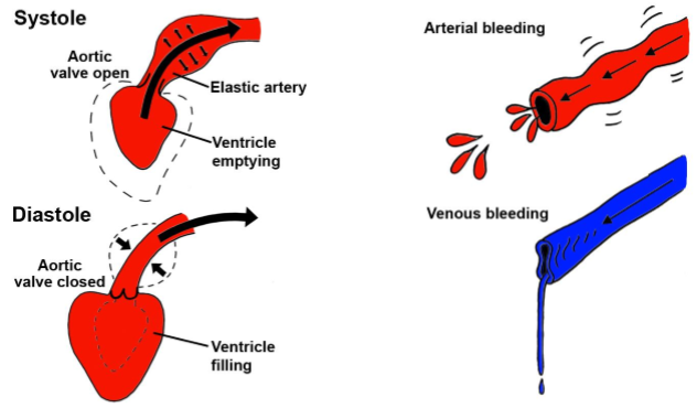
Describe venous bleeding
Larger lumen + v elasticity + v smooth muscles → lumen are largely open just by the amount of blood they have (lots of blood b/c of large lumen)
Laceration → continuous low pressure flow
Happens usually from capillaries & thin-walled veins

Define & describe anastomoses
Links b/w arteries or b/w arterioles
Provide potential detours for blood flow (collateral flow) if usual pathway is obstructed
Adjacent arteries tend to anastomose
Occur around joints, are significant only in muscle belly that crosses the joint
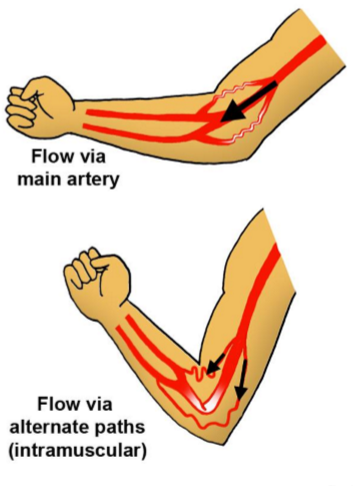
Provide an example of an anastomoses joint
Knee & elbow joints
Joints are flexed → constriction of major blood vessel → anastomoses which occur across the major arteries allow for an alternative path of blood flow
What are end arteries?
Arteries which do not link with other arteries i.e. no anastomoses
Not a ‘dead end’ - still branches into capillary beds drained by veins
What are the 5 principles of arteries of the limb?
Single stem artery
Changes name according to the region it traverses
Travels on the flexor aspect of a joint
Where it crosses a hinge joint the artery will anastomose to avoid compression
Terminal branches are generated as the artery crosses the middle joint
Provide an example of how arteries change names in response to the region it traverses
Brachiocephalic artery → axillary artery → brachial artery → radial & ulnar arteries
Stem artery travels on the flexor aspect of a joint (anterior aspect)
Similar to the knee; external iliac artery → femoral artery
Proceeds down the flexor (posterior side of the knee)
Describe the abundance & structure of arteries
More abundant than arteries
Walls are thinner, but diameter is larger than the corresponding artery
Have larger capacity for expansion than arteries
Typically 80% of blood occupies veins
Usually depicted as a single vessel but are typically double or multiple
Deep arteries accompanied by venae comitantes
~ 2 or more veins : artery
What are venae comitantes?
Paired or multiple veins that closely accompany an artery, with arterial pulsations aiding venous return
How are veins named?
From distal → proximal
Smallest caliber → largest caliber
Describe the structure of venules & small veins
Venules = smallest drain capillary beds
Unnamed b/c there are so many of them)
Small veins unite to form venous plexuses
Also unnamed
Describe the structure & function of medium veins
Similar makeup to large veins
Drain venous plexuses, accompany medium arteries
Often named according to the artery they accompany
Contain valves located where blood flow opposes gravity
Enforces one-way blood flow, preventing accumulation
Describe the structure & function of large veins
Wide bundles of longitudinal smooth muscle
Well-developed tunica adventitia
Found nearer to the heart
Describe the relative tissue makeup of capillaries, venules & veins
Capillaries: 100% endothelium
Venules: endothelium < smooth muscle < fibrous/collagenous tissues
Vein: Elastic tissue < endothelium < smooth muscle < fibrous/cartilaginous tissue
What are the valves in veins made of? Describe their structure
Made of folds of the endothelium lining of the veins
Usually a pair of cups
Often located distal to the entry of the major tributary
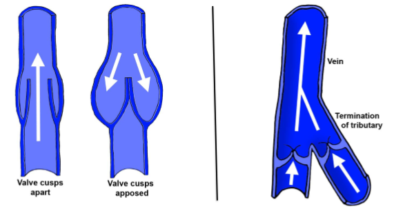
How do valves enforce unidirectional blood flow?
Enforce unidirectional flow from distal → proximal
What are the 4 key principles of veins of the limbs?
Superficial system of veins drains skin & superficial fascia only
Deep system of veins drains deeper structures
Paired venae comitatnes are found distally, whereas single vessels accompany arteries proximally
A set of communicating veins connects superficial & deep veins
Superficial veins outside the deep fascia → communicating veins connect through fascial layers
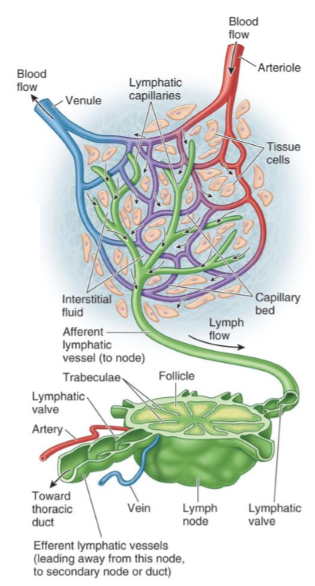
What are capillaries?
Single endothelial tubes connecting arterial & venous sides of circulation
Describe the structure & function of capillaries
Arranged in beds that connect arterioles & venules
Many beds named according to organs & body regions
Allow exchange of materials w/ interstitial & extracellular fluid