BIOL200 Pt 2- Lectures 4,5,6
1/22
There's no tags or description
Looks like no tags are added yet.
Name | Mastery | Learn | Test | Matching | Spaced | Call with Kai |
|---|
No analytics yet
Send a link to your students to track their progress
23 Terms
Identify features shared by all metazoans
Multicellular animals (not like single celled protists)
monoflagellated cells
mitotic aster apparatus (star-shaped tubules coming from centrosomes to anchor the spindle to the pole of the cell for cell division)
have cell junctions to hold cells together into tissue
proteins for movement
likely monophyletic
Evolutionary implications of multicellularity
metazoans = multicellular animals
multicellularity evolved after protists, before basal phyla (sponges, etc.)
multicellularity arose many times, but only evolved once sucessfully in metazoans.
There must be a selective advantage because it evolved many times and stuck once.
implications:
Multicellular organisms have cells that are connected
Cells can specialize, communicate and work together.
Increased size = more mobility to find resources for living, and less likely to be predated
Changes the meaning of an “individual.”
Can do apoptosis (controlled cell death of defective or infected cells) to save the larger organism (as the organism is now bigger than a cell)
reproduction: most multicellular organisms do sexual reproduction; have a small amount of specialized cells in the gonads, which are capable of doing meiosis, The somatic cells have given up sexual reproduction and specialized in their own tissues.
Poriferans
bearers of pores
asymmetrical or radial symmetry
Pinacocyctes, choanocytes, mesenchyme cells, porocytes
pinacocytes = integumentary cells (outside)
porocytes = flat cells that line pores in the skin of animals; openings to connect outside water to inside the sponge
choanocytes = feeding cells; water in via collar of microvilli, food trapped in collar, flagella continuously beating to force filtered water out of osculum, creating negtive pressure that drives more watter in via pores.
mesenchyme cells = unspecialized cells.
Body form and filter feeding:
central cavity, and can have branching chambers (more complex = have diffrent regions)
ascon = just one hole, syncon = sponge wall is folded, leucon = super branched
The pores let water into the cavity (spongocoel)
Choanocytes = cells in cavity wall that have collar of microvill that trap food. Inside the collar is a flagellum that beats, making the filtered water move out through the osculum. This creates negative pressure so more water is drawn in
food stored in food vacuole.
epithelial tissue but no organ system
spicules (sharp, hardened) = hardened calcereous or silicous or protenous/collagenous (spongin) embedded in the coral wall to support.
Reproduction:
asexual reproduction: freshwater. makes gemmules before dying to last over winter. the inside amoeboid cells reorganize into sponge once warm enough
Sexual reproduction: moneceous (male and female parts in one), but asynchronous to prevent selfing. some choanocytes lose collars and rearrange to make flagellated sperm. Other choanocytes do meiosis and become eggs. they then fuse in external fertilization, OR internal fertilization within the female
Choanocytes lose colour and become
Have significant ecological roles:
filtering water = pump water in continuously via porocytes, filters out particles as food,
source of food for others
Cnidarians
Characteristics:
classical jellyfish
radial or biradial symmetry
diploblastic tissue organisation- no mesoderm, 2 layers
inside = gastrodermis (gut lining), cilliated to beat water around.
mesoglea space = jelly like layer
outside = epidermis made of epitheliomuscular cell (contractile- think how they swim)
have internal gastrovascular cavity
nerve net - simple, with specialized nerve cells interacting together like a net (but no brain, ganglia, etc), under the skin of in the gastro cavvity
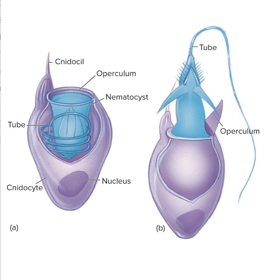
cnidocytes = in epidermis and gastrodermis. Have a modified cilia, that senses movement/chemicals, causing the operculum to open and the nematocyst to fly out with a venamous spikes or a whipping tube/flagella to trap food, ir defend
peristalsis of walls to push food through. cells lining the gastroderm then pick them up, put into vacuoles to be digested.
in the tentacles
Have hydrostatic skeleton to jeep shape:
draw water into gastrovascular cavity, then close mouth.
muscles push against internal water thats pushing out- creates a turgid structure.
Hydrozoa = small, polyp form dominant: in polyp, theres gastrozoid (eating), gonozoan (asexually medusa buds off)
scyphozoa = true jellyfish, medusa phase dominant
cubozoa = box jellyfish- the bell at the top is square shaped and tenacles come from the corner of this square. Highly toxic
Anthozoa = marine, no medusa form, just have a gastrovascular cavity that gets divided by mesentaries with gonads and cnidocytes.
Zooxanthellae live in these corals symbiotically. They’re coourful and get incorporated into the coral.
Choral bleaching- if its too war, coral exposes all zooxanthellae, which die and the coral turns white.
Ctenophorans
diploblastic (but possibly triploblastic because they have true muscle, not just isolated muscular cells, which can only come from mesoderm and speciallized cell)
biradial symmetry
comb jelliers
cellular mesoglia with muscle cells (mesoderm)
gastrovascular cavity with anal opening
nerve net
8 cillia-bearing comb rows
colloblasts = hollow stinging cells
monoecious, external fertilization, cilliated larva
Why are choanoflagellates the sister group of metazoans
choanoflagellates = protists that can come together and form a little colony (a bunch of choanocytes on a stem)
very similar between choanoflagellates and choanocytes, so thought to be the protist-sisters of metazoans
structure of cnidocytes
cnidarian life history + alternation of generations
polyp = has tenticles facing outwards, sits on a solid structure (sessile) and asexual..
Budding (asexual reproduction) occurs to form and release a haploid medusa
medusa = free swimming form (what we see as jelly fish), that then does sexual reproduction
like a polyp flipped over, except its diecious (male or female)
does mitosis to make haploid egg or sperm.
fertilization → zygote
zygote develops into diploblastic blastula
becomes planula (ciliated larva) that settles on a rock and grows into another polyp.
Features of all lophotrochozoans
Have a trochophore larva (not all of them, but if it has it, you KNOW)
small, free-swimming
looks like a dreidle with tufts of cilia around the 2 orifaces (top and bottom) and in a band around the center.
complete digestive system because they have 2 orifaces where food enters and then leaves.
have lophophore = feeding structure with a horse-shoe arrangement of tentacles at the top
like an anemone at the top of the organism, around a mouth
complete digestive tract/system
platyhelminthes: why do we question monophyly of platyhelminthes
flat worms
if its a worm with a flat body → considered a flatworm.
does not contain all descendants
Acoelomate (no cavity, thats why they’re flat. The inside is filled with mesodermally derived parenchyma cells.
bilateral symmetry : can only be divided down one way in symmetry because they are cephalated (concentration of sensory structures at the head)
Finally have organ system-level organization (unlike cnidarians that only had nerves)
but incomplete gut (1 opening to digestive tract)
no respiratory or circulatory systems.
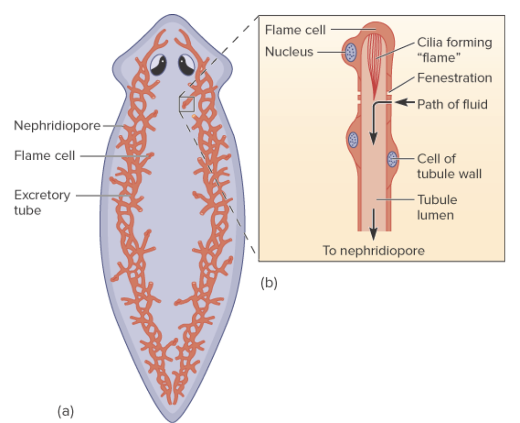
Protonephridia for osmoregulation and excretion
get rid of toxic nitrogenous waste and maintain osmotic balance.
Have nervous system with anterior ganglia (like a brain), longitudinal ventral nerve cords.
Monoecious with internal fertilization, but no self-fertilization, will do copulation
classes:
turbellaria = common planarea
trematoda = internal parasites
monogenea = external parasites
cestoidea = tape worms
nervous and excretory system of tubellarians vs cnedarians
Tubellarians:
excretion = have protonephridia, a collection of cells forming a long tubule
cells make up tubule wall, and absorb nutrients that are needed from the passing water
water enters through fenestrations
the flame cell is at the end, and has a tuft of cilia, which beats around to beat water down the tubule. This creates negative pressure, drawing more water in.
excess water (waste) pushed through to nephridiopore to be excreted
vs. cnidarians have no specialized excretion system; waste just diffuses out.
Nervous system = have 2 main lateral nerve cords, connected with horizontal commisure brances, which often come together on one end to form a cerebral ganglia (like a brain)
some can be net like instead of lateral cmmisure
sensory (input stimulus) and motor nerves present (effect response to stimulus)
vs. cnidarians have a nerve net, with no cerebral ganglia or cephalization.
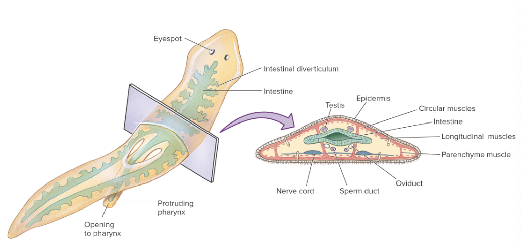
anatomical features of turbellarians
free-living flatworms
live anywhere in water (benthic, marine, freshwater)
move via circular or longitudinal muscle fibres that contract. have cilia on surface to move around
predatory - eat other prey
polyphyletic (not a true taxon)
Digestive system/intestinal region: main tube, with diverticulum pockets out to the side to diffuse nutrients from prey. circular muscle pushes food through, them out the same hole
can range from a simple tube (smaller animals) to. totally branched out with diverticulum when it gets larger.
no circulatory system to move nutrients around: once food is digested, its diffused directly to the cells through highly branched intestinal system
3 kinds of muscle: longitudinal (long/short), circular (wide, thin), parenchymale (round, flat)
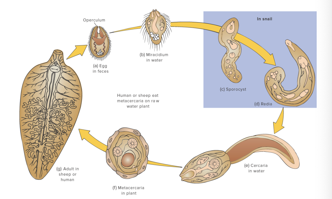
features and life history of digenean trematodes
live in bile duct of hosts: need to withstand enzymatic conditions that can break down fats
flattened and wide (like a whales tail)
Have a mouth and a nephridiopore (excretory)
Have a tough tegument (skin) that is syncytial (or all the cells involved fuse and act like a giant multinucleated cell)
to withstand tough digesting conditiosn
has an outer tegument (glycocalyx = in contact with bile duct), then basement membrane with phospholypids, thennn inner tegument that has nucleus and cytoplasm of the syncitial cells.
Life history:
egg is in feces of host. egg has to end up in aquatic environment, to hatch into miracidium larva that swims around and finds a snail
snail = first intermeediate host. Sporocyst and redia larval forms develop in the soft tissue.
redia produce cercaria, with a tail and digestive system, that swims into water, then lands in a plant = second intermediate host
human/sheep eats it, and the parasite cercaria develops into an adult.
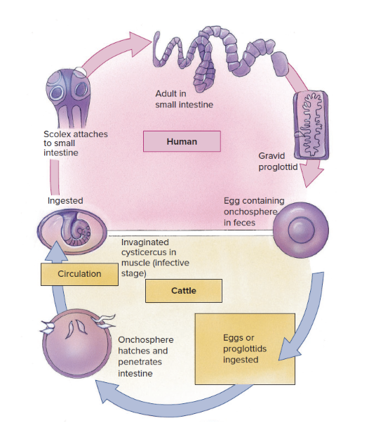
anatomical features of tapeworms (cestoida)
very specialized gut (intestine) parasites of vertebrates
anatomically simple because most of their needs are stolen from the host (like secondary organelle)
no digestive system; they absorb predigested nutrients from the small intestine of host
no excretory system, only absorb what they need.
JUST reproductive systems
has intermediate hosts in life cycles.
Cestode has:
head/scolex: hook to grab on to host intestine, and sucker.
the long body of the animal divides into proglottids. As new proglottids keep getting produced from the neck, older/mature ones are pushed towards the end
mature proglottids have a sac of fertilized eggs, and can break off and release the eggs
life cycle:
proglottid in feces releases gertilized eggs, which are ingested by cattle. the eggs become onchosphere, hatches and penetrates the intestine, and carried around in the circulatory system into the muscle tissue
Humans eat the muscle tissue, where the invaginated cysticercus (infective stage) enters the small intestine, and the scolex attaches to the small intestine. Grows, feeding on food
breaks off mature proglottids for release into feces.
Turbellaria reproductive system
Asexual reproduction: transverse fission
split down the middle (axis of symmetry) to make 2.
Sexual reproduction:
monoecious
eggs released into oviduct by ovary
sperm inserted by copulation into copulary sac, then moves via genital chamber to oviduct
gertilization in oviduct (internal), then mvoes down bast yolk glands that provide a yolk (food source for embryo), then deposited in a protective cocoon.
Describe general molluscan body form
soft body form
snails, squids, octopus, bivalves (oysters)
triploblastic (3 cell layers- have true specialized organ tissue)
coelomates = have a true coelom lined by mesodermal tissue
body form with a musclular head-foot, and dorsally they have a visceral mass with all their organs in it (Can be protected by shell)
mantle - sheet of tissue overlying the visceral mass.
Hardened shell has 3 layers:
nacreous layer = internal smooth layers of CaCo3
prismatic layer = CaCO3
Periostracum = hard protein based material
grows outwards, with mantly expanding outwards
Mantle cavity- space between mantle and head foot at the back of the animal.
walls are muscular, can contract to open/close and circulate water in and out.
respiratory gas exchange with vascularised high SA gills inside he mantle cavity
also gets rid of nitrogenous waste and digestive waste.
reproductive wastes put here for fertilization
Radula - hardened structure of chitin (carbohydrate polymer) by the mouth, protrudes out an used for scraping. Muscles contract to push and pull radula to scrape.
Have trochophore larvae (part of the lophotrochozoans - dreidle shape with tucts of cilia!!)
open circulatory system (going out - blood leaves vessels and fills sinus, bathes the cells lining the sinus in blood for direct diffusion.
EXCPET CEPHALOPODS (SQUIDS AND OCTOPUS)
torsion in gastropod development
gastropods = snails, slugs, limpits
aquatic, intermediate hosts for trematoda
gastropods have torsion, where their headfoot and visceral mass can rotate 180 degrees in the opposite direction so the head is under the mantle cavity
evolved likely for protection of the head: if predation is near, head (most important part of the body) quickly withdraws into cavity first, then foot area afterwards. so head is most internal for protectioin.
how various organ systems are affected by evolution of shell coiling in modern snails
Have a membrane covering the mantle cavity with a small opening, called pneumatostomes.
mantle cavity contains gills, OR it can be highly vascularized in the walls themselves, and do gas exchange like lungs.
The function of the mantle cavity is turning more into a lung.
Coiling can occur in left or right hand direction- asymmetrical shell, and there is space constraints
reduction of some of he organs to fit them all in.
1 gill protruding into the mantle (as opposed to ancestral snails wo coils had 2 gills)
1 nephridium instead of being paired
1 atria of snail heart has been removed.
functions of mantle in gastropods, bivalves and cephalopods
Bivalves: oysters, clams, scallops (marine or FW food source for others, sedentary)
have 2 shells (valves), with a muscular hinge structure .
Adductor muscle scars that attach to both valves and contract to hold them closed, or relax to open them so water can enter.
Mantle lies over the soft body part + holds it to the shell.
movement of water and food through bivalve
water current intaken by inhalant siphon/aperture facing the outside of the shell.
water passes over the gills in the mantle cavity. The surface of the gills is covered in cilia that trap the food and bring it down to the food groove, where it moves towards the mouth opening to the digestve tract.
Food-filtered water moves up through water tubes, in countercurrent exchange, giving oxygen and taking away CO2, turns afferent blood to efferent blood.
fully filtered water moves towards the suprabranchial chamber, where it is moved towards the exhalant siphon/excurrent aperture.
Digestion:
has hardened crystalline style that grinds incoming filtered food
Reproduction of gastropoda vs.bivalvia
Gastropoda:
monoecious or dioecious.
broadcast spawners- release massive amounts of gametes to be fertilized in the water.
sperm intaken and can still be livebearing species (viviparous) = give birth to live young.
monoecious = copulation results in mutual sperm exchange. OR protandrous (start male, then become female)
have trochophore larva, which develop into freeswimming veliger larva that are als free swimming, then settle to a rock to be sedentary.
Bivalvia:
dioecious, some monoecious
gametes expelled via exhalant opening for external fertilization
trochophore larva develop into veliger stages
veliger stages are external parasites on fishes.
some bivalvia have a “lure” in mantle cavity, that looks like a little fish. big fish come closer to eat the “fish”, then the veliger larvae are released and latch on.
Gastropods of note
Banff springs snails - live in hot springs
Cone snails = have cone-shaped shells. Radula is like a venemous mini harpoon, that shoots out and spikes a fish.
Nudibranchs = look like flatworms, but more complex
Bivalvia circulation
anterior aorta- blood towards anterior of animal, then down to foot of animal
can have posterior aorta- pumping blood to exterior (back)
blood sinus in foot, bathes sinus cells linng the walls in blood to diffuse nutrients (o2, etc)