chap 20: the heart
1/95
There's no tags or description
Looks like no tags are added yet.
Name | Mastery | Learn | Test | Matching | Spaced | Call with Kai |
|---|
No analytics yet
Send a link to your students to track their progress
96 Terms
What is the heart?
The heart is a muscular pump to move the blood through the vessels
Where do arteries carry blood?
AWAY from the heart
Where do veins carry blood?
back TOWARD the heart
What are capillaries?
microscopic vessels connecting the smallest arteries and veins
What are the two divisons of the cardiovascular system?
pulmonary and systemic circuit
What is the pulmonary circuit
carries blood to the lungs for gas exchange and returns it to the heart
What is the systemic circuit
supplies blood to every organ of the body, including other parts of the lungs and the wall of the heart itself
what is the pulmonary circuit supplied by
right half of the heart
What blood does the heart recieve from the body and where does it pump it too?
Heart receives deoxygenated blood from body, pumps it to lungs
What is the systemic circuit supplied by
left side of the heart
What type of blood does the heart recieve from the lungs
Heart receives oxygenated blood from lungs, pumps it to body
What does the aorta carry
freshly oxygenate blood leaving the left ventricle to be distributed throughout the body
What are the 3 regions of the aorta
ascending aorta, aortic arch, descending aorta
What does the pulmonary trunk carry
carries deoxygenated blood leaving the right ventricle to be carried to the lungs, it quickly branches into right and left pulmonary arteries to the respective lungs
What are the veins of the heart
pulmonary veins, superior vena cava, inferior vena cava
What do the pulmonary veins return
return freshly oxygenated blood from the lungs to the left atrium, with two of these emptying into the atrium from each side
What does the superior vena cava carry
deoxygenate blood to the heart from the head, upper limbs, and all other regions above the diaphragm
Where does the superior venca cava empty
into the right atrium
what does the inferior vena cava carry
carries deoxygenated blood to the heart from body regions below the diaphragm
where does the inferior vena cava empty
into the right atrium from below
what are the 3 layers of the heart wall from outermost to innermost
epicardium, myocardium, endocardium
what is the epicardium of the heart wall
(visceral layer of serous pericardium)—thin membrane of
simple squamous epithelium overlying elastic connective tissue
What is the myocardium
composed of cardiac muscle; most of the mass of the heart.
What does the myocardium perform
performs the work of the heart & its thickness varies with the workload of the individual chamber.
what is the vortex of the heart?
Cardiac muscle cells (cardiomyocytes) grouped in bundles that coil around heart in a spiral
What does the vortex of the heart cause?
causes heart to contract with a twisting, wringing motion, enhances the ejection of blood.
what is the endocardium
inner lining of heart chambers and covers valves;
simple squamous epithelium and thin layer of connective tissue.
Describe the anatomy of the atria chambers
• Thin, flaccid walls
• Atria separated by interatrial septum
• Right atrium and both auricles contain ridges of pectinate muscles
Describe the anatomy of the ventricle chambers
•Left ventricle wall much thicker than right; reflects workload. It is 4X as thick as it pumps blood through out the entire body
• Ventricles separated by interventricular septum
• Both have trabeculae carneae—internal ridges
• Volume of both ventricle about the same
What is the left ventricle wall much thicker than the right ventricle wall?
reflects workload, it is 2-4X as thick as it pumps blood through out the entire body
What do heart valves ensure?
one-way flow of blood through heart
What do heart valves consist of ?
two or three cusps or leaflets covered with endocardium
What are Atrioventricular (AV) valves?
between atria and ventricles
What is the Right AV Valve
three cusps, so also called tricuspid valve
What is the left AV valve
also called the mitral valve or bicuspid valve
What ate valve cusps anchored to and by what?
anchored to papillary muscles by tendinous cords (chordae tendinae)
What happens when tendinous cords that anchor valve cusps start to slack?
excessive bulging (valvular prolapse) will occur.
What are semilunar valves (SV)
between ventricles and great arteries
Give specific locations of SV valves
aorta, pulmonary trunk-bld vessels leading to the lungs
What are the two SV valves
aortic and pulmonary
Where is the pulmonary valve
between right ventricle and pulmonary trunk
where is the aortic valve
between left ventricle and aorta
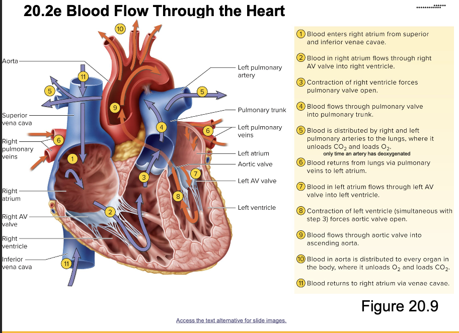
** know blood flow through the heart
T/f: Coronary circulation is critical
True
What happens when blockage occurs in a coronary artery
this can produce a myocardial infarction (MI)
What is a myocardial infarction(MI)
sudden death of cardiac tissue
Where do the LCA and RCA emerge from?
the base of the ascending aorta (proximal end of aorta).
Where does the left coronary artery (LCA) travel?
through coronary sulcus, under left auricle and divides into two branches
What are the two branches of the LCA?
anterior interventricular branch and circumflex branch
Where does the anterior interventricular branch of the LCA pass?
passes down anterior interventricular sulcus, joins posterior interventricular branch.
What is the anterior interventricular branch called clinically?
left anterior descending branch
What does the anterior inter ventricular branch do?
This artery supplies blood to both ventricles and the anterior two-thirds of the interventricular septum.
What occurs with a blockage to the anterior interventricular branch
A blockage here would cause massive damage or death hence it is called the widow maker.
What does the circumflex branch do in the LCA
contuses around left side of heart; gives off left marginal branch
Where does the right coronary artery RCA travel?
travels along coronary sulcus under right article and divides into two branches
What two branches does the RCA divide into
right marginal branch and posterior interventricular branch
Where is the right marginal branch?
along right margin of heart
Where is the posterior interventricular branch
along posterior interventricular sulcus
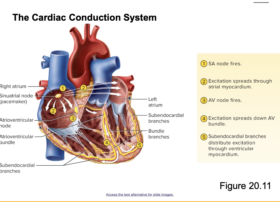
** know the cardiac conduction system
what is the cardiac cycle
one complete cycle of contraction and relaxation of the
heart
what is cardiomyocytes electrically charged
electrically polarized (negative on the inside)
Excitation leads too….
depolarizes (less negative on the inside, as Na+ enters the cells) the cells and cause their contraction; after contraction, the cells repolarize (K+ leaves the cell returning it to more neg on inside) and relax
Define systole
contraction of a heart chamber
Define diastole
relaxation of a heart chamber
What measures electrical activity of the heart
electrocardiogram (ECG or EKG)
What are the major events of ECG
P wave, QRS complex, T wave
What is p-wave?
atrial depolarization
What is QRS complex?
ventricular depolarization
What is t-wave?
ventricular repolarization
What occurs in the cardiac cycle initially?
all four chambers are relaxed; AV valves are open and blood flows from atria to ventricles
What occurs when SA nodes fire?
produces P wave of ECG as atria depolarize; atria contract (atrial systole) to finish filling the ventricles; signal delayed at AV node
Where does the signal delayed at AV node go?
Signal spreads down rest of conduction system, depolarizes ventricles, which produces QRS complex; ventricles contract and AV valves close—first heart sound (S1); blood is ejected
What produces the first heart sound (S1)?
AV valves closing
What happens when ventricles repolarize and relax?
Ventricles repolarize and relax (ventricular diastole), which produces T wave; blood tries to flow backward and closes semilunar valves—second heart sound (S2)
What causes the second heart sound?
SV valves close
How does the cycle start over?
Ventricles begin refilling again and cycle starts over
Why does the heart develop early in the embryo?
so it can distribute nutrients and oxygen
When does the heart start beating?
22-23 days (week 3)
What occurs in week 3 of the embryo?
mesoderm condenses to form cords that hollow out to form parallel endocardial heart tubes
What are the dilated spaces formed from tubes fusing in the embryo?
runcus arteriosus, bulbus cordis (becomes R. atrium), ventricle, atrium, and sinus venosus
What shape does the heart form and how? What else is present?
Heart loops and then forms S shape; primordial atrium and ventricles present
What is the opening between atria?
foramen ovale
What does the foramen ovale allow?
allows the blood to bypass the lungs, goes from right atrium to left atrium.
What allows the most blood to bypass pulmonary circuit in the fetal heart?
foramen ovale and ductus arteriosus
What is caused by lungs inflating at birth?
their resistance to blood flow decreases
A drop in resistance to blood flow causes?
causes tissue flap to seal foramen ovale, becomes a depression—the fossa ovalis
What occurs several hours after birth with the ductus arteriosus?
ductus arteriosus begins to close, fully closed in 2–4 days; remnant is ligamentum arteriosum
What is a ductus arteriosus
a shunt from the base of the left pulmonary artery to the aorta
The ductus arteriosus closes fully in how many days?
2-4 days
What is patent ductus arteriosus?
failure of the ductus arteriosus to close
What does Patent Ductus Arteriosus lead too overtime?
as the lungs become inflated and more functional, pulmonary blood pressure drops below aortic blood pressure. Blood may then begin to flow from the aortic arch back into the pulmonary circuit for an immediate second trip through the lungs. Since this blood returns to the left ventricle, it adds markedly to the left ventricular workload. The lungs sometimes respond to the persistent high blood flower with vascular changes that increase pulmonary resistance and stress the right ventricle as well.
What are some signs of PDA?
poor weight gain, frequent respiratory illnesses, dyspnea, cardiomegaly
When is PDA usually suspected
about 6-8 weeks of age because of persistent “machinery-like” murmur.
What is PDA confirmed by
echocardiogram
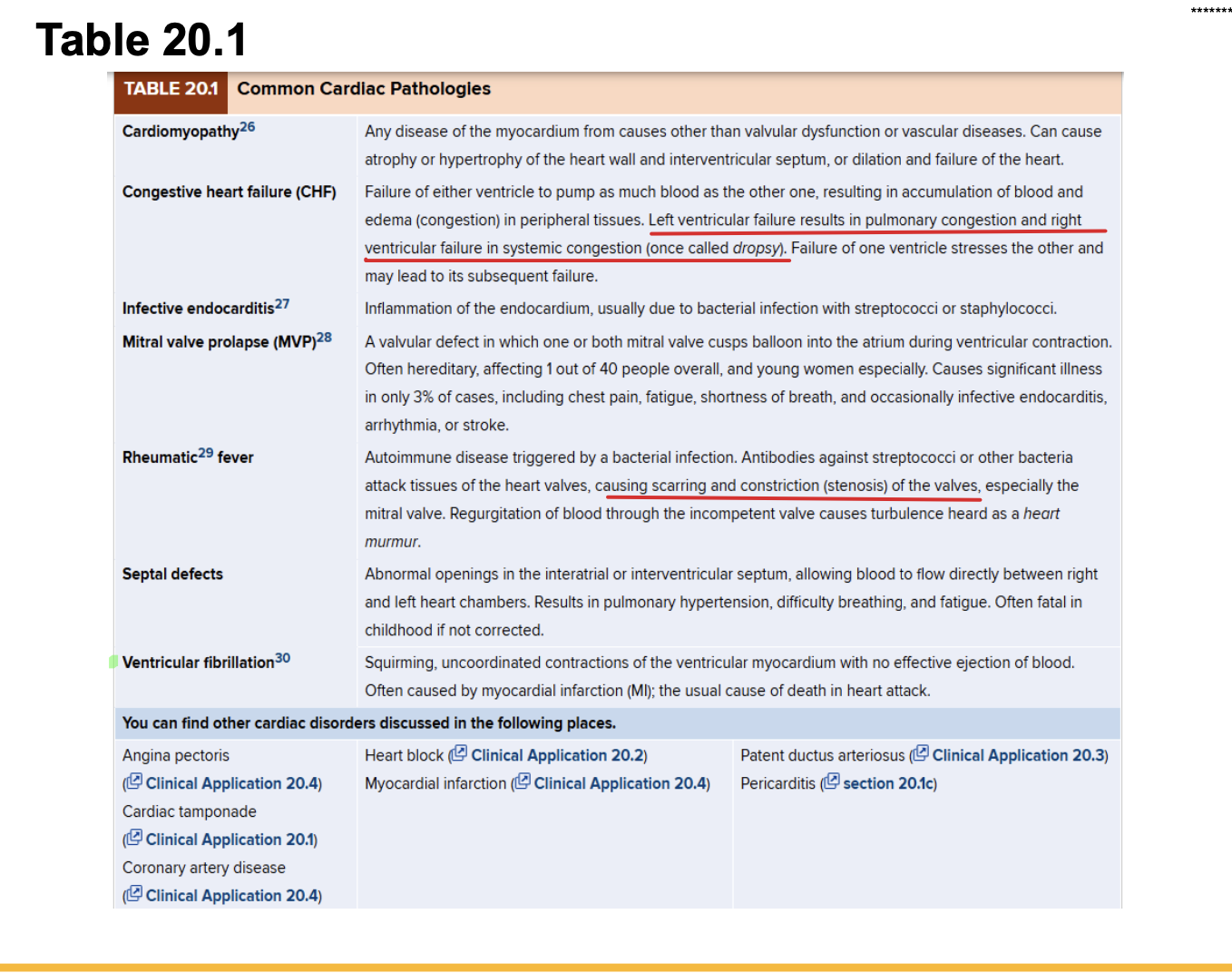
** know the diseases