Human Body Systems HBS PLTW 1.2.2 WCHS Mr. Alasti
1/19
There's no tags or description
Looks like no tags are added yet.
Name | Mastery | Learn | Test | Matching | Spaced |
|---|
No study sessions yet.
20 Terms
Physiatrist
A medical doctor trained in the specialty of physical medicine and rehabilitation. They diagnose illnesses, design treatment protocols, and prescribe medications. They design exercise programs tailored to the patient’s needs and outline a plan of care that physical therapists use
What is a pulled muscle?
A muscle strain. A strain occurs when muscle fibers are under such strong mechanical stress that they are overstretched and can even tear
Endomysium
A delicate, thin layer of connective tissue that surrounds each individual muscle fiber in skeletal muscle
Epimysium
The dense connective tissue that surrounds the entire muscle
Fascicle
A small bundle of fibers wrapped in connective tissue, like a tiny rope made of smaller strings
Perimysium
A connective tissue sheath that surrounds individual muscle fascicles, and separates them from other fascicles within the skeletal muscle
Myofibril
A long, cylindrical organelle within muscle fibers made of repeating protein filaments, primarily actin and myosin, that slide past each other to generate force and produce muscle contraction
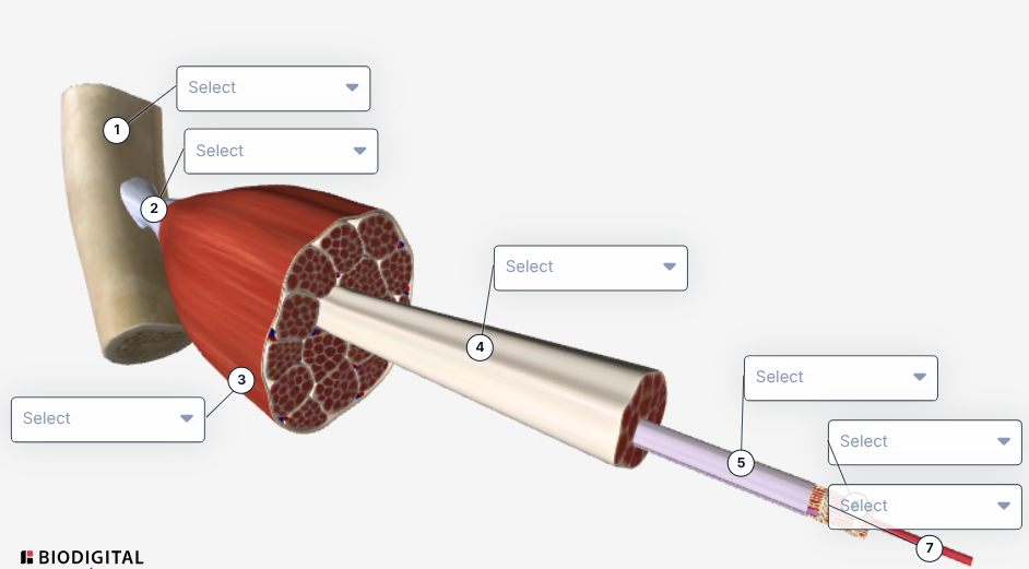
Label the image
Bone
Tendon
Muscle
Fascicle
Sarcolemma
Sarcoplasmic Reticulum
Myofibril
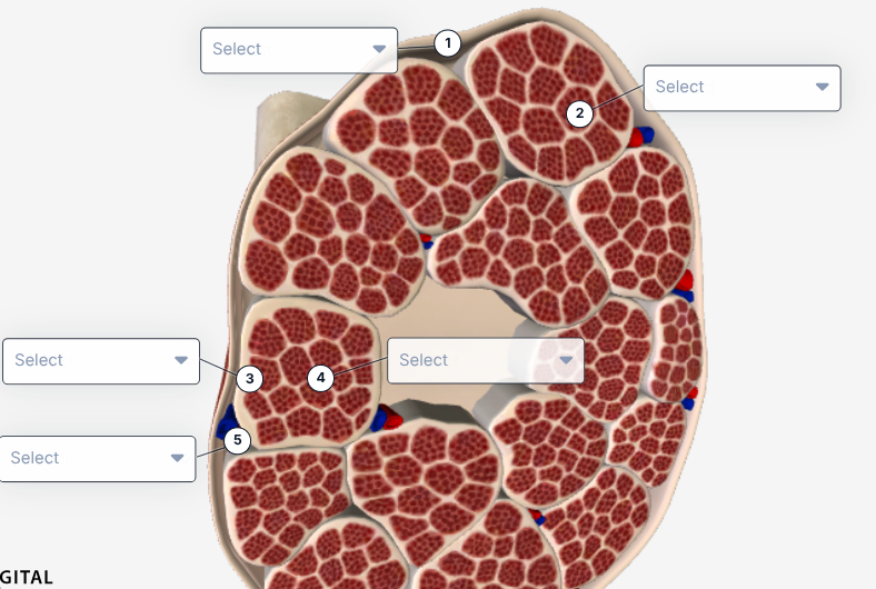
Label the image
Epimysium
Muscle Fiber
Perimysium
Endomysium
Blood Vessels
Where are sarcomeres within our drawings?
Within the myofibrils
What are the six muscle rules?
Muscles must have a minimum of 2 attachment points and must cross at least 1 joint
Muscles always pull and get shorter (they can’t get longer)
An attachment more movable is called the insertion, and the attachment that remains stationary is called the origin (the superior, proximal, or medical part is typically the origin, while the distal, inferior, or lateral part is typically the insertion)
Muscles that decrease the angle between ventral surfaces of the body are known as flexors, while muscles that increase the angle between ventral surfaces of the body are known as extensors
Muscles work in opposing pairs
Muscle striations point to the attachment and show the direction of the pull
For smooth movements to occur, can both extensors and flexors be contracting at the same time?
Yes
Medial head of the triceps
Origin: Proximal half of the dorsal humerus
Insertion: Distal to the elbow on the ulna
Action: Extends elbow
Pectoralis Minor
Origin: Anterior surface of ribs 3-5
Insertion: Coracoid process of the scapula
Action: Stabilizes the scapula by pulling it forward and downward
Clavicular head
Origin: The medial half of the inferior edge of the clavicle
Insertion: Lateral edge of the proximal humerus, inferior to the insertion of the sternal head
Action: Flexes the humerus and helps with the medial rotation of the humerus
Sternocostalis head
Origin: Ribs 1-5 on the lateral edge of the sternum
Insertion: Lateral edge of the humerus, inferior to the insertion of the abdominal head
Action: Draws your forelimb medially (from lateral to medial)
Abdominal head of the pectoralis minor
Origin: Ribs 5-7 (actually attaches to the fascia of the abdominal muscles)
Insertion: Lateral edge of the most proximal part of the humerus
Action: Allows spiking motion- draws the arm up to medial
External intercostals
Origin: Lateral surface of ribs 1-8
Insertion: Medial border of the scapula
Action: Increase the chest cavity, allowing air to rush in
Brachealis
Origin: Halfway down the humerus
Insertion: Proximal ulna
Action: Flexes elbow