PSYC1002: Neuroscience
1/42
There's no tags or description
Looks like no tags are added yet.
Name | Mastery | Learn | Test | Matching | Spaced |
|---|
No study sessions yet.
43 Terms
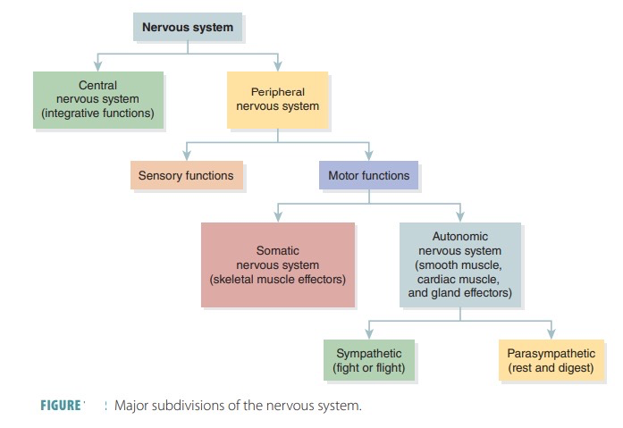
What are the divisions of the nervous system?
How is the central nervous system protected?
CNS consists of the brain and spinal cord
the CNS is protected by:
bone: hard casing around the brain
meninges: 3 membranes that wrap around the brain and protect the brain from the inside surface of the skull
blood brain barrier: a selective semi-permeable border of cells that protects the brain from harmful substances in the blood while allowing essential nutrients to pass through
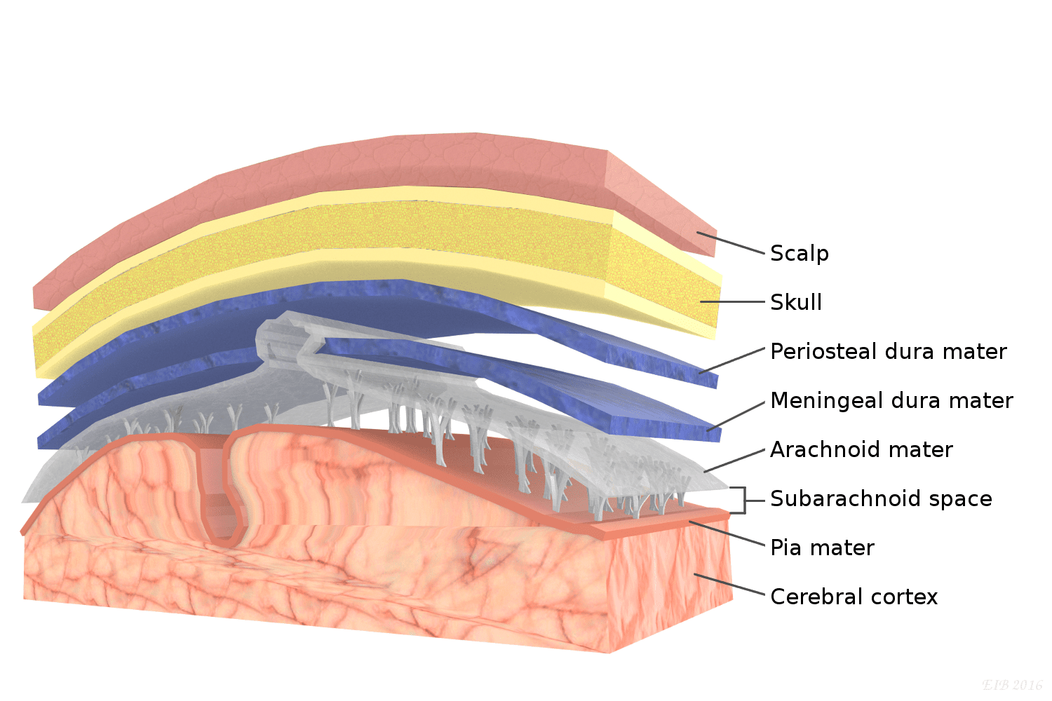
How does the meninges protect the CNS?
It consists of 3 layers which protect the brain:
dura mater: thick outer membrane which surrounds the outside of the brain
arachnoid mater: a spongy membrane for shock absorption (hence the web-like appearance) which is filled with cerebrospinal fluid. large vessels which supply blood to the brain travel through this
pia mater: thinner, fragile, inner membrane which follows the contours of the brain
how does the blood brain barrier protect the CNS?
the brain uses a lot of blood so if there’s toxins in the blood, the brain is highly susceptible to them
the blood brain barrier protects the brain from toxins as a border of capillaries
these capillaries supply blood to the CNS and have special walls which restrict the entry of many chemicals into the CNS
capillaries in the brain are smaller than in any other party of the body, so less material passes through
what is the spinal cord and its role in the nervous system?
the spinal cord is a cable of neural fibres with “roots” branching off
it is the interface between the CNS and PNS
it produces spinal reflexes- movements triggered entirely by the spinal cord receiving a message (rather than through the brain)
e.g. touching a hot surface- you react quickly because the message doesn’t need to be sent to the brain first
how does regeneration differ in the CNS vs PNS?
PNS can regenerate, CNS cant, so damage to the CNS is permanent
if the CNS (brain) could regenerate, we wouldn’t have memory
What are the divisions of the PNS?
PNS is the rest of the nervous system outside of the brain and spinal cord
it’s divided into:
sensory nervous system: nerves which deliver sensory info to the brain (afferent)
motor nervous system: nerves which deliver info from CNS to muscles to generate movement (efferent)
what are the divisions of the motor nervous system?
autonomic: non-voluntary bodily functions
somatic: voluntary bodily functions
what are the divisions of the autonomic nervous system?
sympathetic: activated in a stressful situation, heart beats harder and faster
parasympathetic: activated in a relaxed situation, heart beats more gently and slower, blood vessels dilate
enteric: controls gut movement and digestive processes
why does your mouth get dry in a stressful situation?
Your sympathetic nervous system gets activated and shuts down the parasympathetic situation, which makes you salivate
what is the enteric’s systems role?
located in the walls of the gastrointestinal tract
controls digestive activity (peristalsis and secretion of enzymes)
senses physical and chemical conditions of the gut
interacts with the brain yet can also function independently
uses neurotransmitters including dopamine and serotonin
what are the ventricles in the brain?
the human brain is not solid and consists of ventricles
ventricles are cavities containing cerebrospinal fluid
ventricles are the ‘sewerage system’ of the CNS by collecting waste for removal
what happens if the ventricular system gets blocked?
this system can get blocked in a developing foetus or occasionally in adults
this can be corrected surgically in both foetuses and adults
for a developing foetus, there’s good recovery due to the high neuroplasticity of the developing brain
in adults, blockage causes a build up of cerebrospinal fluid called hydrocephalus which inflates the ventricles and squashes the brain
what are the subdivisions of the human brain?
brain stem
cerebellum
thalamus
hypothalamus
basal ganglia
limbic system
neocortex
corpus callosum
what is the role of the brain stem?
controls life-supporting functions through controlling the autonomic nervous system (heart rate, blood pressure, breathing)
what happens to the brain stem after a head injury?
a head injury breaks the blood vessels, causing intracranial bleeding which raises the pressure inside the skull
there’s only one opening in the head, the foramen magnum, where the spinal cord joins the brain stem
an injury pushes the brain towards this opening, crushing the brain stem
thus, a head injury can lead to a coma or death
treatment involves drilling a hole in the skull to relieve pressure in the brain
what is the role of the cerebellum?
cerebellum means small brain
it controls precise movements, including learned ones
contains a high density of neurons (70% of the brain’s neurons) yet is only responsible for 10% of the brain’s volume
receives sensory input and knows what signals need to be sent out to make the movement accurate and precise, and does this rapidly
what is the role of the thalamus?
located at the centre of the brain on top of the brainstem
is the sensory relay meaning it sends information to the cortex to experience the sensations
thalamus allows humans to participate in selective attention through controlling what sensory input we pay attention to
what is the role of the hypothalamus?
hypothalamus is below the thalamus
it’s involved in hormonal regulation, motivation controls and behaviours such as feeding and sex
it’s connected to and controls the anterior pituitary which controls glands and hormone production
what is the role of the limbic system?
regulates emotional experiences and memories
consists of the hippocampus, amygdala, olfactory bulb and cingulate cortex
memories and emotions are closely linked- you’re more likely to remember more emotional experiences as your brain acknowledges those moments as important
what is the role of the basal ganglia?
consist of the caudate nucleus, globus pallidus and putamen
it’s responsible for action and thought processes, specifically decisions about what action to take
works in conjunction with other brain areas e.g. cortex
what does damage to the basal ganglia cause?
Leads to neurodegenerative diseases which target the basal ganglia:
Parkinson’s disease: results in difficulty making movement as the basal ganglia is not assisting in movement
Huntington’s disease: results in excessive movement as the basal ganglia is not prohibiting certain movements
damage to certain parts can cause dementia
what is the role of the neocortex?
neocortex is a 6 layer 1cm thick convoluted sheet on top of the brain
appears wrinkled due to folds on the cortex which allow the cortex to fit into the head
having lots of cortex is beneficial for processing information and planning movement and action
cortex cannot get thicker so instead gets bigger and folds
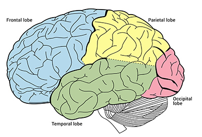
what lobes make up the neocortex?
frontal lobe: planning and executive functions and movement
parietal lobe: representing space for action
temporal lobe: memory and language
occipital lobe: vision
what are the primary motor functions which the cortex performs?
The cortex performance primary motor functions
primary motor cortex: at the back of the frontal lobe, neurons send axons to the spinal cord which send signals to your muscles to move, responsible for fine movement and control (e.g. movement of hands)
somatosensory cortex: at the front of the temporal lobe: relating to tactile senses
primary auditory cortex: in the temporal lobe, receives auditory information from the thalamus, resulting in auditory experiences
what is the corpus callosum?
connects the left and right hemispheres, allowing one unified experience
what is comparative neuroanatomy and its significance?
studies brain structure and function across species
nervous system is almost a defining feature of all animals (except sea sponges)
jellyfish have 5000-10000 neurons organised into networks that allow it to respond to its environment
in insects, the nervous system has a more complex organisation
specialisation of some neurons
how does neuroanatomy differ in vertebrates?
vertebrates have a separate PNS and CNS
nervous system is organised into forebrain, midbrain, hindbrain and spinal cord
telencephalon is a component of the forebrain and varies largely between different animals
among vertebrates, there’s a large difference in relative size of different regions in the brain
only mammals have a neocortex- birds have something similar but it’s not the same 6 layer structure
what does a neuron consists of?
dendrites: thinner filaments extending from the cell body which receive info from other neurons
soma: cell body, including the nucleus
axon: thick filament extending from the cell body
myelin: insulating layer coating the axon
axon terminals: end of the axon which forms connections to other neurons
how does an action potential travel?
neurons are covered in a semipermeable lipid membrane to control ion concentration
polarization: at rest, the inside is negatively charged compared to the outside and the neuron is polarised
this resting potential is maintained by the Na-K pump which deposits 3 Na into extracellular space and 2 K into intracellular space
depolarization: during an action potential, ion channels open so ions can enter and exit
there’s an influx of sodium into the neuron, shifting internal charge to positive
depolarization lasts a few milliseconds
hyperpolarization: potassium channels are opened and membrane potential shifts to more negative
the neuron cannot receive another action potential during this
how are action potentials measured?
neurons contain both digital and analog elements but we process them through digital (discrete) scales meaning an action potential either occurs or doesn’t
once a neuron’s input reaches a certain threshold, an action potential is generated
alternatively, analog scales measure continuously
how are action potentials propagated?
action potentials don’t affect the whole neuron simultaneously
action potentials are localised to a small segment of the membrane then spread along the membrane
signal travels from dendrites to cell body to axon to axon terminal
what is the myelin and what is its purpose?
myelin is an insulating material which coats axons
myelin prevents depolarization by covering ion channels so when they open, sodium cannot flood in and an action potential does not occur at those parts of the membrane
the myelin doesn’t coat the entire axon as if it did, an action potential wouldn’t be possible
there are gaps along the axon, allowing the action potential to jump between these gaps were sodium influx occurs
this jumping increases the speed of transmission
what are the 2 different pain fibres?
we have 2 types of pain fibres- one with myelin and one without
whilst both transmit action potentials, the myelinated a-delta fibres receive the signal faster than the c-fibres
example: touching a hot surface
a-delta fibres receive the signal fast and allow you to react quickly to prevent further harm
c-fibres cause the delayed pain sensation
how do neurons interact?
neurons form small junctions between each other called synapses and communicate across these
synaptic cleft is 10-20nm wide, making transmission fast
synapses require an electron microscope to be seen due to their size
what is the process of neurotransmission?
neurons interact through neurotransmission:
action potential travels along an axon, reaching the axon terminal of the pre-synaptic neuron
arrival of the action potential triggers the opening of calcium channels and the entry of calcium ions
this triggers vesicles containing the neurotransmitter to migrate towards the pre-synaptic membrane
the vesicle fuses with the pre-synaptic membrane, releasing the neurotransmitter into the synapse
the neurotransmitter crosses the synaptic cleft and binds to receptors on the post-synaptic membrane, eliciting a response
binding between the neurotransmitter and receptor is highly specific
this binding interaction generates an electrochemical force which changes the structure of the protein to open the ion channel in the post-synaptic membrane
what are the types of neurotransmitters?
Neurotransmitters can be excitatory or inhibitory
excitatory: increases the likelihood of an action potential occurring e.g. glutamate
inhibitory: decreases the likelihood of an action potential occurring e.g. GABA
this is because GABA binds to a chloride ion channel, opening it during binding
as chloride ions are negatively charged, its influx into the cell hyperpolarizes the membrane, preventing an action potential
why and how are neurotransmitters removed from the synapse?
if neurotransmitters linger in the synapse and continues to stimulate receptors, the synapse wont be able to send any new signals
to stop the neurotransmitter from lingering here:
enzymes in the synapse target and destroy neurotransmitters
OR re-uptake occurs- the pre-synaptic membrane has pumps which suck up the neurotransmitter from the synapse
how do drugs reduce the time of neurotransmitters in the synapse?
psychoactive drugs reduce the amount of time neurotransmitters spend in the synapse by interacting with the mechanisms responsible for this
drugs can target the enzyme which destroys the neurotransmitter, prolonging the effects of the neurotransmitter- e.g. early antidepressant medication
modern antidepressants stop the re-uptake process, prolonging the neurotransmitter’s effect
other drugs act on the receptor itself
how do some drugs action on the receptor?
some drugs mimic the neurotransmitter, as they’re chemically similar to it
these drugs bind to the receptor directly to open the ion channel that the neurotransmitter otherwise would
other chemically similar drugs bind to the receptor but don’t open the ion channel but simply stop the neurotransmitter from accessing that receptor, blocking its action
what are the types of drugs that act on neurotransmitters?
drugs which interact on neurotransmitters are either agonists or antagonists:
agonists: increase the effects of a neurotransmitter
antagonists: decrease the effects of a neurotransmitter
how do recreational drugs impact the brains neurotransmission?
Opiates (heroin, morphine, codeine) mimic the brain's opioid neurotransmitters
Drugs bind to opioid receptors, stimulating them as endogenous NT would
Cocaine, amphetamines and ecstasy promote transmission of dopamine, noradrenaline and serotonin
Drugs promote transmission of dopamine, noradrenaline and serotonin by increasing their release or preventing their reuptake
Nicotine
Stimulates acetylcholine receptors (nicotinic receptors)
Caffeine
Blocks adenosine receptors, stopping adenosine from stimulating the receptor
how do therapeutic drugs impact the brains neurotransmission?
Benzodiazepines (e.g. valium)
Enhance inhibitory effects of GABA
They bind to a different location on the same GABA receptor, amplifying its effect
Barbiturates and alcohol do a similar thing
Anti-schizophrenic drugs
Most work by binding to dopamine receptors and blocking them
Antidepressant drugs
Enhance serotonin and noradrenaline transmission
Modern ones function by blocking reuptake