W5: Cerebral Cortex
1/34
There's no tags or description
Looks like no tags are added yet.
Name | Mastery | Learn | Test | Matching | Spaced |
|---|
No study sessions yet.
35 Terms
Learning objectives
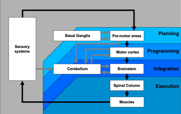
The hierarchical organisation of the motor systems
cortical regions of the motor system
topographic maps in the primary motor cortex
the function of the primary motor cortex
the functional roles played by the other main cortical motor areas
neuroplasticity
Hierarchical sensory-motor organisation
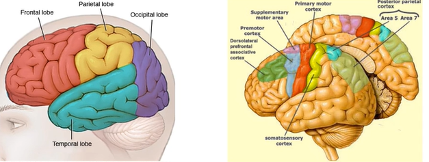
organisation of the cerebral cortex
red = primary motor cortex
somatosensory cortex = involved in processing feedback from the muscles
premotor regions = involved in action selection + planning (dorsal/ventral regions)
dorsal (on top) vs ventral (lower)
supplementary motor area (midline structure) = involved in action planning + sequencing
dorsal lateral prefrontal cortex = decision making + early stages of motor control
cerebral cortex
cortex forms the outer surface of the forebrain (AKA grey matter)
6 distinct layers (laminae)
cortex covers the other sub-cortical forebrain structures (e.g. thalamus, hippocampus)
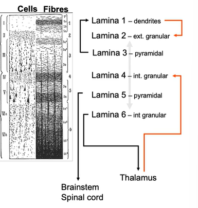
cortex: laminae
inputs and outputs are layer specific
layer 4 = key input layer
5 + 6 = key output layers
3 + 5 = have really large Betz cells = defining feature of primary motor cortex = critical for execution of voluntary movement
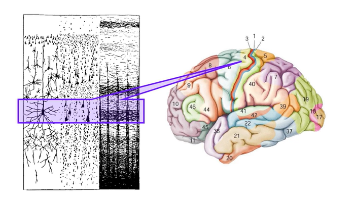
Betz cells
M1 (BA4) contains Betz cells in layer 5 (one of 2 output layers)
Betz cell = large pyramidal cell
they are in layer 5 (one of the two output layers of primary motor cortex)
Cortical Projections
Betz cells - large pyramidal cells
Project from motor cortex to spinal tract (cortical tract neurons)
Only 5% project to motor-neurons + the rest reach spinal interneurons
Betz cells also project to brain stem
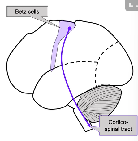
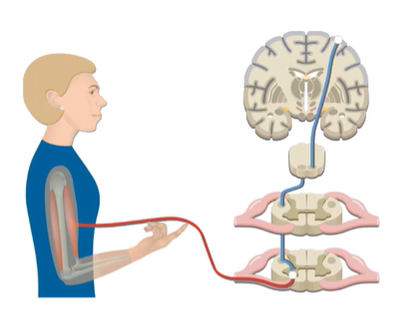
Corticospinal tract
Betz cells from the motor cortex initiate, regulate and control voluntary skilled movements
Done bye innervating alpha + gamma motor neurons in the spinal cord
Tract crosses at medulla, so limb movements are controlled by the contralateral motor cortex
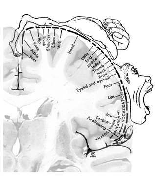
Mapping the motor cortex
Fritsch + Hitzig (1870): electrical stimulation in dog = mapping somatotopic motor representation
somatotopic rep. = the diff parts of the primary motor cortex that send motor commands to diff parts of the body
Penfield (1940): stimulated during surgery on epileptic patients
Discovered electrical stimulation causes simple movements
Map established
hands + feet have largest areas = seems like the more fine motor control needed determines area size in primary motor cortex
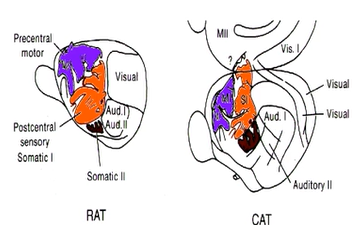
Cortical motor maps
another way of mapping primary cortex
close mirror relationship between sensory + motor maps
multiple maps: maps reflect sensory-motor specialization
purple = motor cortex maps, orange = somatosensory cortex maps
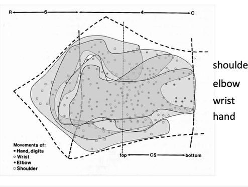
Cortical motor maps are not very realistic
research discovered a lot more overlap between representation of body parts than expected
don’t know what this overlap means - could be how much the body parts have to work together for movement
Study: Effector maps might not be the only organisation principle within the motor cortex
Study - fMRI studies
Argue that there’s a parallel organisational scheme in primary motor cortex
Replicated with lots of very large samples (inc. infants, animals etc)
Found 2 regions
Effector-specific connectivity regions are interdigitated with regions showing different connectivity, structure and function
Inter-effector regions = show high connectivity to each other and to cingulo-opercular network
Inter-effector regions become active during planning (instead of execution) + lack effector specificity
still controversy over whether this is another premotor cortex
What is represented in the motor cortex: muscle or movements?
Electrical stimulation:
Brief micro-stimulation (50ms) = simple movements + contractions of contralateral muscles
Prolonged stimulation (500ms) = complex goal-directed actions
Findings show:
There isn’t just a simple 1-1 mapping between motor cortex + muscle contractions
the region also = represents complex goal-directed actions
What is represented in the motor cortex: precision grip + skilled use of fingers?
study - invasive recording study in monkeys
trained monkeys to execute 2 types of actions
precision grip = more fine control w diff fingers
power grip = apply brute force
recorded muscle activity EMG (electromyography)
similar activity in both movements
more activity before + after in precision task
Primary Motor Cortex summary
The PMC/M1 = a principal brain area involved in motor function + located in frontal lobe
PMC defined anatomically as = the region of cortex that contains large neurons = Betz cells
Betz cells send long axons down the spinal cord to synapse directly onto the alpha motor neurons in the spinal cord which connect to the muscles
Somatotopic contralateral representation but far more integrated
Size is based on precision/fine motor control not size of body part
Unclear specifically what the M1 codes - but individual muscles and complex actions can be ‘stimulated’ = basc, it sends the signals needed for movement
Motor cortical stroke = permanent loss of fine motor control
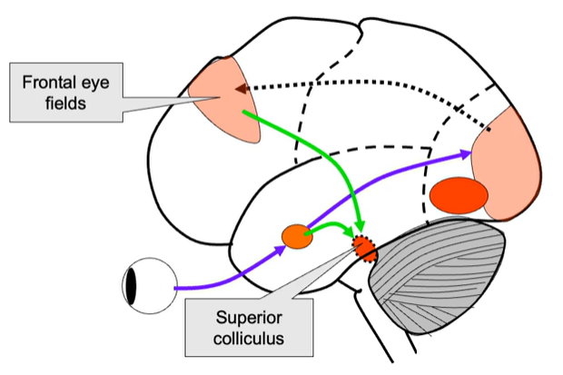
Frontal Eye Fields
it is a region in frontal lobe (a bit anterior so in front of PMC + much smaller)
basc its the equivalent of PMC for controlling eye movements
brain imaging found in monkeys - similar to humans
connects to occipital lobe + receives a lot of bottom-up input about visual surroundings
connects to prefrontal cortex (specifically in dorsolateral prefrontal cortex)
strong connection with superior sulcus (initiates eye movements)
can modulate activity in eye movement
antipsychotic task = classic task used to study control of eye movement
peripheral stimulus appears + tendency is to look towards = in this task you must do opposite (look away)
Secondary motor areas
Supplementary motor area (SMA)
Pre-motor
Dorsal PM (PMd)
Ventral PM (PMv)
Posterior parietal cortex
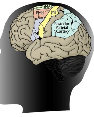
Secondary Areas
Very dense connections between sec. motor areas
Heavily connected to primary cortex = leading to execution of actions
SMA + PMC more involved in planning movements
Brain imaging shows activation when imagining / planning sequence of movements - even if no action is performed
Posterior Parietal Cortex (PPC)
Links frontal cortex (decision-making) with premotor (planning) areas.
Receives info from sensory regions (visual, sensorimotor cortexes)
Important for determining potential actions/goals given the environment (e.g. pick up coffee, continue working)
Frontal cortex more critical = decision about which action to perform + secondary area = develop plan for that actions
Supplementray Motor Area (SMA)
SMA now considered to be 2 areas:
SMA proper = learning
Pre-SMA = execution
Postural stability
Planning + executing complex sequential movements
e.g. Parkinson’s patients usually have decreased activity in sequencing tasks
Initiation of internally generated movements (rather than stimulus driven)
Dorsal premotor (PMd)
Important in preparation of movement
Learning conditional actions (response to external cues/environmental signals)
Red traffic light = foot on brake
green = foot on gas
set related activity e.g. ready, set, go
Ventral premotor (PMv)
Important for sensory guidance of movement - responsive to tactile, visual + auditory stimuli
Visuomotor control during grasping
Mirror neurons
PMv and Mirror Neurons
First reported in ventral premotor cortex (PMv)
MNs show similar activity when monkey makes a goal directed action + when the same action is observed or heard
Thought to be important for learning through observation
Also for understanding other people’s intentions
Neuroplasticity
The ability of the brain to form + reorganise synaptic connections - esp in response to learning following injury
This can occur in all areas of brain - very clear examples in PMC + motor sensory cortex
Very pronounced in childhood + decreases as you get older
Changes in the somatotopic map
Sensory remapping = rapid changes in somatosensory (motor maps) evident after change in inputs training (new skill)
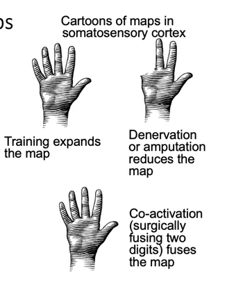
neuroplasticity: example from co-activation
fusion from a lab that uses fMRI-
simple tasks - asked to do things with fingers except thumb
see where in the brain gets recruited
looked at distance in brain space between these hand areas
then index and middle finger glues together for 24 hrs = found changes in motor maps (became more similar)
neuroplasticity: example from amputation
hand amputation
amputees had similar maps to controls = no evidence of reorganisation of adjacent motor areas
people born with only one hand = evidence that some of the face region starts to invade the hand area
Changes in maps reflect neuroplasticity
Long term changes in function connectivity e.g. growth of neurons
Branching (or pruning) of dendritic connections
Neurons appear to ‘compete’ for space in cortex = unused cortex gets overtaken by other inputs
Imaging of the living mouse brain shows changes (growth + pruning) in dendritic branches within hours / days of new task
Learning-based neural changes: synapse efficacy
Synapse enables one neuron to communicate with another
Pre-synaptic:
Increase vesicle volume
Increase availability of vesicles
Increase release probability
Synaptic cleft:
Reduce re-uptake mechanisms
Reduce gap dimenstions
Post-synaptic:
Increase receptor density / area
Growth:
Make new synapses
Long-term synaptic plasticity
Specific times patterns of neuronal activity can = long-term synaptic changes:
Long-term potentiation (LTP) =
is an activity-dependent persistent strengthening of synapses
these produce a long-lasting increase in signal transmission between 2 neurons
Long-term depression (LTD) =
is an activity-dependent reduction in the efficacy of neuronal synapses
these produce long-lasting deceases in signal transmission between 2 neurons
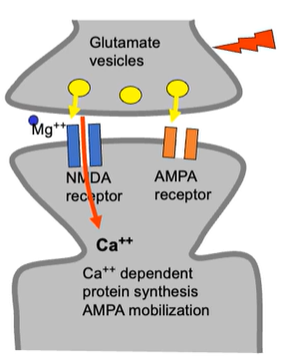
Associative LTP induction
NMDA channel is normally blocked by MG++
Concurrent voltage change to drive out Mg
achieved by glutamate binding to nearby AMPA receptors
equivalent to stimulation with high freq electrical pulses
Glutamate binds to NMDA + AMPA receptors
temporary change in shape of channel = opens up channel
calcium can enter through the open, unblocked NMDA channel
Ca++ entry triggers intra-cellular signalling cascade which results in
migration of AMPA receptors from intracellular stores to the cell membrane
synthesis of more AMPA receptors
Key principles of LTP
Cooperatively:
LTP requires simultaneous activation of large number of axons (due to large depolarisation)
Associative:
When weak synaptic input is paired with strong + long depolarisation = can propagate + cause LTP at synapse with weak input
If 2 neurons fire together (1 weak + 1 strong) then the strong one will allow the weak one to become stronger over time
Synapse specific:
if particular synapse is not activated then LTP will not occur even with strong post synaptic depolarisation
Long Term Depression (LTD)
First identified in hippocampus - thought to be a major component of motor learning in the cerebellum
Cerebellar LTD involves a decrease in AMPA receptors - However, this is NOT NMDA-dependent
Measuring in humans: Transcranial Magnetic Stimulation
A non-invasive method of measuring neuroplasticity
Apply coil on scalp + deliver a brief pulse = causes electrical stimulation of underlying neuron
Cells in PMC = cause involuntary muscle contractions
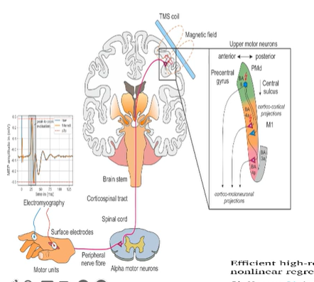
Measuring the somatotopic map with TMS
if you record muscle activity in the middle, index then little finger = you will find spots where simulation will induce activity