BIOL 240 - Midterm 1
1/121
Earn XP
Description and Tags
Topics 1 - 4
Name | Mastery | Learn | Test | Matching | Spaced | Call with Kai |
|---|
No analytics yet
Send a link to your students to track their progress
122 Terms
Robert Hooke’s vs. Antoine van Leeuwenhoek?
Hook was the first to describe fungi.
Leeuwenhoek was the first to provide a written discription of bacteria.
What are the core features of life? How are they achieved?
Metabolism, Growth, & Reproduction
Achieved through genetic variation/evolution, response/adaptation, & homeostasis.
Heterotroph vs autotroph.
Give an example of how microbes get energy and how they can help in biogeochemical cycling.
Heterotrophs ingest organic molecules to get energy, & Autotrophs produce organic molecules.
Ex. organic molecules are broken down by microbes to harness chemical energy (ATP) through fermentation & aerobic respiration. Can help in biogeochemical cycling as they interact with the environment by cycling inorganic to organic molecules and back.
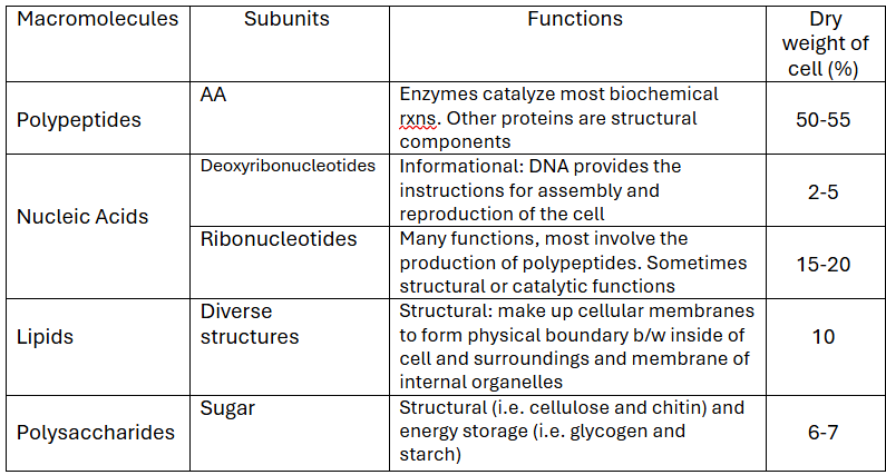
Which macromolecule is more in the cell? Which is the least?
What is the phylogenetic tree divided into?
Who was it made by?
What is it based on?
The phylogenetic tree is divided into Bacteria, Archaea, & Eukarya. It is based on the ribosomal RNA sequences.
The Archaea are more closely related to the Eukarya
It was created by Carl Woese.
What features make SSU rRNA gene sequences ideal for phylogenetic (or evolutionary history) studies?
Compare protein sequences to reveal evolutionary relationships. The more similar the AA sequence = the more related the organisms are. rRNAs have a universal presence in all cells so they can be used to easily compare.
Specifically, SSU rRNA are “molecule chronometer” as their sequences change very slowly b/c of the functional constrains on the molecule. Random mutations are dangerous, so it does not change when passed to further generations.
Founded by Woese
Are viruses considered alive? Why?
Not considered alive as they cannot replicate outside the host cell, have little biochemical activity outside a host cell, and are inert and nonreactive outside the host cell. ARE NOT MADE OF CELLS
What were the early Earth conditions?
Harsh with little O2 in the atmosphere
High temps
Significant amounts of H2O, CH4, HCN, CO2, N2, H2, NH3
Planet surface was a chemical soup w/ a reducing atmosphere
Energy input from various sources, including UV light.
UV irradiation of gaseous mixtures resembling Earth’s early atmosphere can result in the formation of organic macromolecules
What was Miller and Urey’s experiment?
To stimulate the spark that might have started forming organic molecules from the primordial soap. Added their own gasses like CH4, NH3, H2O, H2.
What were the requirements of early life? Give 2 examples that we can see this in.
Genetic info storage, the ability to catalyze biochemical rxns & a way of separating the cell interior from the external environment.
This can be seen in ribozymes (RNA behaving like enzymes) as they catalyze rxns, store genetic info, and can self-replicate & micelles as they may have been an early form of plasma membranes, with a polar head and non-polar tails, creating a bilayer.
The RNA World
Was LUCA part of this world?
The RNA world is a theory of about the origin of life, containing only RNA and lipids to which contain all the basic requirements for life.
LUCA was formed after the RNA world by using the DNA → RNA → protein dogma. It was the many failures & instability that eventually led to LUCA
What are the features of LUCA
Using ATP as chemical energy
DNA to RNA to protein
eating CO2 & fixing N2
Anaerobe (no need for O2)
Thermophile (high temp lover).
Autotroph and H2 oxidizer.
As oxygen was scarce, how could we circumvent the readily oxidized electron donors away from hydrothermal vents? How is this known?
Ancestors of cyanobacteria were able to solve this problem by extracting the e- from the water and creating O2 as a toxic byproduct - oxygenic photosynthesis.
This is known because multicellular fossils from billions of years have been found fossilized into stromatolites due to precipitating carbonate.
How did Earth evolve into so many microbes & microorganisms?
It was the physiology of the existing microorganisms that created the environmental conditions conducive to the rise of multicellular organisms, which then evolved from microorganisms.
What was the early coding material? How did it become to this?
Early coding material was RNA.
Coding material developed the ability to direct peptide synthesis resulting in catalytic activity, which replaced RNA.
The genomic function of RNA → DNA.
What are the 2 different perspectives for examining microbial genomes?
Examining effects of single mutations in DNA individually (microbial genetics)
Studying and comparing pieces of genomes to each other across domains (phylogeny)
a. this makes it like a cobweb of intertwining connections b/w generations
What is the endosymbiotic theory? What is the evidence?
Explains how ancestral archaeal organisms swallowed aerobic bacterial organisms to for a symbiotic relationship through protection and a steady supply of nutrients. Created mitochondrion & chloroplasts.
Evidence is that eukaryotic organisms w/o organelles are near the root of tree & rRNA of mitochondria and chloroplasts are similar to bacterial rRNA than eukaryotic.
Also resumble: cell division w/ FtsZ & circular chromosome
What did Pasteur do?
living organisms discriminate b/w optical isomers
explained biological nature of alcohol fermentation by extending shelf life - pasteurization
vaccines for anthrax, fowl cholera, and rabies
introduced sanitization in hospitals
molecules have chirality
disprove spontaneous generation theory
What did Koch do?
determined Bacilus anthracic was the cause of anthrax & mycobacterium was the cause of tuberculosis
established Koch’s postulates
What are the Koch’s postulates?
What do they prove?
Koch’s postulates can be used to show a specific microbe causes a specific disease. The cause and effect are proven if:
The suspected microbe is identified in every person with the disease, but not those that are healthy
Isolate the suspected microbe/pathogen in a pure culture
Take the pure culture, inculate it into the animals and show that they develop the disease
Get the pathogen that was experimentally inoculated into the animal to prove that the pathogen is the cause of the disease
NOTE: not always possible to apply his postulates
Bacteria shape
spherical = coccus
rod-shaped = bacillus
comma-shaped
spiral
pleiomorphic (varied shapes)
Note: shape is not a good predictor of physiology, ecology, or phylogeny
Morphology may be determined by what selective forces?
Efficient nutrient uptake (surface to volume ratio)
Spirals allow efficient swimming in viscous or turbulent fluids (i.e. near surfaces)
Gliding motility (filaments)
What are the usual sizes of cells?
Prokaryotes are 0.2 - 700 um in length/ diameter.
Rod-shaped bacteria b/w 0.5 um - 4.9 um wide and 1-15 um long.
The min size is simply due to minimum space requirements for genome, proteins, and ribosomes.
What are exceptions to the general size of bacterial cells.
Thiomargarita namibiensis - up to 700 um
Namibiensis species wraps around a vacuole containing water (and nitrate)
Epulopiscium fishelsoni - up to 600 um
Have larger genomes
Thiomargarita magnifica - > 9000 um
magnifica is a hollow tube
Note: Thiomargarita have high SA-V ratios because of a thin cytoplasm layer, despite a very large size.
What are the advantages of being small?
How about being big?
Being small allows for high SA-V ratio, the greater rate of nutrient/ waste exchange per unit volume, supports higher metabolic rate, and supports faster growth rate and faster evolution. Good size for low-nutrient environments.
Being big is favorable in high-nutrient environments as it can save up food (in inclusion bodies) and have space for important structures (i.e. ribosomes)
What are the limits of being small?
Can only contain few ribosomes and proteins but can’t do a lot of things. Most are parasitic and rely on the host to live.
What is the largest area inside the bacteria?
The nucleoid region is the largest area. It houses chromosomes and DNA replication machinery.
It DOES NOT contain a membrane surrounding the nucleoid and no histone proteins.
How and why is DNA compressed in the bacteria.
DNA repels one another due to its structure and charge. It reduces space by using small, positively charged proteins to bind to the phosphate backbone to maintain the condensed structure.
Topoisomerases modify the structure of DNA to enable supercoiling.
What is inside the cytoplasm of a bacterium?
It contains macromolecules (i.e. tRNA, rRNA, etc.), inclusion bodies & microcompartments
Sulfur globules: sulfur storage for energy - IB
PHB granules & Glycogen: carbon storage - IB
Gas vesicle: buoyancy control
Carboxysomes: location of carbon fixation rxns (RUBISCO)
Magnetosomes: only found in bacterial organelles associated w/ direction finding
What are magnetosomes?
They are only found in bacterial organelles and is a membrane enclosed structure w/ a cell wall. It acts as a compass that points towards the north pole, which is to the ground, as there is low O2 w/ lots of nutrients.
What is the bacterial cytoskeleton?
What is it made out of?
It is a series of internal proteins that assist in keeping everything in the right place at the right time. Some cytoskeleton proteins are involved in cell wall synthesis during cell division:
MreB provides structure (homolog of actin - microfilaments)
Not found in Staphylococcus
FtsZ aids & organizes the cell division (homolog of tubulin - microtubules)
The ring around a dividing cell is a Z ring & made out of FtsZ
ParM proteins (homolog of actin) direct plasmids by pushing them into dividing cells
Chromosomes are pushed by another type of unknown homolog
PARTITION PROTEIN
Takes ATP to polymerize and de-polymerize these things
What does the bacterium's cell envelope include?
The cell envelope is all the layers surrounding the cytoplasm of a cell, including the cell membrane (plasma membrane), the cell wall, and the outer membrane (if present).
What is the role of the cytoplasmic membrane in Bacteria?
What is it made out of?
All cells have a plasma membrane that separates the interior from the external environment.
Captures energy: using ETC to create a PMF, which can be used for respiration, photosynthesis, and can drive energy for motion (flagella)
Sensory Systems are embedded proteins that can detect env. changes and alter gene expression
Permeability barrier, but not structural, as it is sensitive to sheer forces
The membrane has a hydrophobic core and hydrophilic surfaces. Modifying the fatty acids w/ double bonds can affect fluidity. It is attached through ester linkages.
No sterols for stability w/ high abundance of proteins.
What are hopanoids?
What do they do?
Hopanoids are sterol-like molecules that are found in some bacteria. They help w/ stability across temp ranges.
They are only found in bacteria.
Facilitated diffusion vs co-transport
Both are protein channels that move particles by using a [ ] gradient and do not use ATP.
Facilitated diffusion moves molecules DOWN the [ ] gradient & requires no ATP.
Co-transport is a form of active transport and can be symport or antiport: Symport moves 2 molecules in the same direction. Antiport drives 2 molecules AGAINST one another.
BOTH have one going along and one against [ ] gradient. Uses energy released from going from high to low [ ].
![<p>Both are protein channels that move particles by using a [ ] gradient and do not use ATP.</p><p>Facilitated diffusion moves molecules DOWN the [ ] gradient & requires no ATP.</p><p>Co-transport is a form of active transport and can be symport or antiport: <strong>Symport</strong> moves 2 molecules in the same direction. <strong>Antiport</strong> drives 2 molecules AGAINST one another.</p><p>BOTH have one going along and one against [ ] gradient. Uses energy released from going from high to low [ ].</p>](https://knowt-user-attachments.s3.amazonaws.com/9c44a4ac-4adb-46a8-81e0-43833428f6fe.png)
What is the ABC transporter?
ATP-Binding Cassette is a protein transporter that moves particles AGAINST a [ ] gradient & requires energy in the form of ATP.
Use a solute-binding protein and a channel complex
![<p>ATP-Binding Cassette is a protein transporter that moves particles AGAINST a [ ] gradient & requires energy in the form of ATP.</p><p><span>Use a solute-binding protein and a channel complex</span></p>](https://knowt-user-attachments.s3.amazonaws.com/952d0ae5-dc41-4f21-937c-f300588b635d.png)
What is protein secretion?
It ships proteins outside the cell using ATP. The protein is threaded through the membrane and folded outside of the cell.
Portein is synthesized → SecB prevents new protein from folding → secE, secG, and SecY form a channel → signal peptide is removed by peptidase → protein folds outside of the membrane
What is the cell wall of bacteria made out of?
What does it do?
It is made out of a thin sheet called peptidoglycan, found only in bacteria. Composed of a glycan backbone connected by peptide cross-links.
The cell wall gives cells their shape, protects them from osmotic lysis/mechanical forces, and forms a matrix of crosslinked strands of peptidoglycan subunits = polysaccharides connected to peptides.
Note: not a permeable barrier
What do peptidoglycan subunits include?
G+ vs G-
NAG attaches to NAM, which connects w/ a small peptide chain. NAM & NAG are sugars & B-1,4 linkage.
These crosslinks vary by species, as G+ have a pentaglycine inter-bridge, causing it to make a bigger cell wall. G- has DAP while G+ does not.
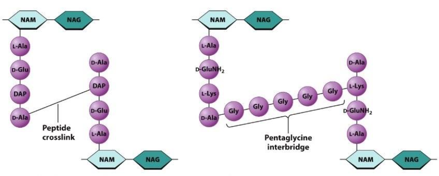
NAM & NAG & peptide chain
NAM & NAG have NHCOCH3! SUGARS!
Also contains a B-1,4 linkage.
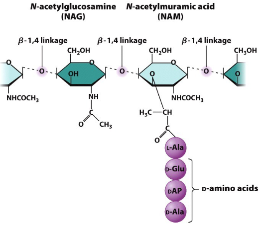
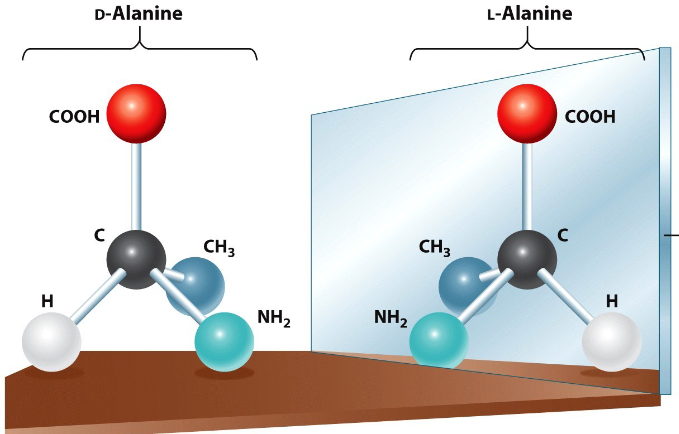
Explain NAM and the it’s 2 forms
Several of the AA associated w/ NAM in peptidoglycan are unusual D forms. D forms are stereoisomers (mirror images) of the L form normally found in biological proteins.
Bacterium cell walls include both D & L forms.
What are the 2 main enzymes associated w/ the bacterial cell wall?
What is its role?
What do they act on?
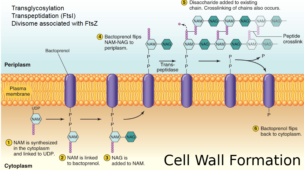
FtsI catalyzes transpeptidation (formation of peptide crosslinks), and transglycosylase catalyzes the joining of NAM and NAG together via 1,4 glycosidic bonds.
Both of these enzymes act on this Z ring b/c it forms the cell wall.
What happens specifically in transpeptidation?
NAM is synthesized in the cytoplasm and linked to UDP before being linked to bactoprenol. NAG is added to NAM before bactoprenol flips them into the periplasm. Transpeptidase cleaves the D-Ala (N-terminus) in NAM (5C → 4C) b/c its bond provides energy for cross-linking. Then, the disaccharide is added to the existing chain, and crosslinking of chains occurs.
How is the cell wall degraded in bacteria?
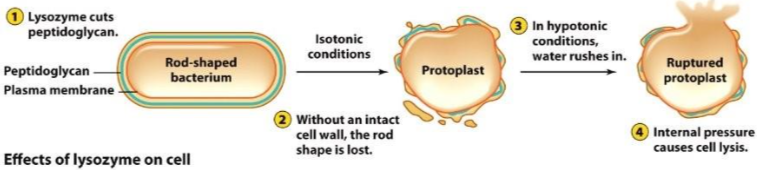
It is degraded by lysozyme secretions as it cleaves the backbones of peptidoglycans and affects glycosidic bonds.
Lysostaphin acts on the crossbridge of certain Staphylococcus species only & affects the pentaglycine in G+ cells.
What happens when a bacteria cell loses its cell wall?
The cell cannot resist osmotic pressure changes and will end up rupturing, but pushing the cytoplasm out of the cell.
Osmosis can cause a cell to swell w/ water or shrivel as water leaves.
What do antibiotics do? What do they target?
What is antibiotic resistance? What does it do?
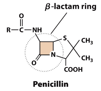
β-lactam antibiotics prevent peptidoglycan crosslinking by inhibiting FtsI transpeptidation.
Antibiotic resistance is when some bacteria produce an enzyme (i.e. carbapenem) to destroy the critical β-lactam ring structure. A second drug must be added to inhibit the enzyme.
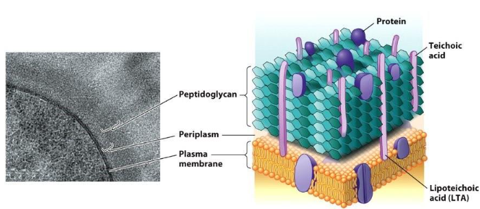
What are G+ cell membranes made of?
G+ cells have a thick outer layer of peptidoglycan with a narrow periplasmic space. It has negatively charged teichoic acids (polysaccharides) embedded into the peptidoglycan. Those attached to the membrane are lipoteichoic acid (LTA), which anchors the cell wall into the membrane.
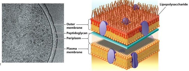
What are G- cell membranes made of?
What is it’s outer membrane made of?
G- cells have a more complex cell envelope as it includes an outer membrane exterior to the thin peptidoglycan layer. This space b/w them includes the periplasm.
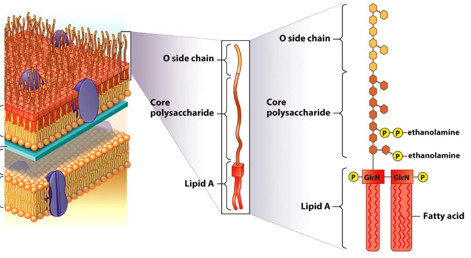
The outer membrane consists of a phospholipid bilayer w/ lipopolysaccharide (LPS) & have DAP. Direct peptide links.
How is the LPS in G- cell harmful?
Lipid A is immunogenic, meaning it can initiate an immune response in the body upon antibiotic treatment.
The O side chain can be changed by the bacterium and these changes can cause different effects/reactions during infection which can be harmful.
How does nutrients get into G+ & G- cells?
G+ have peptidoglycan layers w/ large pores so it can go inside the cell.
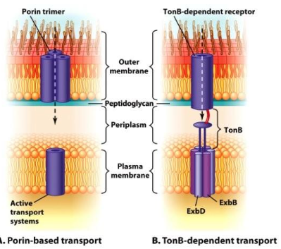
G- cells have porins & TonB proteins on the outer membrane. Porin transfers molecules into the periplasmic space, PASSIVE.
TonB receptors has a TonB-dependent receptor on outer membrane. it has a high affinity to grab nutrients and sends signal to TonB. TonB uses a PMF to undergo confirmational change which pulls nutrient down the receptor and into cell. ACTIVE.
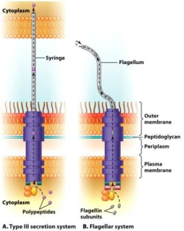
How do molecules leave the G- cell?
Some move from the periplasm to the outside directly, by autotransporters (rare), while some use single-step transport systems (never entering the periplasm).
What are the 2 single-step transport systems?
Type II secretion passes polypeptides through a syringe to the outside of the cell.
The flagellar system secretes flagellin subunits using the flagellum. These subunits are then used to build the flagellum from the tip up.
What are the 4 types of organelles found on the bacterial cell surface?
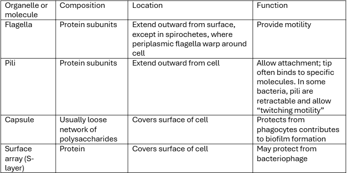
What are flagella?
What are it’s polar arrangements and what are not?
Spiral, hollow, rigid filaments extending from the cell surface. Location and # vary across species w/ protein anchors for flagella in the cell membrane, cell wall, and outer membrane.
3 polar arrangements:
Monotrichous: 1 hair
Amphitrichous: 2 hairs (at each end)
Lophotrichous: many flagella at the end of the cell
Peritrichous: multiple flagella spread over the surface of the cell - not polar arrangement
What are the zones of stability of a flagella in a G- cell.
Filament of multiple flagellin proteins
Hook protein portion connects filament to the basal body
Basal bodies are disk-like structures that turn filaments like a propeller
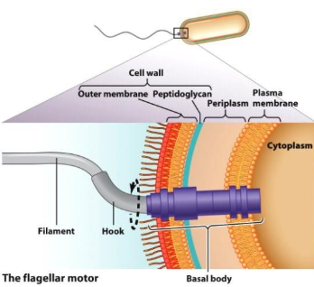
How do flagella move?
How about non-flagellar motility?
The energy to spin a flagellum derived from PMF is expensive.
It is a complex structure, and the spinning creates runs and tumbles w/ no steering. It moves through random orientations of turning and tumbling before it knows it goes to the right way.
Non-flagellar motility has gliding motility to slide over the surface of the cell & twitching motility slow, jerky process using pili that extend, attach to, and pull along a surface.
What is the chemical movement called?
What causes this to occur?
Chemical movement is chemotaxis. Chemoreceptor proteins temporally sense changes in the [ ] of attractants or repellents.
A random walk has alternative runs and tumbles resulting in no direction. Positive chemotaxis is prolonged runs that move a bacterium TOWARDS a chemical. Negative chemotaxis is prolonged runs that move a bacterium AWAY from a chemical.
This occurs as flagella.
What is phototaxis, aerotaxis, and osmotaxis?
Phototaxis is light. Aerotaxis is O2. Osmotaxis is osmotic strength.
What are adherence molecules?
They are located outside to cell which allow cells to stick to surface. Pili, fibers of pilin protein (subunit), possess other proteins on their tips for sticking.
Function: conjugation, attachment to host cell, motility.
What is stalk?
Stalk is when some microbes will use an extension of the cell envelope, tipped by a holdfast of polysaccharides.
Proves extra SA-V for nutrient absorption & contains cytoplasm.
What are Capsules?
Capsules are thick layers of polysaccharides surrounding some cells, in both G+ and G-.
It provides adhesion, defense against host immunity, & protection against desiccation. It can help bacteria from biofilms which provide protection and enhanced survivability in harsh environments.
What are Surface arrays (S-layers)
They have crystalline array of interlocking proteins. It can protect a cell against predation or infection w/ bacteriophages. Found in G+ and G- & can sometimes be the only thing archaea have.
How are microbes named?
What are the types of classification?
Named according to the standard binomial system:
Species: group of strains sharing common features, while differing considerably from other strains
Genus: group of closely related species
Phylum, class, order, family, genus, species.
What do classifications depend on?
Classifications depends on many features: DNA sequence data, size/ shape, Gram types, colony morphology, presence of structures such as capsules or endospores, physiological/ metabolic traits
What are endospores?
Why is it limited to specific phylogenetic groups?
Endospores are inside vegetative cells. They have thickened cell envelopes w/ more proteins covering the peptidoglycan layer, making them resistant to heat & other unfavorable conditions.
Spores are dormant for a long-time w/ no metabolism and compress chromosomal DNA using protective proteins.
Hundreds of genes are involved in making spores, which explains why the trait is limited to very specific phylogenetic groups. It is common in soils and germination occurs very rapidly when conditions improve.
Who found the archaea?
Woese and Fox were the ones that found archaea (or archaebacteria) through comparisons of rRNA gene sequences from methanogens.
What is the size & shape of archaea?
size: 0.5 - 5 um
Shapes are similar to bacteria: rods, cocci, spirals, squares, etc.
What is the cytoplasm of archaea?
What does the nucleoid include?
It is similar to bacteria, which include microcompartments & inclusion bodies.
They have single circular chromosomes and lack a membrane-bound nucleus. Many of the DNA replication enzymes look like those of Eukarya.
Histones in Archaea
The development of histones may have been an early branch point event in the evolution for Archaea and Eukarya, and histones are protein structures that DNA wraps around to form nucleosomes, but it is different in Archaea and Eukarya.
Eukarya: 160-nucleotide-pair length of DNA w/ octamer of histone proteins
Archaea: 60-nucleotide-pair length of DNA w/ teramer of histone proteins
The charge for the histone proteins are +.
What is the cytoskeleton in Archaea?
Cytoskeletal homologues are found in both domains. Comparing MreB and actin, some of the sequences look more like eukaryotic actin while others seem more similar to MreB. THEREFORE, it is not sure which Archaea looks more like when looking at the cytoskeleton.
What is the cell envelope of an Archaea?
All poses a plasma membrane & most have a cell wall but most do not have an outer membrane (making it look like G+ rather than G-)
These structures are diff from their equivalents in the other domains.
What is the plasma membrane of the Archaea made of?
A unique bilayer construction containing glycerol-1-phosphate, phytanyl side chains (branched isoprene units), & ether linkages, which makes it more stable and can resist high temp conditions.
Do not have sterols for membrane stability, but have a high abundance of proteins.
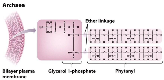
Why are the isoprene phytanyl side chains more stable that the fatty acid chains?
FA: Branching reduces the flexibility of the lipid chain, leading to a more rigid and stable membrane structure.
Isoprene: the chains pack more tightly cause can der Waals forces to be more effective which is good for high temp stability.
What can form from Archaea cell membranes.
The bilayer can turn into a monolayer by having 2 phytanyl covalently linked in the middle, forming tetra-ether lipids.
The monolayer is really resistant and does not break easily which is good for STABILITY. There are phosphoglycerol molecules on both ends and this is very stable, often seen in archaea living in high temps.
Also have membrane fluidity b/c Archaea have rings
What is an Ignicoccus?
Ignicoccus is an exception to all rules for archaea. It has an outer membrane and periplasm similar to the arrangement in G- cells. ATP synthase enzymes are housed in the outer membrane.
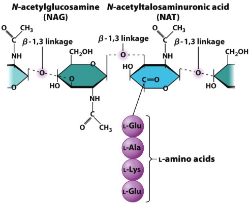
What is the cell wall made of in Archaea?
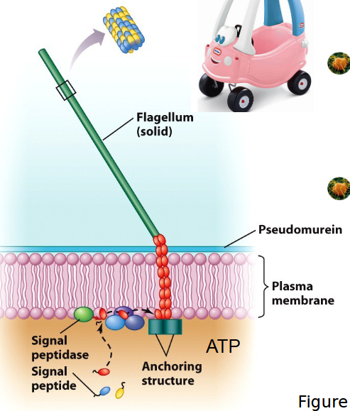
Pseudomurein (pseudo-peptidoglycogan) – polysaccharides similar to peptidoglycan - NAT, NAG, & B-1,3 linkages that are lysozyme insensitive.
Contains only L amino acids & NAT can create create pseudo-peptidoglycogan cross-links
What does the surface of an Archaea cell contain?
S-layers protect against predation and viruses, and mediate adhesion, while cannulae are hollow glycoprotein tubes that link cells together to form a complete network.
Flagellum vs Archaellum
Archaellum is an Archaea flagella that grows from the base rather than the tip and uses ATP rather than a PMF like in bacteria flagella.
What are the 4 major phyla in Archaea?
Euryarchaeota
Crenarchaeota
Thaumarchaeota
Nanoarchaeota.
But many other phyla have been proposed.
What are Crenarchaeota?
Creanarchaeota are thermophiles or hyperthermophiles (high temp lover). Can be acidophiles (thrive in low pH) or barophiles (thrive in high pressures).
They have a lipid monolayers w/ modified proteins (more A/T & less C/S), strong chaperone protein complexes, thermostable DNA-binding proteins & reverse DNA gyrase enzyme to increase DNA supercoiling.
What are Halophiles?
What is an example of a halophile?
Which phyla do they belong to?
A type of Euryarchaeota that lives in high salt environment & requires a high NaCl [ ] of over 1.5M. An example is Halobacterium.
But b/c they need that much Na+, it is possible that too much of it in the cytoplasm will cause osmotic shock so it pumps K+ into the cytoplasm to live in an isotonic environment. A high intracellular [K+] offset a high extracellular [Na+].
K+ is a compatable solute b/c it has a bigger ionic radium & does not grab onto water like Na+ does
What is the consequence for having too high of a [K+] in Halobacterium?
How is this solved?
Too high of an intracellular K+ [ ] can cause denaturing of proteins and split dsDNA.
DNA denaturing → higher GC contents (stronger bonds)
Protein denaturing → highly acidic proteins that remain more stable in high salt environments.
How do Halobacterium get energy?
Halobacterium pumps proteins across the membrane using light energy, creating a PMF that creates ATP = phototrophic.
Bacteriorhodopsin is a protein pump with a retinal pigment that is used to capture light energy and gives off the red color. Can adapt to low O2 conditions by generating a PMF.
What are Methanogens?
Where are they found/produced?
It is a type of Euryarchaeota that carries out redox rxns by reducing CO2 and H2 to produce CH4 & H2O. The energy released can be used to fix C.
Strict anaerobes
Found in anoxic (no O2) sediments and environments
The methane produced forms gas in humans & combusitble air in swamps - shown in the volta experiment.
H2 + CO2 = CH4 → hydrogenoclastic
acetate = CH4 → acetoclastic
methanol = CH4 → methylotrophic
What is the volta experiment?
Captured methane in bottle by disturbing the sediment in swamps than inverted it and it ignites into a flame.
What does the TACK superphylum contain?
Thaumarhcaeota
Aigarchaeota
Crenarchaeota
Korarchaeota
What are Thaumarchaeota/ Nitrososphaerota?
It separates phylum for many mesophilic crenarchaeota. They are ammonium-oxidizing & abundant in oceans and soil.
Mesophiles: 15 - 40 °C
Psychrophile < 15
Are important for the biogeochemical cycling of C and N in the ocean.
What are the emerging phyla?
Korarchaeota w/ distinct 16S rRNA sequences obtained from hydrothermal environments. There are no species that have been cultivated and only one genome is available.
Aigarchaeota also have no cultivated species and have one genome available – thermophile.
What is DPANN?
What are its common features?
A superphylum of backpack organism → small cells growing in association to something else, ultrasmall archaea.
Very small cell sizes (< 1 um), small genomes, restricted metabolism – unable to generate basic building blocks, interspecies interactions, and can be mutualistic or parasitic.
What is Nanoarchaeota?
What is its relationship w/ Ignicoccus?
Key member of DPANN. Nanoarchaeum equitans is the only isolated member so far w/ 2 other genomes available. They have distinct 16S rRNA gene sequences.
Ignicoccus and Nanoarchaum grow together in hyperthermal vents and the nanoarchaeum is the obligate parasite of Ignicoccus. It is 0.4 um w/ a 0.49 Mbp genome, which has no metabolic genes and only carries genes for replication, transcription and translation. The nanoarchaeum is dependent on the host for everything except replication.
How do backpack organisms benefit the host and vice versa?
B/c of their high SA-V ratio, they have an inflex of nutrients which is a viable source & can be protected from predators.
What are the Asgard superphylum?
What do they include?
It is a proposed superphylum consisting of a group of uncultivated Archaea. It includes Lokiarchaeota, Thorarchaeota, Odinarchaeota, Heimdollarchaeota.
They represent the closet prokaryotic relatives of eukaryotes.
Within Likarchaeota/ Thorarchaeota, they are thermophilic archaea that are distinct from Crenarcheota. Grouped w/ eukaryotes on some phylogenis b/c genomes show eukaryote-like proteins for cell compartmentalization.
What is the morphology of a typical eukaryote?
Membrane-bound nucleus, larger than bacterial & archaeal cells, contain membrane-bounded organelle, and possess a cell wall w/ a complex internal cytoskeleton.
Which organelles are double-membrane bound?
What do they do?
Nucleus contains pores& has an outer membrane that is continuous w/ the ER.
Mitochondria contain DNA, ribosomes, & can independently replicate.
Chloroplasts conain DNA, independent replication, & are unique to photosynthetic organisms.
Hydrogenosomes produce H2 & ATP. Found in some amitochondriate and cells w/o mitochondria. Have similar genes, remnants, & metabolism to mitochondria.
What does the nucleus do?
What do they hold?
Plays a role in the storage and expression of info, has a double membrane, and contains big, linear chromosomes. A non-membrane-bound nucleolus is responsible for ribosome synthesis.
It has spatial separation as transcription occurs in the nucleus and translation occurs in the cytoplasm.
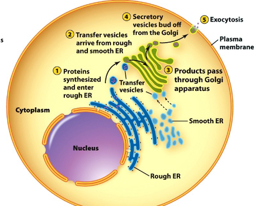
What is the secretory pathway?
A series of multiple organelles that direct newly made proteins to their proper destinations. Made up of the ER & Golgi apparatus.
What is the mitochondria?
Plays a role in cell metabolism as the Krebs cycle occurs. It uses ETC to produce ATP (chemiosmosis via PMF).
What are chloroplasts?
It plays a role in cell membrane as it contains ETC to produce ATP (chemiosmosis via PMF) and uses the ATP produced to fix C into organic compounds.
Thylakoids are membrane bound sacks w/ pigments that capture energy.
What organelles are semi-autonomous?
Mitochondria and chloroplasts are semi-autonomous. Each has DNA genomes, ribosomes, and transcription machinery. They can replicate independently of the rest of the cell. But b/c most of their proteins originate from the DNA in the nucleus of the cell, there is a mutualistic relationship b/w the cell and these 2 organelles.
What is the plasma membrane made of in the eukaryote?
The plasma membrane is a phospholipid bilayer w/ embedded proteins that allow molecule transport through facilitated diffusion and active transport.
It plays a role in homeostasis & contains sterols for membrane stability.
Made of a low abundance of proteins with the lipid structure (fluidity) of ester linkages & straight fatty acid chains.