M1(1) The Sensory System (Taste)
1/56
There's no tags or description
Looks like no tags are added yet.
Name | Mastery | Learn | Test | Matching | Spaced | Call with Kai |
|---|
No study sessions yet.
57 Terms
Sensory System
the part of the nervous system that detects internal and external stimuli through sensory receptors
converts them into neural signals
processes this information so the body can perceive and respond to its environment.
Sensory System Steps
Stimulus (e.g. flower scent)
Stimulation of a sensory receptor
Generation of nerve signals
Travel of signals
Interpretation of the signals in the brain
Perceiving of the stimulus (e.g. flower scent)
General Senses
Sensory systems that detect basic physical stimuli from many parts of the body rather than being limited to one specialized organ
Touch
Pain
Temperature
Proprioception
Crude pressure
Two Ways to Classify Receptors
Receptors may be classified based on
Type of stimulus
Function / location
Type of Stimulus
Includes:
Mechanoreceptors
Thermoreceptors
Nociceptors
Chemoreceptors
Mechanoreceptors
Respond to changes in pressure or body movement
e.g. pressure receptors in skin
Theremoreceptors
Respond to changes in temperature
e.g. those in skin and internal organs
Nociceptors
Responds to damage (pain) or oxygen deprivation to tissues
e.g. those in skin
Chemoreceptors
Responds to changes in the chemical concentration of substances
e.g. taste buds, olfactory receptors
Photoreceptors
Responds to changes in light energy
Located only in eye
Location and Function
Includes
Proprioceptors
Cutaneous receptors
Pain receptors
Proprioceptors
Mechanoreceptors involved in reflex actions that maintain muscle tone
helps us know the position of our limbs in space
e.g. those that maintain body’s equilibrium and posture
Proprioceptor Types
Two forms:
Muscle spindles
Golgi tendon organs
Muscle Spindles
A type of proprioceptor that detects change in muscle length and speed of stretching
“Over-stretching of muscle”
Parallel to muscle fibers (inside muscle belly)
Knee-Jerk Reflex
Tapping of patellar tendon stretches quadriceps muscle
Muscle spindle detects stretching of muscle
Generation of action potential in associated sensory nerve (afferent)
In grey matter of spinal cord, sensory neuron synapses with motor neuron (efferent)
Motor signal to muscle
Muscle contraction
Golgi Tendon Organ
A type of proprioceptor that detects changes in muscle tension/force
“Over-contraction of muscle”
Located in tendon
Cutaneous Receptors
Sensory receptors located in the skin that detect touch / pressure
Fine Touch Cutaneous Receptors
Pressure Cutaneous Receptors
Fine Touch Cutaneous Receptors
Meissner Corpuscles
Merkel Disks
Root Hair Plexus
Meissner Corpuscles
Cutaneous receptor sensitive to fine touch
concentrated in the dermal papillary layer of hairless skin
Merkel Disks
Cutaneous receptor sensitive to fine touch
found in the deepest epidermal layer
Root Hair Plexus
Cutaneous receptor sensitive to fine touch
winds around the base of a hair follicle
Pressure Cutaneous Receptors
Pacinian Corpuscles
Ruffini Endings
Krause End Bulbs
Pacinian Corpuscles
Cutaneous receptor sensitive to pressure
lie deep inside dermis
Ruffini Endings
Cutaneous receptor sensitive to pressure
found in the dermis and hypodermis
Krause End Bulbs
Cutaneous receptor sensitive to pressure
located in the superficial layers of the dermis
Pain Receptors
Also known as nociceptors
Located in skin and many internal organs
Pain Receptor Types
There are two types:
Somatic Nociceptors
Visceral Nociceptors
Somatic Nociceptors
Nociceptors sensitive to mechanical, thermal, electrical, or chemical damage
e.g. those in the skin and skeletal muscles
Visceral Nociceptors
Nociceptors sensitive to excessive stretching of internal organs, oxygen deprivation, or chemicals released by damaged tissues’
e.g. the pain sensation when stomach is too full
e.g. crushing pain of a heart attack when blood supply to heart is reduced
Referred Pain
Pain that is felt in a different area of the body from where it actually originates
Happens because some somatic nociceptors converge along the same nervous pathway
Brain cannot distinguish the two
Referred Pain Example
Pain from the heart that occurs during a heart attack is often accompanied by referred pain the the left shoulder and arm
Receptor Potential
System by which sensory receptors start signal transmission
A small, local change in the electrical charge of a sensory receptor’s membrane in response to a stimulus
If strong enough, can trigger an action potential in the sensory neuron
Receptor Potential Mechanism
Begins with a stimulus (e.g. light for receptors in the eye)
Can be weak or strong (unlike action potentials that act on the all-or-nothing principle)
Can add together
Part of neurons or synapse with neurons that can create action potentials
Taste
What: gustatory epithelial cells
Stimulus: molecules of the food we eat
Where: taste buds
Taste Buds
The overall sensory unit for taste
Contains gustatory epithelial cells and supporting cells (e.g. basal epithelial cells)
Gustatory Epithelial Cell
The receptor cell for taste
Gustatory Epithelial Cell Structure
Gustatory hairs (microvilli) extend through taste pores into oral cavity
bathed by saliva
Dendrites are coiled around the cell
Pathway to the brain
Gustatory Epithelial Cell Types
Type I Gustatory Epithelial Cell
Type II Gustatory Epithelial Cell
Type III Gustatory Epithelial Cell
Type I Gustatory Epithelial Cell
Function mainly as supporting cells.
Help regulate the taste bud environment.
Do not form traditional synapses and do not release classical neurotransmitters.
Type II Gustatory Epithelial Cell
Detect sweet, bitter, and umami tastes.
Lack synaptic vesicles and do not form conventional synapses.
Release ATP as a neurotransmitter through ion channels to activate sensory neurons.
Type III Gustatory Epithelial Cell
Detect primarily sour (and some salty) tastes.
Form traditional synapses with sensory nerve fibers.
Release the neurotransmitter serotonin
Basal Epithelial Cell
Act as stem cells, dividing and differentiating into new gustatory epithelial cells
Essential because taste receptors have a short lifespan (7-10 days)
Papillae
Small, raised projections on the surface of the tongue
Ripping and moving food during chewing

Fungiform Papillae
Mushroom-shaped.
Scattered over the entire surface of the tongue
Each papilla contains 1–5 taste buds.

Vallate Papill
Largest and least numerous papillae.
Contain many taste buds.
Typically 8–12 papillae arranged in an inverted V at the back of the tongue.

Foliate Papillae
Located on the lateral (side) edges of the tongue.
Contain many taste buds in childhood.
Decrease in number with age.
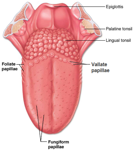
Filiform Papillae
Do not contain taste buds
Mainly help grip and move food
Covers dorsal side of the tongue
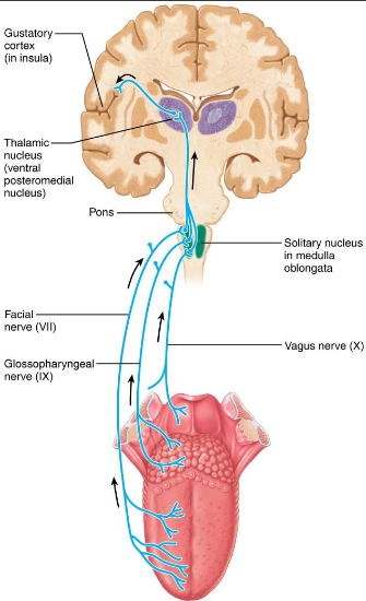
Gustatory Pathway Afferent Fibers
Chorda tympani (branch of Facial Nerve, VII)
Transmits impulses from taste receptors in the anterior 2/3rds of the tongue
Lingual branch (branch of Glossopharyngeal Nerve, IX)
Transmits impulses from taste receptors in the posterior 1/3rd of the tongue
Vagus nerve (X)
Transmits impulses from the few taste receptors in the epiglottis and lower pharynx

Gustatory Pathway Synapse
These afferent fibers synapse in the solitary nucleus of the medulla
Impulses stream to the thalamus and proceed to the gustatory cortex in the insula
Basic Taste Sensations
Sweet
Sour
Salty
Bitter
Umami
Sweet
Detected from many organic substances, including sugars, saccharin, alcohols, some amino acids.
Can also be triggered by certain lead salts (like in lead paint — toxic!).
Sour
Caused by acids, specifically the hydrogen ions (H⁺) in solution.
Salty
Caused by metal ions, mainly sodium ions.
Table salt (NaCl) is the most common salty taste.
Bitter
Detected from alkaloids such as quinine, caffeine, nicotine, morphine, and strychnine.
Also triggered by some non-alkaloid substances, e.g., aspirin.
Umami
Caused by amino acids like glutamate and aspartate.
Responsible for:
“Beef taste” in steak
Tangy flavor of aged cheese
Flavor of monosodium glutamate (MSG)
Perception of Taste
The brain does not rely on just one taste receptor.
It surveys the overall pattern of sensory input from all taste buds.
The perceived taste is like a “weighted average” of the signals from sweet, sour, salty, bitter, and umami receptors.
Possible Sixth Taste
Evidence suggests humans can detect long-chain fatty acids with taste receptors.
This may explain our preference for fatty foods.