Unit 3 Muscles OINF
1/52
There's no tags or description
Looks like no tags are added yet.
Name | Mastery | Learn | Test | Matching | Spaced | Call with Kai |
|---|
No analytics yet
Send a link to your students to track their progress
53 Terms
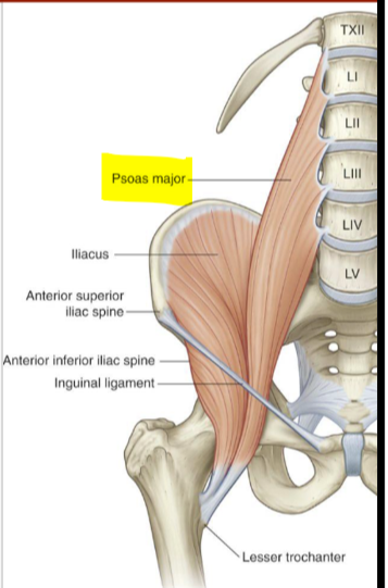
Psoas Major
Origin: vertebral bodies and intervertebral discs of T12-L5, transverse processes of lumbar vertebrae
Insertion: lesser trochanter of the femur
Innervation: anterior rami L1-L3
Function: Flexes the thigh at the hip joint
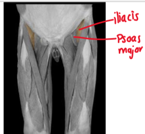
Iliacus
Origin: iliac crest, sacrum, superior 2/3 of the iliac fossa
Insertion: lesser trochanter of the femur
Innervation: femoral N
Function: flex the thigh at the hip joint
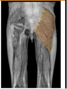
Gluteus Maximus
Origin: posterior side of the iliac crest, lateral edge of sacrum and coccyx
Insertion: gluteal tuberosity of the femur, iliotibial tract
Innervation: inferior gluteal N
function: laterally rotate and ABDuct the thigh, extend flexed femur at the hip joint, lateral stabilizer of hip and knee
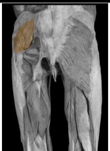
Gluteus medius
Origin: posterior side of the iliac blade
Insertion: greater trochanter of the femur
Innervation: superior gluteal N
Function: medially rotate and ABDuct the thigh at hip joint
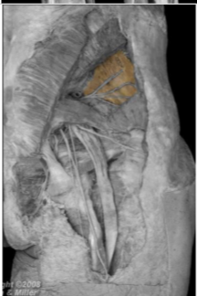
Gluteus minimus
Origin: posterior side of the iliac blade
Insertion: greater trochanter of the femur
Innervation: superior gluteal N
Function: medially rotate and ABDuct the thigh at hip joint
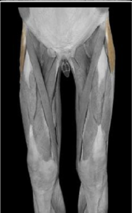
Tensor Fascia Latae
Origin: outer lip of iliac crest
Insert: lateral condyle of the tibia via the iliotibial tract
Innervation: superior gluteal N
Function: stabilize the knee in extension
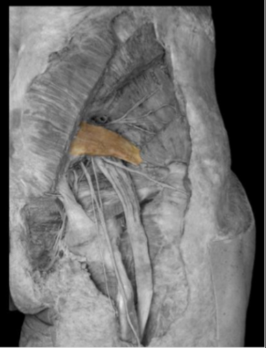
Piriformis
Origin: anterior surface of the sacrum
Insertion: greater trochanter of the femur
Innervation: branches of spinal nerves S1 and S2
Function: laterally rotate extended femur, ABDuct flexed femur at hip joint
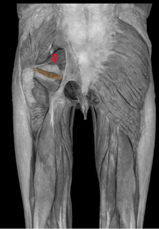
Gemellus Superior
Origin: ischial spine
Insertion: greater trochanter of the femur
Innervation: nerve to obturator internus
Function: laterally rotate extended femur, ABDuct flexed femur at hip joint
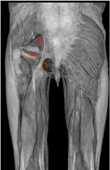
obturator internus
Origin: deep surface of obturator membrane, anterolateral wall of true pelvis, superior ramus of pubis and bone surrounding obturator foramen
Insertion: greater trochanter of the femur
Innervation: nerve to obturator internus
Function: laterally rotate extended femur, ABDuct flexed femur at hip joint
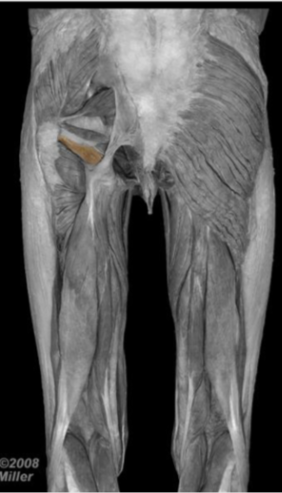
gemellus inferior
Origin: ischial tuberosity
Insertion: greater trochanter of the femur'
Innervation: nerve to quadratus femoris
Function: laterally rotate extended femur, ABDuct flexed femur at hip joint
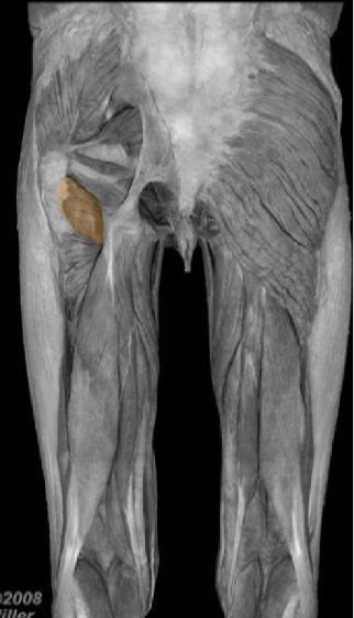
quadratus femoris
Origin: lateral aspect of ischium, anterior to the ischial tuberosity
Insertion: intertrochanteric crest of femur
Innervation: Nerve to quadratus femoris
Function: laterally rotate femur at the hip joint
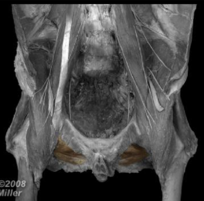
Obturator externus
Origin: external surface of obturator membrane and obturator foramen
Insertion: trochanteric fossa of femur
Innervation: Obturator N
Function: laterally rotate femur at the hip joint
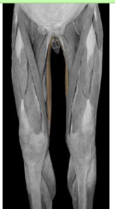
Gracilis
Origin: external surface of body of pubis, ischiopubic ramus
Insertion: medial surface of proximal shaft of tibia
Innervation: obturator N
Function: ADDuct thigh at hip joint, flexes leg at knee joint
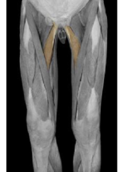
Adductor Longus
Origin: external surface of body of pubis
Insertion: linea aspera of femur
Innervation: Obturator N
Function: ADDuct and medially rotate thigh at hip joint
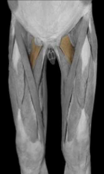
Pectineus
Origin: pectineal line of pubis
Insertion: posterior surface of femur, inferior to the lesser trochanter of the femur
Innervation: femoral N
Function: ADDucts and flex thigh at hip joint
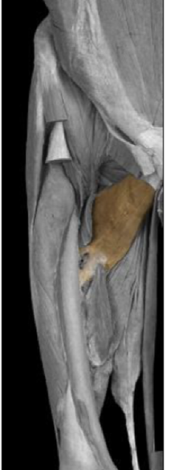
adductor brevis
Origin: external surface of pubis, inferior pubic ramus
Insertion: Linea aspera of femur, posterior surface of femur
Innervation: obturator N
Function: ADDuct and medially rotate thigh at hip joint
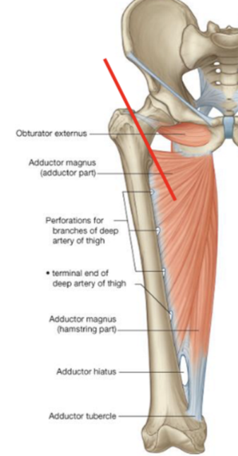
adductor magnus (adductor component)
Origin: ischiopubic ramus
Insertion: posterior surface of femur, linea aspera of femur
Innervation: obturator N
Function: ADDuct and medially rotate thigh at hip joint
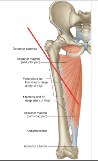
Adductor Magnus (hamstring component)
Origin: ischial tuberosity of ischium
Insertion: adductor tubercle of femur
Innervation: tibial portion of sciatic N
Function: ADDuct and medially rotate thigh at hip joint
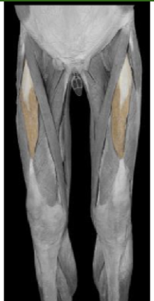
Rectus Femoris
Origin: anterior inferior iliac spine, ilium superior to acetabulum
Insertion: patella via quadriceps femoris tendon
Innervation: femoral N
Function: extends leg at knee joint, flex thigh at hip joint
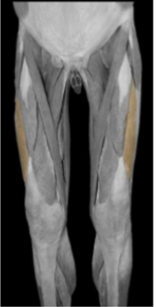
Vastus Lateralis
Origin: intertrochanteric line of femur, linea aspera of femur, greater trochanter of femur
Insertion: patella via quadriceps femoris tendon
Innervation: femoral N
Function: extends leg at the knee joint
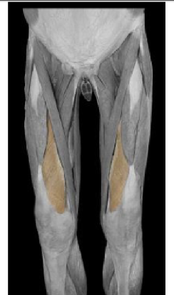
Vastus Medialis
Origin: intertrochanteric line of femur, linea aspera of femur, pectineal line of femur
Insertion: patella via quadriceps femoris tendon
Innervation: femoral N
Function: extends leg at knee joint
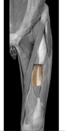
Vastus intermedius
Origin: upper 2/3 of femoral shaft
Insertion: patella via quadriceps femoris
Innervation: Femoral N
Function: extends leg at the knee joint
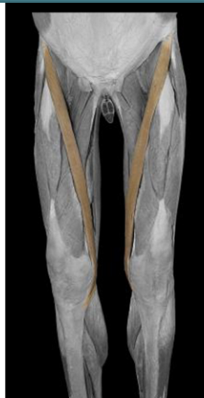
sartorius
Origin: anterior superior iliac spine
Insertion: medial surface of the tibia inferior and medial to the tibial tuberosity
Innervation: femoral N
Function: flexes thigh at the hip joint, flexes leg at the knee joint
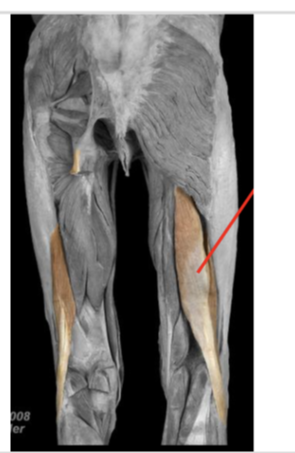
biceps femoris (long head)
Origin: ischial tuberosity
Insertion: head of fibula
Innervation: tibial portion of sciatic N
Function: Flex leg at knee joint, laterally rotate leg at knee joint, extend and laterally rotate thigh at hip joint
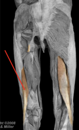
Biceps femoris (short head)
Origin: linea aspera of femur, lateral supracondylar ridge of femur
Insertion: head of fibula
Innervation: common fibular portion of sciatic N
Function: flex leg at the knee joint
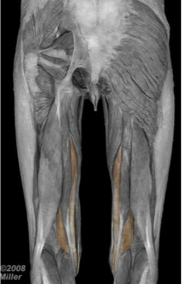
Semimembranosus
Origin: ischial tuberosity
Insertion: medial and posterior surface of medial tibial condyle
Innervation: tibial portion of sciatic N
Function: flex leg at the knee joint, extend thigh at hip joint, medially rotate thigh at the hip, medially rotate leg at the knee
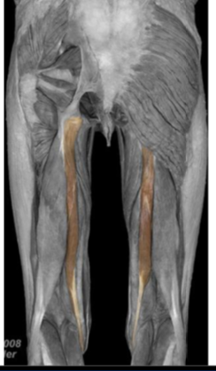
semitendinosus
Origin: ischial tuberosity
Insertion: medial surface of proximal tibia
Innervation: tibial portion of sciatic N
Function: flex leg at the knee joint, extend thigh at hip joint, medially rotate thigh at the hip, medially rotate leg at the knee
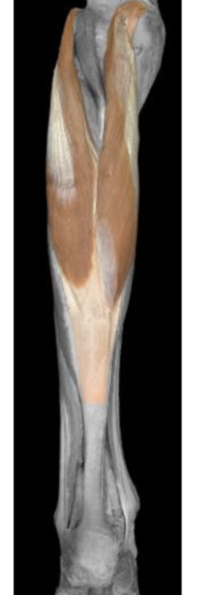
gastrocnemius medial head
Origin: posterior surface of femur superior to the medial condyle
Insertion: calcaneus via the Achilles tendon
Innervation: tibial N
Function: plantarflex foot, flex the knee
gastrocnemius lateral head
Origin: posterolateral surface of the lateral condyle of the femur
Insertion: calcaneus via the Achilles tendon
Innervation: tibial N
Function: plantarflex foot, flex the knee
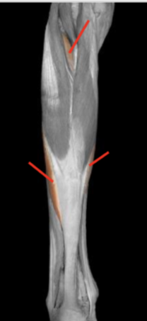
soleus
Origin: soleal line and medial border of tibia, head of fibula
Insertion: calcaneus via the Achilles tendon
Innervation: tibial N
Function: plantarflex foor
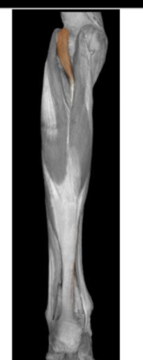
plantaris
Origin: supracondylar line of femur, popliteal ligament of knee
Insertion: calcaneus via Achilles tendon
Innervation: tibial N
Function: plantarflex foot, flex the knee

popliteus
Origin: lateral condyle of femur
Insertion: posterior surface of tibia
Innervation: tibial N
Function: stabilize the knee joint by resisting lateral rotation of tibia on femur, unlocks knee joint by laterally rotating femur on fixed tibia
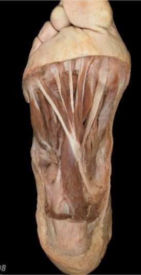
flexor digitorum longus
Origin: medial side of posterior surface of tibia
Insertion: plantar surfaces of distal phalanges 2-5
Innervation: tibial N
Function: flexes digits 2-5

tibialis posterior
Origin: interosseus membrane, tibia and fibula near the interosseus membrane
Insertion: plantar surface of navicular, plantar surface of medial cuneiform
Innervation: tibial N
Function: inverting foot, plantarflexion of foot, supports medial arch of foot during walking
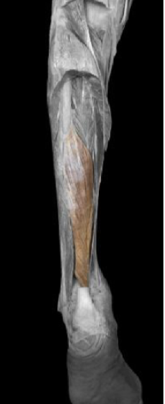
Flexor hallucis longus
Origin: posterior surface of fibula, interosseus membrane
Insertion: plantar surface of distal phalanx of hallux
Innervation: tibial N
Function: flexes hallux
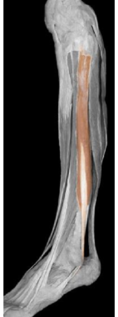
Fibularis (peroneus) longus
Origin: lateral surface of fibula, head of fibula
Insertion: plantar surface of medial cuneiform, plantar surface of base of metatarsal 1
Innervation: superficial fibular N
Function: everts foot, plantarflexes foot, supports arches of foor

Fibularis (peroneus) brevis
Origin: lower 2/3 of shaft of fibula
Insertion: base of metatarsal 5
Innervation: superficial fibular N
Function: everts foot
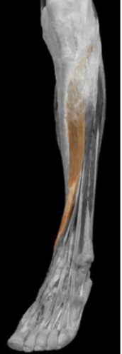
Tibialis anterior
Origin: lateral surface of the tibia, interosseus membrane
Insertion: dorsal surface medial cuneiform, dorsal surface base of metatarsal 1
Innervation: deep fibular N
Function: dorsiflex foot at ankle joint, inverts foot, supports medial arch of foot
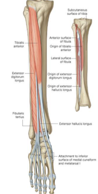
Extensor hallucis longus
Origin: medial surface of fibula, interosseus membrane
Insertion: Dorsal surface of distal phalanx of hallux
Innervation: deep fibular N
Function: extends hallux, dorsiflex foot at the ankle joint
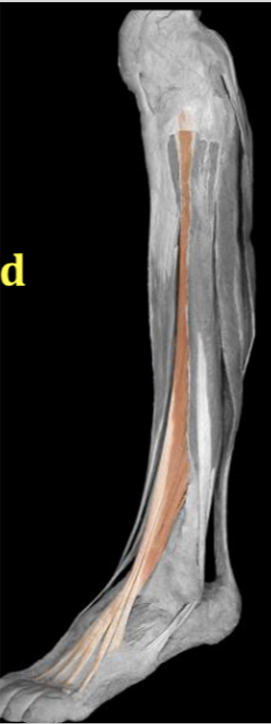
Extensor digitorum longus
Origin: proximal half of fibula, lateral condyle of tibia
Insertion: dorsal surfaces of bases of distal and middle phalanges of digits 2-5
Innervation: deep fibular N
Function: extends digits 2-5, dorsiflex at ankle joint
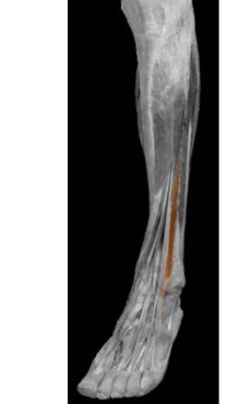
Fibularis tertius
Origin: medial surface of fibula
Insertion: dorsal surface of base of metatarsal 5
Innervation: deep fibular N
Function: dorsiflex foot at ankle joint, everts foot
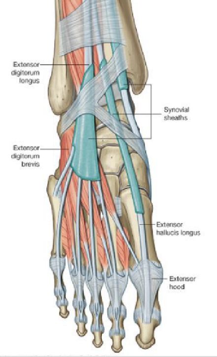
extensor digitorum brevis
Origin: calcaneus
Insertion: tendons of extensor digitorum longus
Innervation: deep fibular N
Function: extends toes 2-4
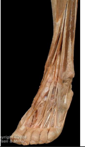
Extensor Hallucis brevis
Origin: calcaneus
Insertion: base of proximal phalanx of hallux
Innervation: deep fibular N
Function: extend hallux at metatarsophalangeal joint
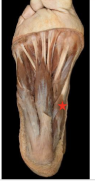
abductor hallucis
Origin: calcaneal tuberosity
Insertion: medial side of base of proximal phalanx of hallux
Innervation: medial plantar N
Function: ABDuct and flex hallux at metatarsophalangeal joint
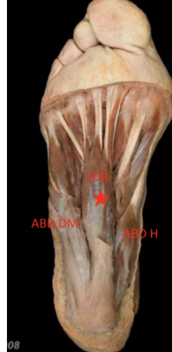
flexor digitorum brevis
Origin: calcaneal tuberosity, plantar aponeurosis
Insertion: plantar surfaces of middle phalanges of toes 2-5
Innervation: medial plantar N
Function: flexes toes 2-5 at proximal interphalangeal joint
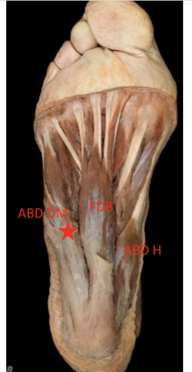
Abductor digiti minimi
Origin: calcaneal tuberosity, connective tissue between calcaneus and metatarsal 5
Insertion: lateral side of base of proximal phalanx of digit 5
Innervation: lateral plantar N
Function: ABDuct little toe at metatarsophalangeal joint
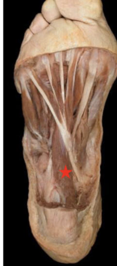
Quadratus plantae
Origin: medial surface of calcaneus, calcaneal tuberosity
Insertion: tendon of flexor digitorum longus in sole of foot
Innervation: lateral plantar N
Function: assists flexor digitorum longus tendon in flexing toes 2-5
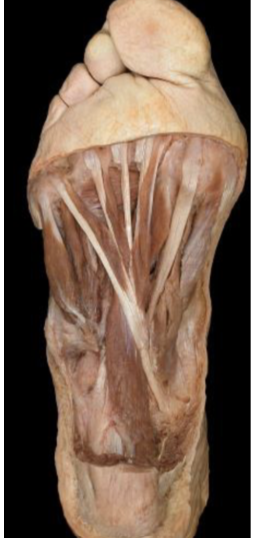
Lumbricals
Origin: 1st = medial side of tendon of flexor digitorum longus associated with toe 2
2nd to 4th = adjacent surfaces of adjacent tendons of flexor digitorum longus
Insertion: medial margins of extensor hoods of toes 2-5
Innervation: 1st = medial plantar N
2nd to 4th = lateral plantar N
Function: flexion of metatarsophalangeal joint and extension of interphalangeal joints
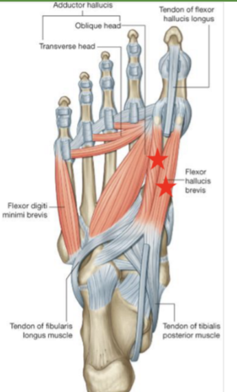
Flexor hallucis brevis
Origin: plantar surface of cuboid and lateral cuneiform, tendon of tibialis posterior
Insertion: lateral and medial sides of the base of proximal phalanx of hallux
Innervation: medial plantar N
Function: flexes hallux at metatarsophalangeal joint
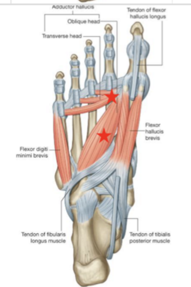
adductor hallucis
Origin: ligaments of metatarsophalangeal joints of toes, bases of metatarsals 2-4, sheath covering fibularis longus
Insertion: lateral side of base of proximal phalanx of hallux
Innervation: lateral plantar N
Function: ADDcuts hallux at metatarsophalangeal joint
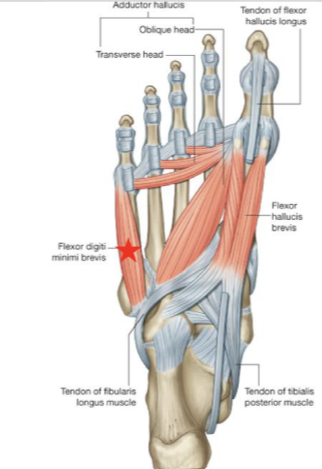
flexor digiti minimi brevis
Origin: base of metatarsal 5, tendon sheath of fibularis longus
Insertion: lateral side of base of proximal phalanx of little toe
Innervation: lateral plantar N
Function: flexes little toe at metatarsophalangeal joint
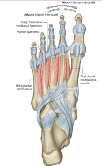
Plantar interossei
Origin: medial sides of metatarsals 3-5
Insertion: extensor hoods and bases of proximal phalanges of toes 3-5
Innervation: lateral plantar N
Function: ADDuction of toes 3-5 at MTP, resist extension of metatarsophalangeal joint and flexion of interphalangeal joints
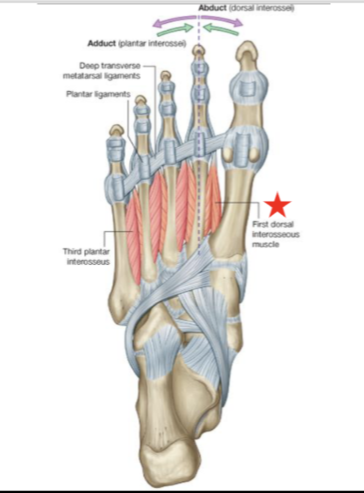
Dorsal interossei
Origin: sides of adjacent metatarsals
Insertion: extensor hoods of toes 2-4, bases of proximal phalanges of toes 2-4
Innervation: 1&2 = deep fibular N and lateral plantar N
3&4 = lateral plantar N
Function: ABDucts toes 2-4 at MTP, resists extension of metatarsophalangeal joint and flexion of interphalangeal joints