Structural Kinesiology Exam 1
1/108
There's no tags or description
Looks like no tags are added yet.
Name | Mastery | Learn | Test | Matching | Spaced | Call with Kai |
|---|
No study sessions yet.
109 Terms
Structural Kinesiology
Study of muscles, bones, and joints as they are involved in the science of movement (how these structural vary in size shape, and location determines their function)
600+ muscles
206 bones
Anatomical Position
Standing in an upright posture, facing straight ahead, feet parallel and close, palms facing forward (all questions regarding plane should be done in this position)
Fundamental Position - same but with arms at the side and palms facing body
Sagittal Plane
Flexion/ Extension
Frontal Axis
Ankle Plantarflexion and dorsiflexion
Flexion
Bending movement that results in a decrease of angle in joint
Extension
Straightening movement that results in an increase of angle in a joint
Frontal Plane
Abduction/Adduction
Sagittal Axis
Scapular movements - elevation, depression, protraction, retraction, downward rotation, trunk flexion, ankle inversion/eversion
Abduction
Lateral movement away from midline of trunk in frontal plane
Adducation
Movement medially toward midline of trunk in lateral plane
Transverse Plane
Internal/External Rotation
Longitudinal Axis
Wrist pronation/supination, horizontal shoulder abduction/adduction, transverse pelvic rotation
Internal Rotation
Rotary movement around longitudinal axis of bone toward midline of body (medial rotation)
External Rotation
Rotary movement around longitudinal axis of a bone away from midline of body (lateral rotation)
Diagonal/Oblique Plane
Most movements in daily life in a combo of planes
Combination of sagittal, frontal, transverse plane
Any combination of movements
Diagonal Abduction/Adduction
Circumduction
Diagonal Abduction
Movement by a limb through a diagonal plane away from the midline of the body
Diagonal Adduction
Movement by a limb through a diagonal plane toward and across the midline of body
Circumduction
Circular movement of a limb that delineates an arc or describes a cone
When shoulder joint and hip joint move in a circular fashion around a fixed point
Combination of flexion/extension, abduction/adduction
Axes of Rotation
90 deg. angle to the plane
Sagittal Plane - Frontal Axis (flexion/extension)
Frontal Plane - Sagittal Axis (Abduction/adduction)
Transverse Plane - Longitudinal Axis (internal/external rotation)
Diagonal Plane - Diagonal Axis
Anatomical Terminology
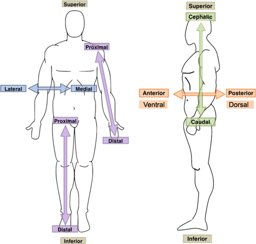
Anterior/Posterior
Superior/Inferior
Deep/Superficial
Proximal/Distal
Lateral/Medial
Dorsal/Ventral
Plantar/Palmar
Bilateral
Contralateral - same side of the body
Ipsilateral - opposite side of body
Skeletal System
206 bones
Axial Skeleton - 80 bones
Appendicular Skeleton - 126 bones
Axial Skeleton
Skull, vertebral column, ribs, sternum
Appendicular Skeleton
Extremities and the pelvic and shoulder girdles
5 Main function of Bone
Protection of vital soft tissues
Support to maintain posture
Movement; points of attachment for muscles and acting as levers
Mineral storage such as calcium and phosphorus
Hematopoiesis which occurs in vertebral bodies, femur, humerus, ribs, sternum
Process of blood cell formation in the red bone marrow
Osteoblasts
Primary function is new bone formation - build bone
Osteoclasts
Primary function is bone restoration - breakdown/absorb bone
important in the development, growth, maintenance, repair of bone
Bone Composition
60-70% of bone weight is made of mineral
Ca, P, Na - Give bone its strength
20-30% of bone weight is water
Collagen / ground substance - Give bone its flexibility
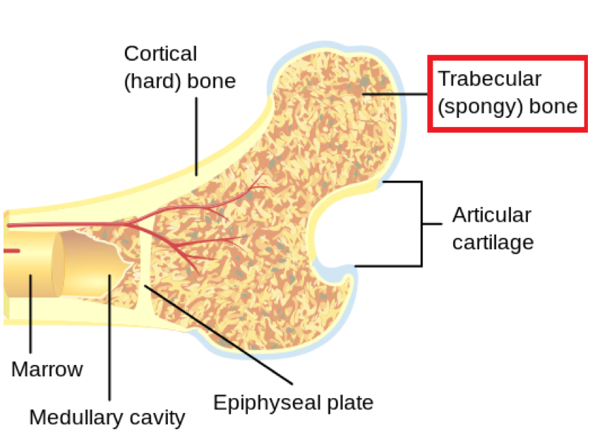
Cortical / Compact Bone
5-30% Porous
Stiff/Dense
Provides protection and support
Can withstand greater stress
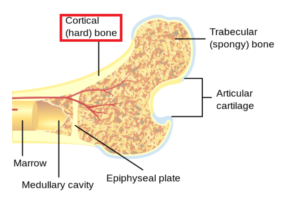
Cancellous / Trabecular Bone
30-90% Porous
Spongy/Permeable
Makes up most of bone tissue epiphysis of long bones, and the other majority of other bone types
More elastic/flexible than cortical bone
Bone Properties
Bone is living tissue and is active though life
Remodeling (ongoing replacement of old bone with new bone) occurs continuously
Bone size and shape are due to the stresses applied to them
Wolff’s Law
Bone is laid down where needed and resorbed where not needed
Bone becomes stronger/thicker with stress
When loading is decreased, bone weakens
Add bone to where there is stress and leaves w/ absent stress
Osgood Slaughter - Extra bone under patella
Bone Types
Size, Shape, Location of bone - Determine its function
Long Bones
Short Bones
Flat Bones
Sesamoid Bones
Irregular Bones
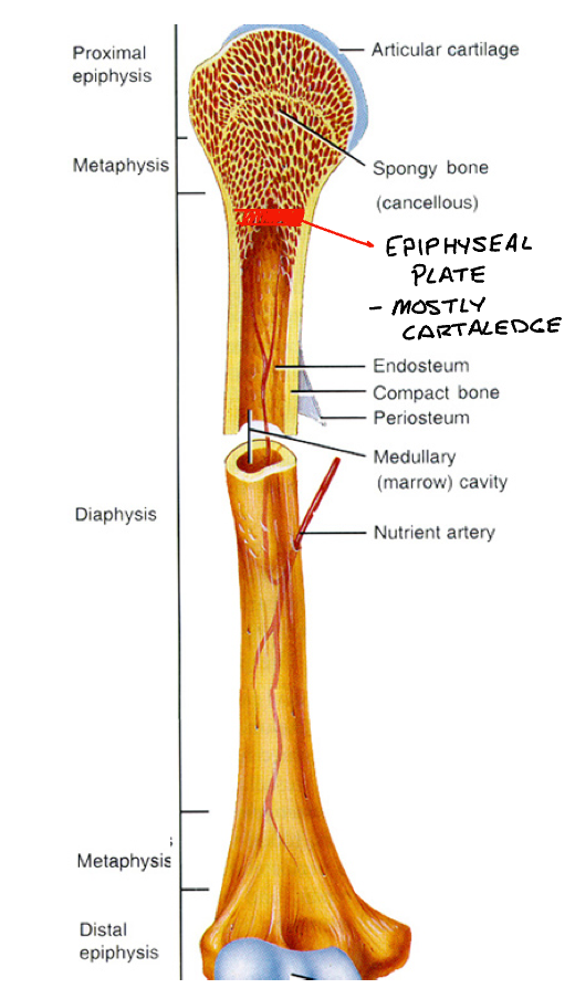
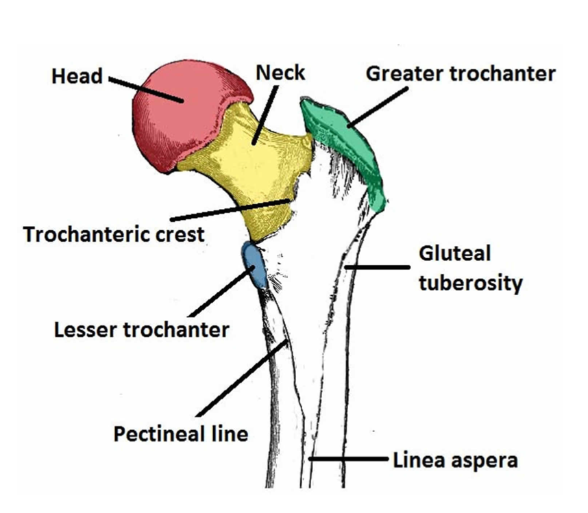
Long Bones
Long cylindrical shaft with relatively wide, protruding ends
Shaft contains the medullary canal
Ex: Phalanges, metatarsals, metacarpals, tibia, fibula, fumur, radius, ulna, humerus
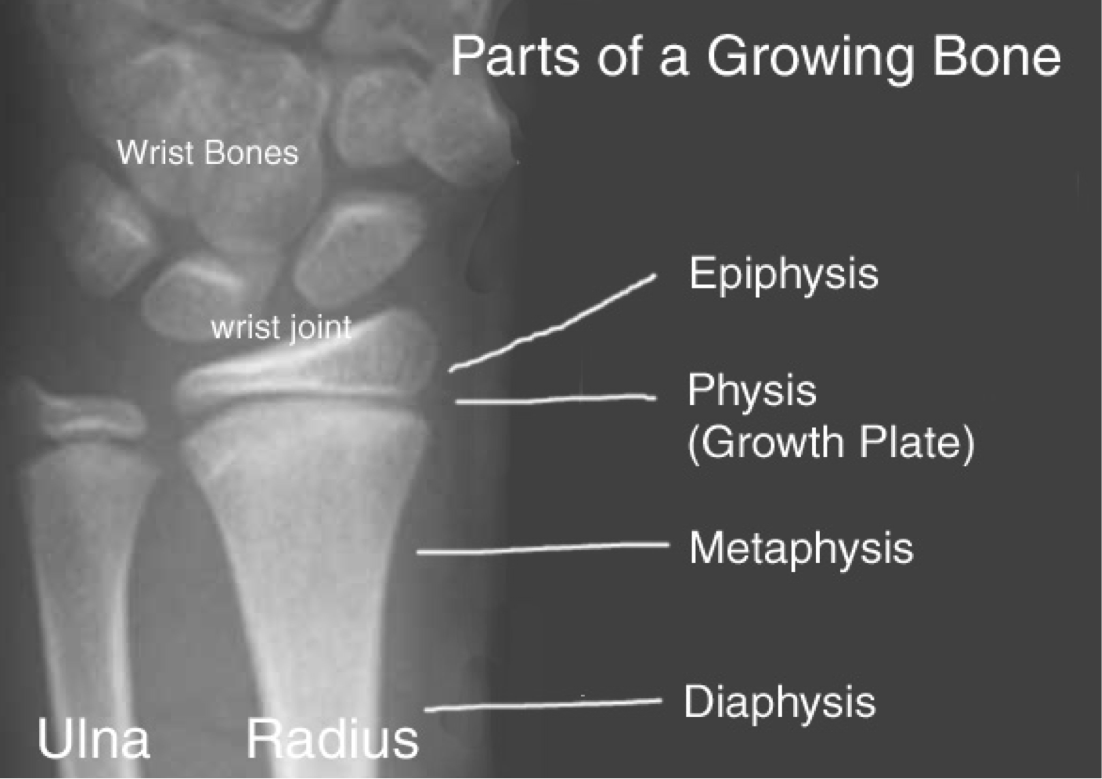
Diaphysis
Long cylindrical shaft - main portion

Epiphysis
Ends of long bone
Made of cancellous bone
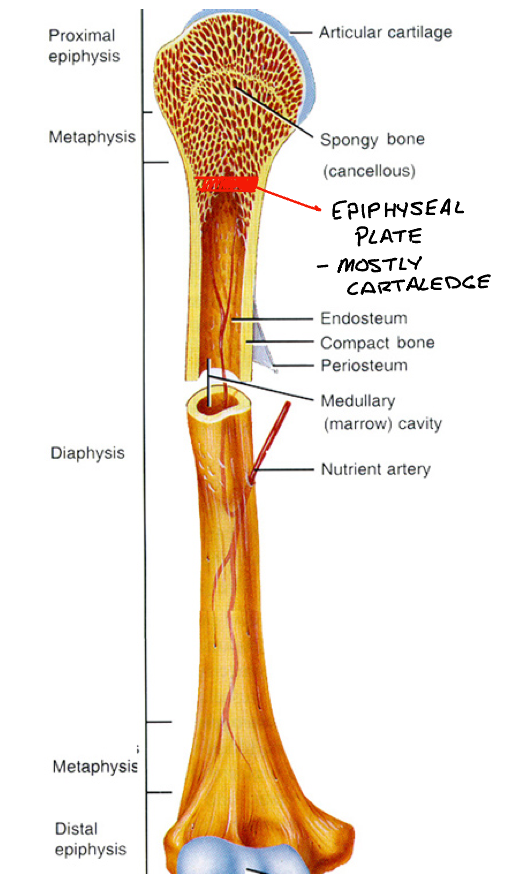
Epiphyseal Plate
Referred to as growth plate
Thin plane of cartilage
Plates close with maturity, time varies
Long bone Xray
Short Bones
Small, cubed-shaped, solid bones
Proportionally large articular surface to articular with more than one bone
EX: carpals, tarsals, talus (base of hands/foot)
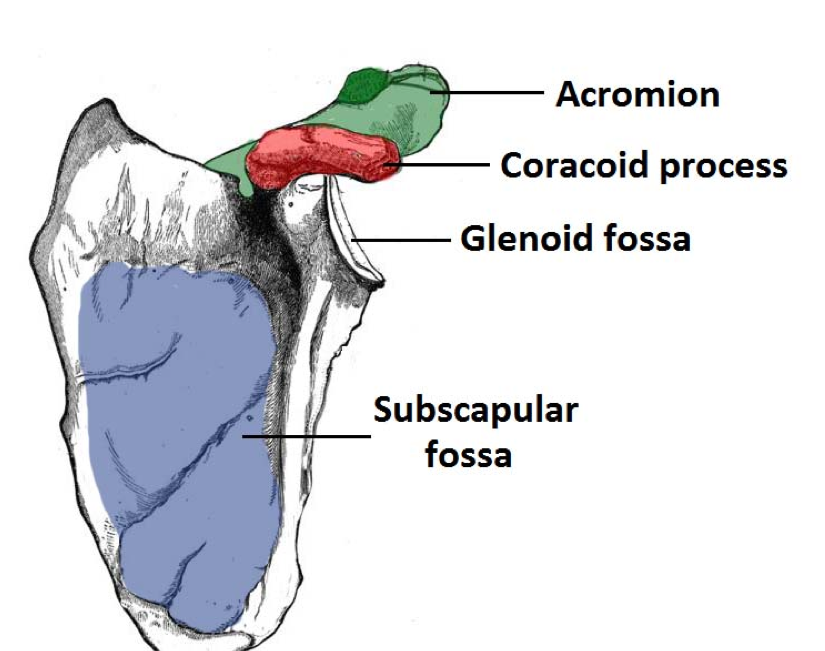
Flat Bones
Usually have a curved surface, vary from thick (where tendons attach) to thin
EX: ilium, ribs, sternum, clavicle, scapula
Sesamoid Bones
Embedded within tendons, Improve mechanical advantage of joint
Improve mechanical advantage of movement
Within patellar tendon and flexor tendon in 1st metatarsophalangeal joint (knee cap and big toe)
Irregular Bones
Variety of shapes and sizes for variety of purposes
Include bones throughout the entire spine, ischium, pubis, maxilla
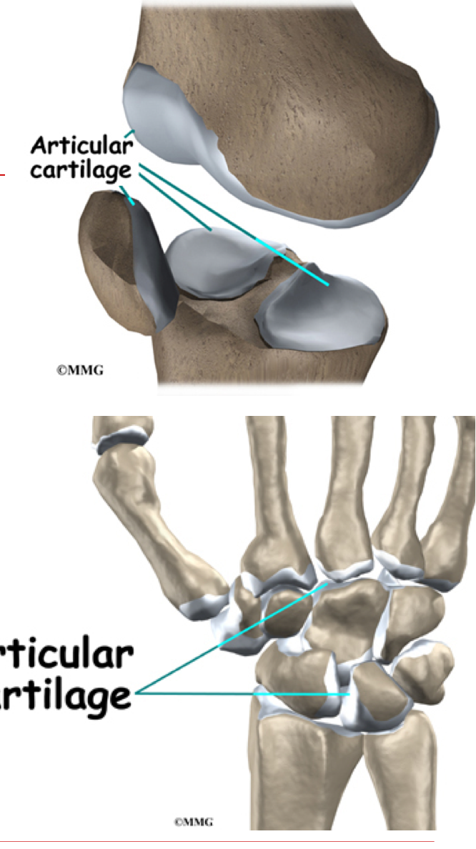
Articular Cartilage
Thin layer of hyaline cartilage covering articular surface of bone
Cushions - absorbs compressive forces
Reduces friction on underlying bone
Periosteum
Membrane around surface of bone not covered by articular cartilage
Important for bone growth, repair, nutrition
Point of attachment for ligament and tendons
Bony Landmarks can Indicate. . .
Muscular attachment
Joint Function
Space for soft tissue
Processes
Elevations and Projections
Process that form joints:
Condyle, Facet, Head
Processes to which ligaments, muscles or tendons attach:
Crest, Epicondyle, Line, Process, Spine (spinous process), Suture, Trochanter, Tubercle, Tuberosity
Cavities
Depressions - Opening and Groves
Facet, Foramen, Fossa, Fovea, Metus, Sinus, Sulcus
Articulation
Connection of bones at a joint usually to allow movement between surfaces of bone
Freedom or range of motion is limited by ligaments and muscles
Articulations may be classified according to the structure and function of joints
Classification of Joints
Classification Categories by Structure
Fibrous —> Cartilaginous —> Synovial (Most Movement)
Classification Categories by Function
Synarthrodial —> Amphiarthrodial —> Diarthrodial (Most Movement)
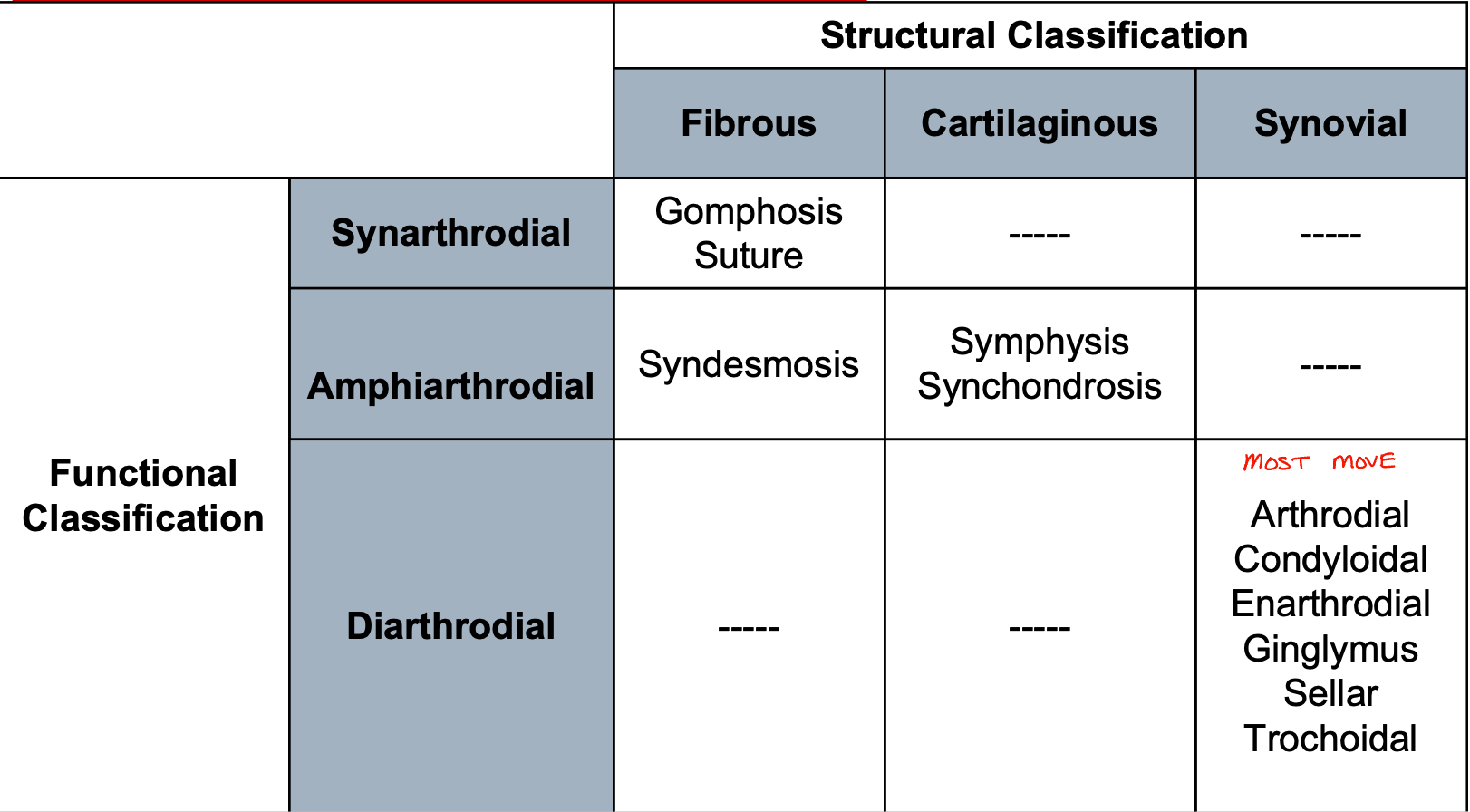
Synarthrodial Joints
Immovable Joints
2 Groups
Suture - Cranial bones
Gomphosis - Sockets of teeth
Amphiarthrodial Joints
Slightly Moveable Joints
Syndesmosis
Held by strong ligaments
EX: inferior tib/fib
Symphysis
Separated by fibrocartilage
EX: Symphysis Pubis
Synchondrosis (Most Movement)
Separated by hyaline cartilage '
EX: Costochondral Joints
Diarthrodial Joints
Freely Movable
Synovial Joints
Composed of a sleeve-like joint capsule
Secretes synovial fluid to lubricate the joint cavity (area w/ in cavity)
Ligaments (result from thicker areas of joint capsule)
Bone to bone
Provide static stability to joints
Intraarticular Ligaments - Within the joint capsule (ACL)
Extraarticular Ligaments - Outside of the joint capsule (LCL)
Fibrocartilage Discs - Some have, used for additional cushioning and stability
EX: glenohumeral Labrum, Meniscus of knee
6 Types of Diarthrodial Joints
Arthrodial - Gliding joint
Condyloid - Biaxial ball and socket, ovoid, ellipsoidal
Enarthrodial - Multiaxial ball and socket
Ginglymus - Hinge joint
Sellar - Saddle joint
Trochoid - Pivot, screw joint
Arthrodial (gliding) Joint
2 plane or flat bony surfaces which butt against each other
little motion possible in 1 joint articulation
Permits limited gliding
EX: Vertebral Column, Carpal Bones on Hand
Motions - Flexion/Extension, Abduction/Adduction, Internal/External Rotation, Circumduction
Condyloid (Ellipsoidal) Joint
Biaxial ball and socket joint
2 axes and 2 planes
one bone with oval concave surface received by another bone with an oval convex surface
EX: 2-5 metacarpophalangeal or knuckle joints, Base of Hand into wrist
Motions - Flexion/Extension, Abduction/Adduction, Circumduction
Enarthrodial (ball and socket) Joint
Multiaxial / triaxial ball and socket joint
Bony rounded had fitting into a concave articular surface
Ex: Hip and GH shoulder Joint
Motion - All movements
Ginglymus (hinge) Joint
Uniaxial Articulation
Articular surfaces allow motion in only one plane
EX: Elbow, Knee, Ankle
Motions - Flexion / Extension
Trochoid (Pivot) Joint
Uniaxial Articulation
Articualr surafces allow motion in only one plane
EX: Atlantoaxial joint - odontoid, which turns in a bony ring, proximal and distal radioulnar joints (Atlas/Axis, radioulnar joint)
Motions - Internal and External Rotation
Sellar (saddle) Joint
Unique multiaxial / Triaxial joint
2 reciprocally concave and convex articular surfaces
EX: 1st carpometacarpal joint at the thumb, sternoclavicular joint
Motions - Flexion/Extension, Abduction/Adduction, Internal/External Rotation, Circumduction
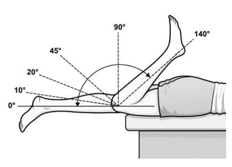
Range of Motion
Area through which a joint may normally be freely and painlessly moved
Normal ROM varies
Measured with a goniometer
Skeletal Muscles
600+ skeletal muscles compose 40-50% of body weight
215 pairs of skeletal muscles usually work together to produce actions at the joints that they cross
Purpose of Skeletal Muscles
Responsible for movement of body at all joints - muscle contraction produces forces that cause joint movement
Provide:
Protection
Posture and support
produce a major portion of total body heat (shivering thermogenesis)
Motor Neurons
Transmit impulses from CNS to muscles
Sensory Neurons
Transmit impulses from body to CNS
Motor Unit
Motor Neuron + Muscle it innervates
Innervation
Refers to the nerves responsible for stimulating muscle fibers
Muscle may be innervated by one or more nerves and one nerve mau innervate several muscle groups
Proprioception
Subconscious mechanism by which the body regulates posture and movement
Neutral feedback from joints, skin, muscles
Allows quick reaction time and prevents injury
Kinesthesis
Conscious awareness of the position and movement of the body in space
Walking downstairs without looking at each step
Closing eyes and touching nose
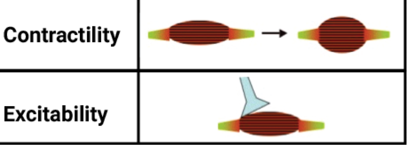
4 Properties of Skeletal muscle tissue to produce force and movement of joints
Irritability or Excitability
Contractility
Extensibility
Elasticity
Irritability or Excitability
Property of muscle being sensitive or responsive to chemical, electrical, or mechanical stimuli

Contractility
Ability of muscle to contract and develop tension or internal force against resistance when stimulated
Extensibility
Ability of muscle to be stretched beyond its normal resting length without tissue damage
Elasticity
Ability of muscle to return to its original length following stretching or contraction
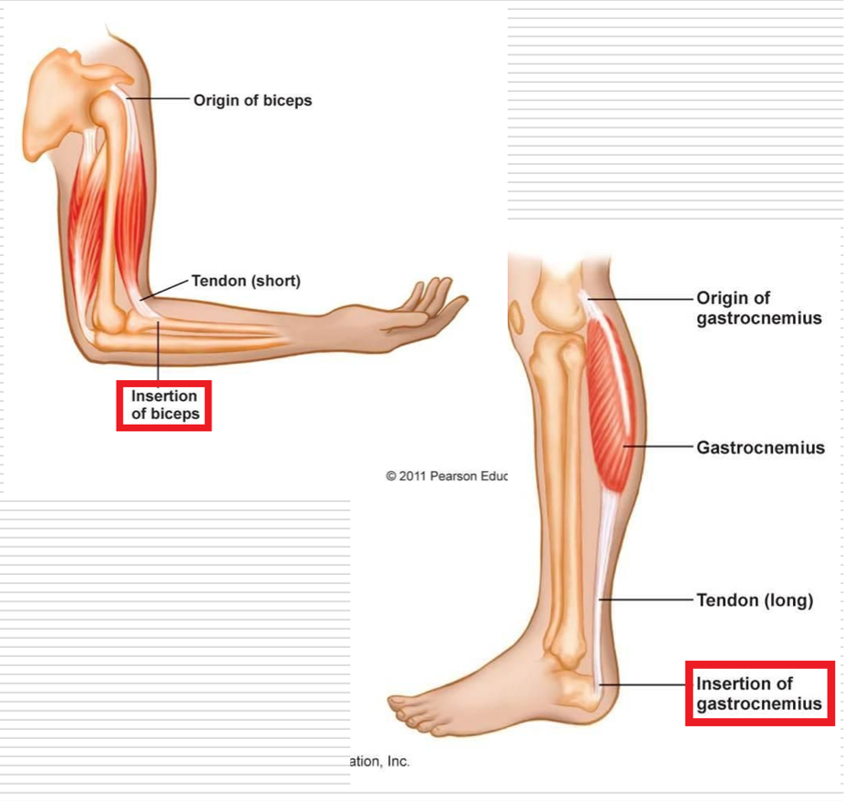
Tendon
Fibrous connective tissue that connects muscles to bones and other structures
2 muscles may share a common tendon (achilles tendon)
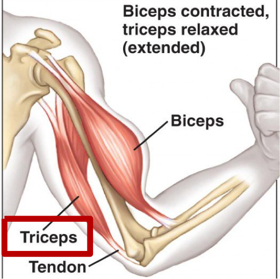
A muscle may have multiple tendons connecting it to one or more bones (triceps brachii)
Origin
Structurally - Proximal attachment of a muscle or the part that attaches closest to the midline or center of the body
Functionally - Least moveable part or attachment of the muscle
Insertion pulled toward origin (typically)
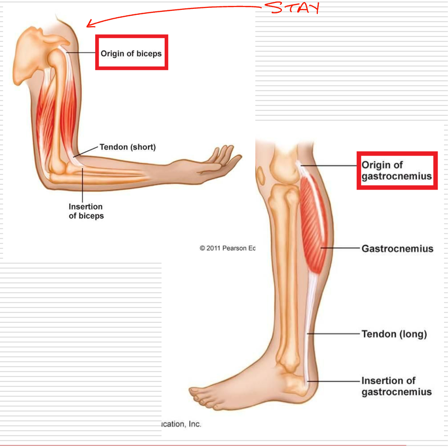
Insertion
Structurally - Distal attachment or the part that attaches farthest from the midline or center of body
Functionally - Most moveable part is generally considered the insertion
Insertion → Origin
Isotonic Contractions
Involve muscle developing tension to either cause or control joint movement
Dynamic contractions
Contractions are either concentric or eccentric on basis of shortening/lengthening under tension
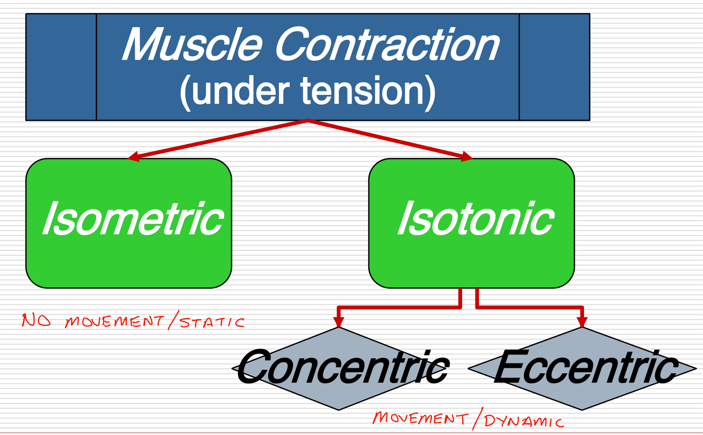
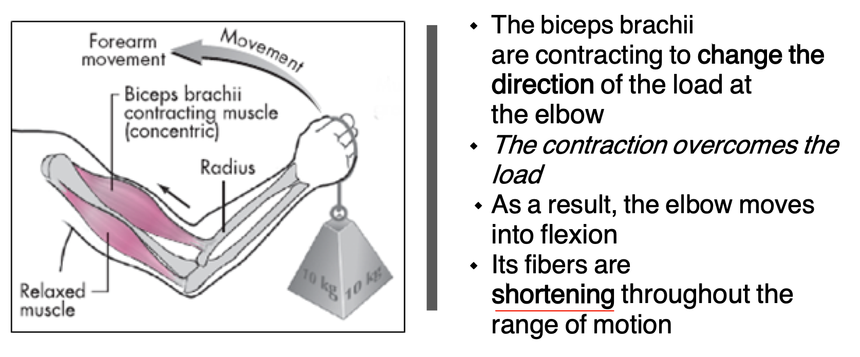
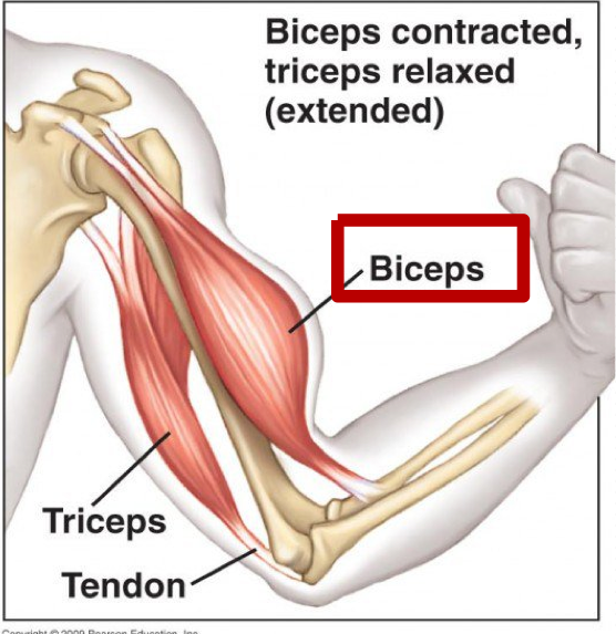
Concentric Contractions
Muscle fibers shorten throughout the range of motion to generate force to overcome the load and move the object
Curl on the way up
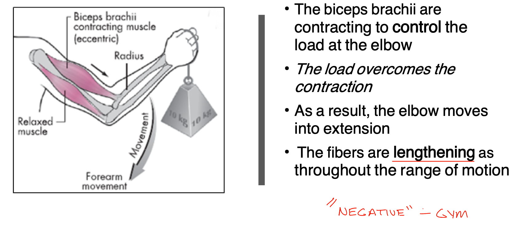
Eccentric Contraction
Muscle fibers lengthen throughout the range of motion to control the load of the object, load overcomes contraction
Curl on the way down
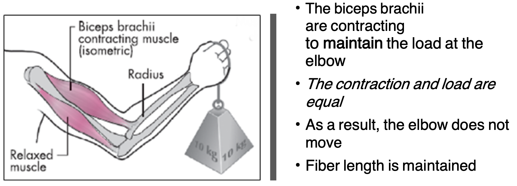
Isometric Contraction
Muscle fibers contract without movement, muscle is not shortened or lengthened, contraction and load are equal - fiber length is maintained
Plank
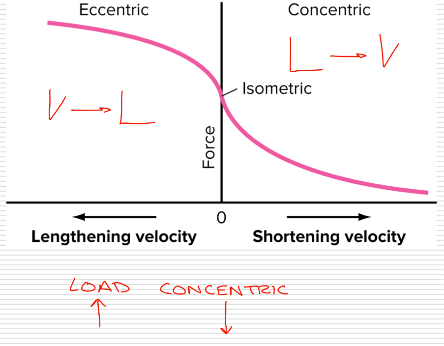
Force Velocity Relationship
Velocity of a muscle contracting concentrically is inversely related to the amount of the force of applied
Contraction occurs faster as load decreases
Contraction occurs slower as load increases
In relation to concentric types
Concentric: Load < Contraction
Eccentric: Load > Contraction
Isometric: Load = Contraction
Passive Movement
Movement at a given joint without any muscle contraction
Due to external forces such as those applied by another person, resistance of force, or gravity in the presence of muscle relaxation
Stretching
Action
Specific movement of joint resulting form concentric contraction of a muscle which crosses joint
Biceps brachii has the action of flexion at elbow
Multiple muscles have common action
Actions are inherent to the muscle - based on location, size, and shape
Agonist Muscle
Muscles causing a certain joint motion through a specified plane of motion - know as prime movers
Dependent on the type of contraction and circumstances of the activity
Antagonist Muscles
When contracting concentrically perform the opposite joint motion of agonist
Usually located on the opposite side of the joint from the agonist
Work in cooperation with agonists by relaxing and allowing motion
Must relax in order for prime mover to move
Synergist
Assist in action of agonist
Not necessarily prime movers
Know as guiding muscles
Assist in refined movement and rule out undesired motions
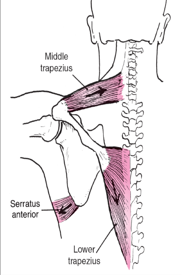
Force Couples
Occur when two or more forces are pulling in different directions on an object, causing the object to rotate about its axis
Coupling of muscular forces together in the body can result in a more efficient movement
Force-Tension Relationship
The force/tension that a muscle exerts varies with the length it is at when stimulated
The greatest force / tension is developed when there is optimal overlap between the actin and myosin fibers
Maximal tension is exerted when the muscle is at its resting length
100 - 130% of a muscles resting length is optimal
>130% decreases the amount of force muscle can exert
50 - 60 % of resting length = cannot contract effectively (too much overlap of myosin and actin)
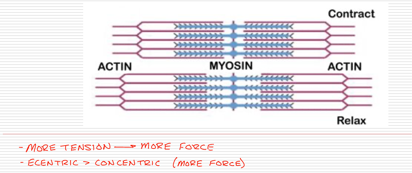
What is force-tension relationship?
Muscles can produce movement most efficiently when the muscle fibers are at 100-130% of their resting length (optimal overlap)
Force-Tension Relationship Example
EX: 1 - Increasing ability to exert force
Squat slightly to stretch the calf, hamstrings, and quadriceps before contracting same muscles concentrically to jump (slightly contracted - more force)
EX: 2 - Reducing ability to jump
Isolate the gluteus maximus by maximally shortening the hamstrings with knee flexion (all the way down - less force)
Muscles are usually named due to . . .
Visual Appearance, Anatomical Location, Function
Shape - deltoid, rhomboid
Size - gluteus maximus, teres minor
Number of Divisions - triceps brachii
Direction of its Fibers - external oblique
Location - rectus femoris, palmaris longus
Point of Attachment - coracobrachialis, extensor hallucis longus
Action - erector spinae, supinator, extensor digiti minimi
Action & Size - adductor magnus
Shape & Location - serratus anterior
Location & Attachment - brachioradialis
Location & Number of Divisions - biceps brachii
Parallel Muscles
Fibers arranged parallel to length of muscle
Produce a greater range of movement than smaller-sized muscles with pennate arrangement
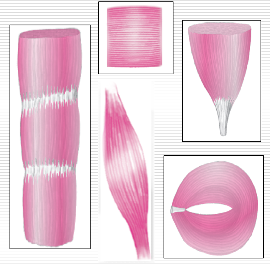
Parallel Muscles Categorized as
Flat - abdominal
Fusiform - biceps brachii
Strap - sartorius
Radiate - pec. major
Pennate Muscles
Arranged obliquely to their tendons in a manner similar to a feather
Have shorter fibers
Arrangement increases the cross-sectional area of the muscle - increasing the power
More strength - not as broad as range of motion
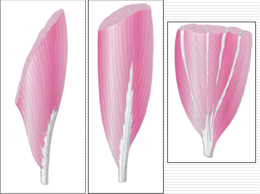
Pennate Muscles Categorized as
Unipennate - gastrocnemius
Bipennate - rectus femoris
Multipennate - deltoid
Fascia
Sheet or band of fibrous connective tissue that envelopes, separates, or binds together parts of the body such as muscles, organs, and other parts of the body
In certain places around the body such as around joints like the wrist and ankle, fascial tissue forms a retinaculum (band-like) to retain/hold tendons close to the body
EX: Foscia
Intrinsic
Pertaining usually to muscles within or belonging solely to the body part upon which they act
EX: small intrinsic muscles found entirely within the hand or feet
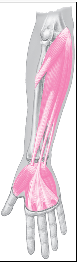
Extrinsic
Pertaining usually to muscles that arise or originate outside of (proximal to) body part upon which they act
EX: forearm muscles that attach proximally on distal humerus and insert on fingers
Uniarticular Muscles
Cross and act on one joint only
EX: brachialis
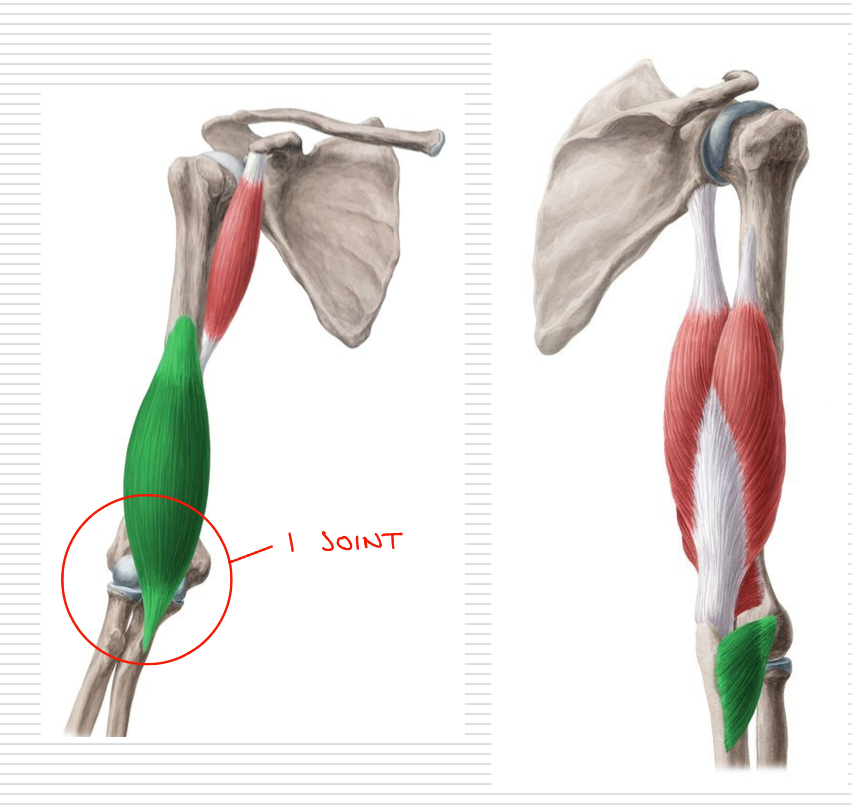
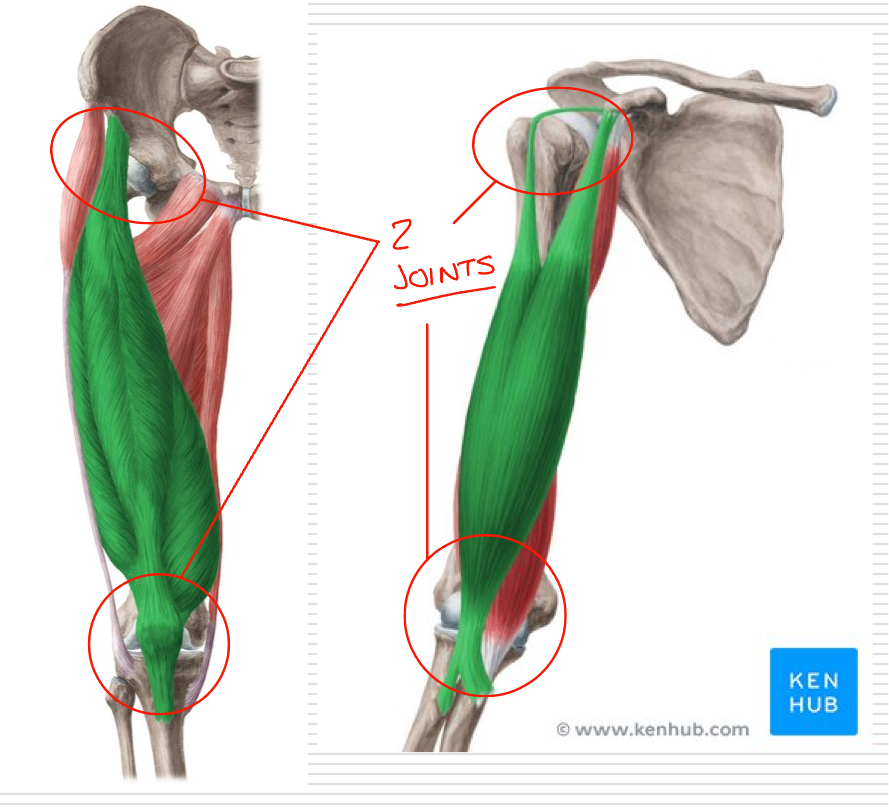
Biarticular Muscles
Cross and act on 2 different joints
EX: rectus femoris can act either one or both of its joints - maintain a relatively consistent length due to shortening at one joint and lengthening at another joint